Show PDB file:   
         Plain Text   HTML   (compressed file size)
QuickSearch:   
by PDB,NDB,UniProt,PROSITE Code or Search Term(s)  
(-)Asym./Biol. Unit
(-)Asym./Biol. Unit - sites
collapse expand < >
Image Asym./Biol. Unit
Asym./Biol. Unit  (Jmol Viewer)
Image Asym./Biol. Unit - sites
Asym./Biol. Unit - sites  (Jmol Viewer)

(-) Description

Title :  CRYSTAL STRUCTURE OF BACTEROIDES FRAGILIS TRXP
 
Authors :  S. R. Shouldice
Date :  22 Jun 09  (Deposition) - 12 Jan 10  (Release) - 12 Jan 10  (Revision)
Method :  X-RAY DIFFRACTION
Resolution :  2.00
Chains :  Asym./Biol. Unit :  A,B
Keywords :  Thioredoxin, Electron Transport (Keyword Search: [Gene Ontology, PubMed, Web (Google))
 
Reference :  S. R. Shouldice, S. H. Cho, D. Boyd, B. Heras, M. Eser, J. Beckwith, P. Riggs, J. L. Martin, M. Berkmen
In Vivo Oxidative Protein Folding Can Be Facilitated By Oxidation-Reduction Cycling
Mol. Microbiol. V. 75 13 2010
PubMed-ID: 19968787  |  Reference-DOI: 10.1111/J.1365-2958.2009.06952.X
(for further references see the PDB file header)

(-) Compounds

Molecule 1 - THIOREDOXIN
    ChainsA, B
    EngineeredYES
    Expression SystemESCHERICHIA COLI
    Expression System PlasmidPMALP2X
    Expression System StrainNEB EXPRESS
    Expression System Taxid562
    Expression System Vector TypePLASMID
    FragmentRESIDUES IN UNP 43-161
    GeneTRXP
    Organism ScientificBACTEROIDES FRAGILIS
    Organism Taxid817
    Strain638R
    SynonymTRXP

 Structural Features

(-) Chains, Units

  12
Asymmetric/Biological Unit AB

Summary Information (see also Sequences/Alignments below)

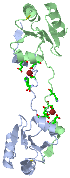
(-) Ligands, Modified Residues, Ions  (1, 2)

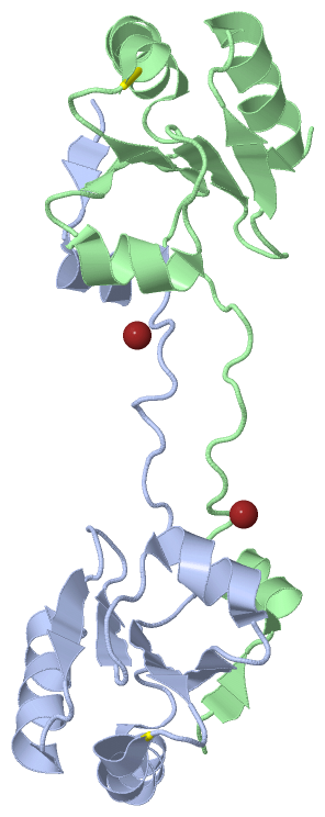
Asymmetric/Biological Unit (1, 2)
No.NameCountTypeFull Name
1ZN2Ligand/IonZINC ION

(-) Sites  (2, 2)

Asymmetric Unit (2, 2)
No.NameEvidenceResiduesDescription
1AC1SOFTWAREGLU A:40 , HIS B:42 , GLU B:96 , ASP B:100 , HOH B:211BINDING SITE FOR RESIDUE ZN A 142
2AC2SOFTWAREHIS A:42 , GLU A:96 , ASP A:100 , TYR B:39 , GLU B:40BINDING SITE FOR RESIDUE ZN A 143

(-) SS Bonds  (2, 2)

Asymmetric/Biological Unit
No.Residues
1A:63 -A:66
2B:63 -B:66

(-) Cis Peptide Bonds  (2, 2)

Asymmetric/Biological Unit
No.Residues
1Ile A:106 -Pro A:107
2Ile B:106 -Pro B:107

 Sequence-Structure Mapping

(-) SAPs(SNPs)/Variants  (0, 0)

(no "SAP(SNP)/Variant" information available for 3HXS)

(-) PROSITE Motifs  (0, 0)

(no "PROSITE Motif" information available for 3HXS)

(-) Exons   (0, 0)

(no "Exon" information available for 3HXS)

(-) Sequences/Alignments

Asymmetric/Biological Unit
   Reformat: Number of residues per line =  ('0' or empty: single-line sequence representation)
  Number of residues per labelling interval =   
  UniProt sequence: complete  aligned part    
   Show mapping: SCOP domains CATH domains Pfam domains Secondary structure (by author)
SAPs(SNPs) PROSITE motifs Exons
(details for a mapped element are shown in a popup box when the mouse pointer rests over it)
Chain A from PDB  Type:PROTEIN  Length:117
 aligned with Q64SV7_BACFR | Q64SV7 from UniProtKB/TrEMBL  Length:161

    Alignment length:117
                                    52        62        72        82        92       102       112       122       132       142       152       
         Q64SV7_BACFR    43 GTIHLTRAEFLKKIADYENHSKEWKYLGDKPAIVDFYADWCGPCKMVAPILEELSKEYAGKIYIYKVNVDKEPELARDFGIQSIPTIWFVPMKGEPQVNMGALSKEQLKGYIDKVLL 159
               SCOP domains d3hxsa_ A: automated matches                                                                                          SCOP domains
               CATH domains --------------------------------------------------------------------------------------------------------------------- CATH domains
               Pfam domains --------------------------------------------------------------------------------------------------------------------- Pfam domains
         Sec.struct. author ..ee.hhhhhhhhh.................eeeeee...hhhhhhhhhhhhhhhhhh....eeeeee...hhhhhhhh......eeeee......eeee...hhhhhhhhhhhh.. Sec.struct. author
                 SAPs(SNPs) --------------------------------------------------------------------------------------------------------------------- SAPs(SNPs)
                    PROSITE --------------------------------------------------------------------------------------------------------------------- PROSITE
                 Transcript --------------------------------------------------------------------------------------------------------------------- Transcript
                 3hxs A  23 GTIHLTRAEFLKKIADYENHSKEWKYLGDKPAIVDFYADWCGPCKMVAPILEELSKEYAGKIYIYKVNVDKEPELARDFGIQSIPTIWFVPMKGEPQVNMGALSKEQLKGYIDKVLL 139
                                    32        42        52        62        72        82        92       102       112       122       132       

Chain B from PDB  Type:PROTEIN  Length:119
 aligned with Q64SV7_BACFR | Q64SV7 from UniProtKB/TrEMBL  Length:161

    Alignment length:119
                                    52        62        72        82        92       102       112       122       132       142       152         
         Q64SV7_BACFR    43 GTIHLTRAEFLKKIADYENHSKEWKYLGDKPAIVDFYADWCGPCKMVAPILEELSKEYAGKIYIYKVNVDKEPELARDFGIQSIPTIWFVPMKGEPQVNMGALSKEQLKGYIDKVLLKQ 161
               SCOP domains d3hxsb_ B: automated matches                                                                                            SCOP domains
               CATH domains ----------------------------------------------------------------------------------------------------------------------- CATH domains
               Pfam domains ----------------------------------------------------------------------------------------------------------------------- Pfam domains
         Sec.struct. author ..ee.hhhhhhhhh.................eeeeee...hhhhhhhhhhhhhhhhhh....eeeeee...hhhhhhhh......eeeee......eeee...hhhhhhhhhhhh.... Sec.struct. author
                 SAPs(SNPs) ----------------------------------------------------------------------------------------------------------------------- SAPs(SNPs)
                    PROSITE ----------------------------------------------------------------------------------------------------------------------- PROSITE
                 Transcript ----------------------------------------------------------------------------------------------------------------------- Transcript
                 3hxs B  23 GTIHLTRAEFLKKIADYENHSKEWKYLGDKPAIVDFYADWCGPCKMVAPILEELSKEYAGKIYIYKVNVDKEPELARDFGIQSIPTIWFVPMKGEPQVNMGALSKEQLKGYIDKVLLKQ 141
                                    32        42        52        62        72        82        92       102       112       122       132         

   Legend:   → Mismatch (orange background)
  - → Gap (green background, '-', border residues have a numbering label)
    → Modified Residue (blue background, lower-case, 'x' indicates undefined single-letter code, labelled with number + name)
  x → Chemical Group (purple background, 'x', labelled with number + name, e.g. ACE or NH2)
  extra numbering lines below/above indicate numbering irregularities and modified residue names etc., number ends below/above '|'

 Classification and Annotation

(-) SCOP Domains  (1, 2)

Asymmetric/Biological Unit

(-) CATH Domains  (0, 0)

(no "CATH Domain" information available for 3HXS)

(-) Pfam Domains  (0, 0)

(no "Pfam Domain" information available for 3HXS)

(-) Gene Ontology  (6, 6)

Asymmetric/Biological Unit(hide GO term definitions)
Chain A,B   (Q64SV7_BACFR | Q64SV7)
molecular function
    GO:0046872    metal ion binding    Interacting selectively and non-covalently with any metal ion.
    GO:0015035    protein disulfide oxidoreductase activity    Catalysis of the reaction: a protein with reduced sulfide groups = a protein with oxidized disulfide bonds.
biological process
    GO:0045454    cell redox homeostasis    Any process that maintains the redox environment of a cell or compartment within a cell.
    GO:0006662    glycerol ether metabolic process    The chemical reactions and pathways involving glycerol ethers, any anhydride formed between two organic hydroxy compounds, one of which is glycerol.
    GO:0055114    oxidation-reduction process    A metabolic process that results in the removal or addition of one or more electrons to or from a substance, with or without the concomitant removal or addition of a proton or protons.
cellular component
    GO:0005623    cell    The basic structural and functional unit of all organisms. Includes the plasma membrane and any external encapsulating structures such as the cell wall and cell envelope.

 Visualization

(-) Interactive Views

Asymmetric/Biological Unit
  Complete Structure
    Jena3D(integrated viewing of ligand, site, SAP, PROSITE, SCOP information)
    WebMol | AstexViewer[tm]@PDBe
(Java Applets, require no local installation except for Java; loading may be slow)
    STRAP
(Java WebStart application, automatic local installation, requires Java; full application with system access!)
    RasMol
(require local installation)
    Molscript (VRML)
(requires installation of a VRML viewer; select preferred view via VRML and generate a mono or stereo PDF format file)
 
  Ligands, Modified Residues, Ions
    ZN  [ RasMol | Jena3D ]  +environment [ RasMol | Jena3D ]
 
  Sites
    AC1  [ RasMol ]  +environment [ RasMol ]
    AC2  [ RasMol ]  +environment [ RasMol ]
 
  Cis Peptide Bonds
    Ile A:106 - Pro A:107   [ RasMol ]  
    Ile B:106 - Pro B:107   [ RasMol ]  
 

(-) Still Images

Jmol
  protein: cartoon or spacefill or dots and stick; nucleic acid: cartoon and stick; ligands: spacefill; active site: stick
Molscript
  protein, nucleic acid: cartoon; ligands: spacefill; active site: ball and stick

 Databases and Analysis Tools

(-) Databases

Access by PDB/NDB ID
  3hxs
    Family and Domain InformationProDom | SYSTERS
    General Structural InformationGlycoscienceDB | MMDB | NDB | OCA | PDB | PDBe | PDBj | PDBsum | PDBWiki | PQS | PROTEOPEDIA
    Orientation in MembranesOPM
    Protein SurfaceSURFACE
    Secondary StructureDSSP (structure derived) | HSSP (homology derived)
    Structural GenomicsGeneCensus
    Structural NeighboursCE | VAST
    Structure ClassificationCATH | Dali | SCOP
    Validation and Original DataBMRB Data View | BMRB Restraints Grid | EDS | PROCHECK | RECOORD | WHAT_CHECK
 
Access by UniProt ID/Accession number
  Q64SV7_BACFR | Q64SV7
    Comparative Protein Structure ModelsModBase
    Genomic InformationEnsembl
    Protein-protein InteractionDIP
    Sequence, Family and Domain InformationInterPro | Pfam | SMART | UniProtKB/TrEMBL
 
Access by Enzyme Classificator   (EC Number)
  (no 'Enzyme Classificator' available)
    General Enzyme InformationBRENDA | EC-PDB | Enzyme | IntEnz
    PathwayKEGG | MetaCyc
 
Access by Disease Identifier   (MIM ID)
  (no 'MIM ID' available)
    Disease InformationOMIM
 
Access by GenAge ID
  (no 'GenAge ID' available)
    Age Related InformationGenAge

(-) Analysis Tools

Access by PDB/NDB ID
    Domain InformationXDom
    Interatomic Contacts of Structural UnitsCSU
    Ligand-protein ContactsLPC
    Protein CavitiescastP
    Sequence and Secondary StructurePDBCartoon
    Structure AlignmentSTRAP(Java WebStart application, automatic local installation, requires Java; full application with system access!)
    Structure and Sequence BrowserSTING
 
Access by UniProt ID/Accession number
  Q64SV7_BACFR | Q64SV7
    Protein Disorder PredictionDisEMBL | FoldIndex | GLOBPLOT (for more information see DisProt)

 Related Entries

(-) Entries Sharing at Least One Protein Chain (UniProt ID)

UniProtKB/TrEMBL
        Q64SV7_BACFR | Q64SV73hyp

(-) Related Entries Specified in the PDB File

(no "Related Entries Specified in the PDB File" available for 3HXS)