Show PDB file:   
         Plain Text   HTML   (compressed file size)
QuickSearch:   
by PDB,NDB,UniProt,PROSITE Code or Search Term(s)  
(-)Asymmetric Unit
(-)Asym. Unit - sites
(-)Biological Unit 1
(-)Biol. Unit 1 - sites
collapse expand < >
Image Asymmetric Unit
Asymmetric Unit  (Jmol Viewer)
Image Asym. Unit - sites
Asym. Unit - sites  (Jmol Viewer)
Image Biological Unit 1
Biological Unit 1  (Jmol Viewer)
Image Biol. Unit 1 - sites
Biol. Unit 1 - sites  (Jmol Viewer)

(-) Description

Title :  MURINE UNGLYCOSYLATED IGG FC FRAGMENT
 
Authors :  M. J. Feige, S. Nath, S. R. Catharino, D. Weinfurtner, S. Steinbacher, J. Buchner
Date :  23 May 09  (Deposition) - 10 Nov 09  (Release) - 10 Nov 09  (Revision)
Method :  X-RAY DIFFRACTION
Resolution :  2.50
Chains :  Asym. Unit :  A
Biol. Unit 1:  A  (2x)
Keywords :  Antibody, Glycosylation, Stability, Immune System (Keyword Search: [Gene Ontology, PubMed, Web (Google))
 
Reference :  M. J. Feige, S. Nath, S. R. Catharino, D. Weinfurtner, S. Steinbacher, J. Buchner
Structure Of The Murine Unglycosylated Igg1 Fc Fragment
J. Mol. Biol. V. 391 599 2009
PubMed-ID: 19559712  |  Reference-DOI: 10.1016/J.JMB.2009.06.048
(for further references see the PDB file header)

(-) Compounds

Molecule 1 - IGH PROTEIN
    ChainsA
    EngineeredYES
    Expression SystemESCHERICHIA COLI
    Expression System PlasmidPET
    Expression System Taxid562
    Expression System Vector TypePLASMID
    FragmentUNP RESIDUES 252-463
    GeneIGH-4, IGH
    Organism CommonMOUSE
    Organism ScientificMUS MUSCULUS
    Organism Taxid10090

 Structural Features

(-) Chains, Units

  1
Asymmetric Unit A
Biological Unit 1 (2x)A

Summary Information (see also Sequences/Alignments below)
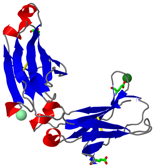
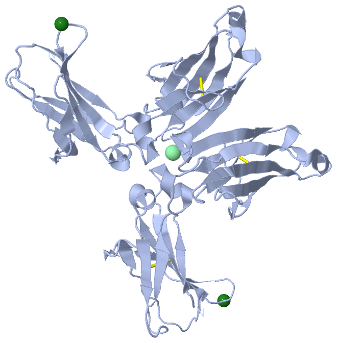
(-) Ligands, Modified Residues, Ions  (2, 2)

Asymmetric Unit (2, 2)
No.NameCountTypeFull Name
1CL1Ligand/IonCHLORIDE ION
2MG1Ligand/IonMAGNESIUM ION
Biological Unit 1 (0, 0)
No.NameCountTypeFull Name
1CL-1Ligand/IonCHLORIDE ION
2MG-1Ligand/IonMAGNESIUM ION

(-) Sites  (1, 1)

Asymmetric Unit (1, 1)
No.NameEvidenceResiduesDescription
1AC1SOFTWAREGLU A:286 , GLU A:297 , GLN A:416BINDING SITE FOR RESIDUE MG A 1

(-) SS Bonds  (2, 2)

Asymmetric Unit
No.Residues
1A:264 -A:324
2A:370 -A:428

(-) Cis Peptide Bonds  (1, 1)

Asymmetric Unit
No.Residues
1Phe A:376 -Pro A:377

 Sequence-Structure Mapping

(-) SAPs(SNPs)/Variants  (0, 0)

(no "SAP(SNP)/Variant" information available for 3HKF)

(-) PROSITE Motifs  (0, 0)

(no "PROSITE Motif" information available for 3HKF)

(-) Exons   (0, 0)

(no "Exon" information available for 3HKF)

(-) Sequences/Alignments

Asymmetric Unit
   Reformat: Number of residues per line =  ('0' or empty: single-line sequence representation)
  Number of residues per labelling interval =   
  UniProt sequence: complete  aligned part    
   Show mapping: SCOP domains CATH domains Pfam domains Secondary structure (by author)
SAPs(SNPs) PROSITE motifs Exons
(details for a mapped element are shown in a popup box when the mouse pointer rests over it)
Chain A from PDB  Type:PROTEIN  Length:205
 aligned with Q99LC4_MOUSE | Q99LC4 from UniProtKB/TrEMBL  Length:463

    Alignment length:205
                                   263       273       283       293       303       313       323       333       343       353       363       373       383       393       403       413       423       433       443       453     
         Q99LC4_MOUSE   254 SSVFIFPPKPKDVLTITLTPKVTCVVVDISKDDPEVQFSWFVDDVEVHTAQTQPREEQFNSTFRSVSELPIMHQDWLNGKEFKCRVNSAAFPAPIEKTISKTKGRPKAPQVYTIPPPKEQMAKDKVSLTCMITDFFPEDITVEWQWNGQPAENYKNTQPIMDTDGSYFIYSKLNVQKSNWEAGNTFTCSVLHEGLHNHHTEKSLS 458
               SCOP domains d3hkfa1 A:241-344 automated matches                                                                     d3hkfa2 A:345-445 automated matches                                                                   SCOP domains
               CATH domains 3hkfA01 A:241-340 Immunoglobulins                                                                   3hkfA02 A:341-445 Immunoglobulins                                                                         CATH domains
               Pfam domains ------------------------------------------------------------------------------------------------------------------------------------------------------------------------------------------------------------- Pfam domains
         Sec.struct. author ..eeee..hhhhhhh.....eeeeeeeee..........eee...ee...ee...ee.....eeeeeeee.hhhhhhh...eeeee........eeeee..........eeeee..hhhhh....eeeeeeeeee.....eeeeee.......eee...ee.....eeeeeeeee.hhhhh....eeeeee...hhh.eeeeee. Sec.struct. author
                 SAPs(SNPs) ------------------------------------------------------------------------------------------------------------------------------------------------------------------------------------------------------------- SAPs(SNPs)
                    PROSITE ------------------------------------------------------------------------------------------------------------------------------------------------------------------------------------------------------------- PROSITE
                 Transcript ------------------------------------------------------------------------------------------------------------------------------------------------------------------------------------------------------------- Transcript
                 3hkf A 241 SSVFIFPPKPKDVLTITLTPKVTCVVVDISKDDPEVQFSWFVDDVEVHTAQTQPREEQFNSTFRSVSELPIMHQDWLNGKEFKCRVNSAAFPAPIEKTISKTKGRPKAPQVYTIPPPKEQMAKDKVSLTCMITDFFPEDITVEWQWNGQPAENYKNTQPIMDTDGSYFVYSKLNVQKSNWEAGNTFTCSVLHEGLHNHHTEKSLS 445
                                   250       260       270       280       290       300       310       320       330       340       350       360       370       380       390       400       410       420       430       440     

   Legend:   → Mismatch (orange background)
  - → Gap (green background, '-', border residues have a numbering label)
    → Modified Residue (blue background, lower-case, 'x' indicates undefined single-letter code, labelled with number + name)
  x → Chemical Group (purple background, 'x', labelled with number + name, e.g. ACE or NH2)
  extra numbering lines below/above indicate numbering irregularities and modified residue names etc., number ends below/above '|'

 Classification and Annotation

(-) SCOP Domains  (1, 2)

Asymmetric Unit

(-) CATH Domains  (1, 2)

Asymmetric Unit
(-)
Class: Mainly Beta (13760)

(-) Pfam Domains  (0, 0)

(no "Pfam Domain" information available for 3HKF)

(-) Gene Ontology  (0, 0)

Asymmetric Unit(hide GO term definitions)
    (no "Gene Ontology" information available for 3HKF)

 Visualization

(-) Interactive Views

Asymmetric Unit
  Complete Structure
    Jena3D(integrated viewing of ligand, site, SAP, PROSITE, SCOP information)
    WebMol | AstexViewer[tm]@PDBe
(Java Applets, require no local installation except for Java; loading may be slow)
    STRAP
(Java WebStart application, automatic local installation, requires Java; full application with system access!)
    RasMol
(require local installation)
    Molscript (VRML)
(requires installation of a VRML viewer; select preferred view via VRML and generate a mono or stereo PDF format file)
 
  Ligands, Modified Residues, Ions
    CL  [ RasMol | Jena3D ]  +environment [ RasMol | Jena3D ]
    MG  [ RasMol | Jena3D ]  +environment [ RasMol | Jena3D ]
 
  Sites
    AC1  [ RasMol ]  +environment [ RasMol ]
 
  Cis Peptide Bonds
    Phe A:376 - Pro A:377   [ RasMol ]  
 
Biological Unit
  Complete Structure
    Biological Unit 1  [ Jena3D ]

(-) Still Images

Jmol
  protein: cartoon or spacefill or dots and stick; nucleic acid: cartoon and stick; ligands: spacefill; active site: stick
Molscript
  protein, nucleic acid: cartoon; ligands: spacefill; active site: ball and stick

 Databases and Analysis Tools

(-) Databases

Access by PDB/NDB ID
  3hkf
    Family and Domain InformationProDom | SYSTERS
    General Structural InformationGlycoscienceDB | MMDB | NDB | OCA | PDB | PDBe | PDBj | PDBsum | PDBWiki | PQS | PROTEOPEDIA
    Orientation in MembranesOPM
    Protein SurfaceSURFACE
    Secondary StructureDSSP (structure derived) | HSSP (homology derived)
    Structural GenomicsGeneCensus
    Structural NeighboursCE | VAST
    Structure ClassificationCATH | Dali | SCOP
    Validation and Original DataBMRB Data View | BMRB Restraints Grid | EDS | PROCHECK | RECOORD | WHAT_CHECK
 
Access by UniProt ID/Accession number
  Q99LC4_MOUSE | Q99LC4
    Comparative Protein Structure ModelsModBase
    Genomic InformationEnsembl
    Protein-protein InteractionDIP
    Sequence, Family and Domain InformationInterPro | Pfam | SMART | UniProtKB/SwissProt
 
Access by Enzyme Classificator   (EC Number)
  (no 'Enzyme Classificator' available)
    General Enzyme InformationBRENDA | EC-PDB | Enzyme | IntEnz
    PathwayKEGG | MetaCyc
 
Access by Disease Identifier   (MIM ID)
  (no 'MIM ID' available)
    Disease InformationOMIM
 
Access by GenAge ID
  (no 'GenAge ID' available)
    Age Related InformationGenAge

(-) Analysis Tools

Access by PDB/NDB ID
    Domain InformationXDom
    Interatomic Contacts of Structural UnitsCSU
    Ligand-protein ContactsLPC
    Protein CavitiescastP
    Sequence and Secondary StructurePDBCartoon
    Structure AlignmentSTRAP(Java WebStart application, automatic local installation, requires Java; full application with system access!)
    Structure and Sequence BrowserSTING
 
Access by UniProt ID/Accession number
  Q99LC4_MOUSE | Q99LC4
    Protein Disorder PredictionDisEMBL | FoldIndex | GLOBPLOT (for more information see DisProt)

 Related Entries

(-) Entries Sharing at Least One Protein Chain (UniProt ID)

UniProtKB/Swiss-Prot
        Q99LC4_MOUSE | Q99LC41afv
UniProtKB/TrEMBL
        Q99LC4_MOUSE | Q99LC41ae6 1e6j 1f58 1gig 1jhk 1jnl 1jnn 2a1w 2ntf 2vir 2vis 2vit 4q0x

(-) Related Entries Specified in the PDB File

(no "Related Entries Specified in the PDB File" available for 3HKF)