Show PDB file:   
         Plain Text   HTML   (compressed file size)
QuickSearch:   
by PDB,NDB,UniProt,PROSITE Code or Search Term(s)  
(-)Asymmetric Unit
(-)Asym. Unit - sites
(-)Biological Unit 1
(-)Biol. Unit 1 - sites
(-)Biological Unit 2
collapse expand < >
Image Asymmetric Unit
Asymmetric Unit  (Jmol Viewer)
Image Asym. Unit - sites
Asym. Unit - sites  (Jmol Viewer)
Image Biological Unit 1
Biological Unit 1  (Jmol Viewer)
Image Biol. Unit 1 - sites
Biol. Unit 1 - sites  (Jmol Viewer)
Image Biological Unit 2
Biological Unit 2  (Jmol Viewer)

(-) Description

Title :  CRYSTAL STRUCTURE OF GLUTAREDOXIN DOMAIN OF HUMAN THIOREDOXIN REDUCTASE 3
 
Authors :  A. Chaikuad, C. Johansson, E. Ugochukwu, A. K. Roos, F. Von Delft, E. Pi W. Yue, C. H. Arrowsmith, A. M. Edwards, J. Weigelt, C. Bountra, U. Oppe Structural Genomics Consortium (Sgc)
Date :  29 Apr 09  (Deposition) - 12 May 09  (Release) - 13 Jul 11  (Revision)
Method :  X-RAY DIFFRACTION
Resolution :  2.21
Chains :  Asym. Unit :  A,B
Biol. Unit 1:  A  (1x)
Biol. Unit 2:  B  (1x)
Keywords :  Oxidoreductase, Structural Genomics, Structural Genomics Consortium, Sgc, Developmental Protein, Differentiation, Disulfide Bond, Electron Transport, Endoplasmic Reticulum, Fad, Flavoprotein, Microsome, Nadp, Nucleus, Phosphoprotein, Redox-Active Center, Selenium, Selenocysteine, Spermatogenesis, Transport (Keyword Search: [Gene Ontology, PubMed, Web (Google))
 
Reference :  A. Chaikuad, C. Johansson, E. Ugochukwu, A. K. Roos, F. Von Delft, E. Pilka, W. Yue, C. H. Arrowsmith, A. M. Edwards, J. Weigelt, C. Bountra, U. Oppermann
Crystal Structure Of Glutaredoxin Domain Of Human Thioredoxin Reductase 3
To Be Published
PubMed: search

(-) Compounds

Molecule 1 - THIOREDOXIN REDUCTASE 3
    ChainsA, B
    EC Number1.8.1.9
    EngineeredYES
    Expression SystemESCHERICHIA COLI
    Expression System PlasmidPNIC-CTHF
    Expression System StrainORIGAMI2(DE3)
    Expression System Taxid562
    Expression System Vector TypePLASMID
    FragmentGLUTAREDOXIN DOMAIN, RESIDUES 162-267
    GeneTGR, TRXR3, TXNRD3, TXNRD3
    Organism CommonHUMAN
    Organism ScientificHOMO SAPIENS
    Organism Taxid9606
    SynonymTHIOREDOXIN REDUCTASE TR2, THIOREDOXIN AND GLUTATHIONE REDUCTASE

 Structural Features

(-) Chains, Units

  12
Asymmetric Unit AB
Biological Unit 1 (1x)A 
Biological Unit 2 (1x) B

Summary Information (see also Sequences/Alignments below)

(-) Ligands, Modified Residues, Ions  (2, 4)

Asymmetric Unit (2, 4)
No.NameCountTypeFull Name
1CL1Ligand/IonCHLORIDE ION
2SO43Ligand/IonSULFATE ION
Biological Unit 1 (1, 1)
No.NameCountTypeFull Name
1CL-1Ligand/IonCHLORIDE ION
2SO41Ligand/IonSULFATE ION
Biological Unit 2 (1, 2)
No.NameCountTypeFull Name
1CL-1Ligand/IonCHLORIDE ION
2SO42Ligand/IonSULFATE ION

(-) Sites  (4, 4)

Asymmetric Unit (4, 4)
No.NameEvidenceResiduesDescription
1AC1SOFTWAREHOH B:22 , HOH B:26 , HIS B:189 , PRO B:232 , GLY B:243 , CYS B:244 , ASP B:245BINDING SITE FOR RESIDUE SO4 B 1
2AC2SOFTWAREHOH A:15 , HOH A:28 , HIS A:189 , CYS A:244 , ASP A:245BINDING SITE FOR RESIDUE SO4 A 2
3AC3SOFTWARELYS A:238 , LEU A:264 , HIS B:169 , LYS B:258BINDING SITE FOR RESIDUE SO4 B 3
4AC4SOFTWAREHOH A:17 , HOH A:18 , ASP A:213 , ASP A:214BINDING SITE FOR RESIDUE CL A 4

(-) SS Bonds  (0, 0)

(no "SS Bond" information available for 3H8Q)

(-) Cis Peptide Bonds  (2, 2)

Asymmetric Unit
No.Residues
1Val A:231 -Pro A:232
2Val B:231 -Pro B:232

 Sequence-Structure Mapping

(-) SAPs(SNPs)/Variants  (0, 0)

(no "SAP(SNP)/Variant" information available for 3H8Q)

(-) PROSITE Motifs  (1, 2)

Asymmetric Unit (1, 2)
 PROSITEUniProtKBPDB
No.IDACDescriptionIDLocationCountLocation
1GLUTAREDOXIN_2PS51354 Glutaredoxin domain profile.TRXR3_HUMAN56-156
 
  2A:167-267
B:167-267
Biological Unit 1 (1, 1)
 PROSITEUniProtKBPDB
No.IDACDescriptionIDLocationCountLocation
1GLUTAREDOXIN_2PS51354 Glutaredoxin domain profile.TRXR3_HUMAN56-156
 
  1A:167-267
-
Biological Unit 2 (1, 1)
 PROSITEUniProtKBPDB
No.IDACDescriptionIDLocationCountLocation
1GLUTAREDOXIN_2PS51354 Glutaredoxin domain profile.TRXR3_HUMAN56-156
 
  1-
B:167-267

(-) Exons   (0, 0)

(no "Exon" information available for 3H8Q)

(-) Sequences/Alignments

Asymmetric Unit
   Reformat: Number of residues per line =  ('0' or empty: single-line sequence representation)
  Number of residues per labelling interval =   
  UniProt sequence: complete  aligned part    
   Show mapping: SCOP domains CATH domains Pfam domains Secondary structure (by author)
SAPs(SNPs) PROSITE motifs Exons
(details for a mapped element are shown in a popup box when the mouse pointer rests over it)
Chain A from PDB  Type:PROTEIN  Length:108
 aligned with TRXR3_HUMAN | Q86VQ6 from UniProtKB/Swiss-Prot  Length:643

    Alignment length:108
                                    59        69        79        89        99       109       119       129       139       149        
          TRXR3_HUMAN    50 EAREELRRHLVGLIERSRVVIFSKSYCPHSTRVKELFSSLGVECNVLELDQVDDGARVQEVLSEITNQKTVPNIFVNKVHVGGCDQTFQAYQSGLLQKLLQEDLAYDY 157
               SCOP domains d3h8qa_ A: automated matches                                                                                 SCOP domains
               CATH domains ------------------------------------------------------------------------------------------------------------ CATH domains
               Pfam domains ------------------------------------------------------------------------------------------------------------ Pfam domains
         Sec.struct. author ..hhhhhhhhhhhhhhh.eeeee...hhhhhhhhhhhhhh....eeee.....hhhhhhhhhhhhhh.....eeee..eeeehhhhhhhhhhhhhhhhhhhh...... Sec.struct. author
                 SAPs(SNPs) ------------------------------------------------------------------------------------------------------------ SAPs(SNPs)
                    PROSITE ------GLUTAREDOXIN_2  PDB: A:167-267 UniProt: 56-156                                                       - PROSITE
                 Transcript ------------------------------------------------------------------------------------------------------------ Transcript
                 3h8q A   0 MAREELRRHLVGLIERSRVVIFSKSYCPHSTRVKELFSSLGVECNVLELDQVDDGARVQEVLSEITNQKTVPNIFVNKVHVGGCDQTFQAYQSGLLQKLLQEDLAYDA 268
                            ||     170       180       190       200       210       220       230       240       250       260        
                            ||                                                                                                          
                            0|                                                                                                          
                           162                                                                                                          

Chain B from PDB  Type:PROTEIN  Length:106
 aligned with TRXR3_HUMAN | Q86VQ6 from UniProtKB/Swiss-Prot  Length:643

    Alignment length:106
                                    61        71        81        91       101       111       121       131       141       151      
          TRXR3_HUMAN    52 REELRRHLVGLIERSRVVIFSKSYCPHSTRVKELFSSLGVECNVLELDQVDDGARVQEVLSEITNQKTVPNIFVNKVHVGGCDQTFQAYQSGLLQKLLQEDLAYDY 157
               SCOP domains d3h8qb_ B: automated matches                                                                               SCOP domains
               CATH domains ---------------------------------------------------------------------------------------------------------- CATH domains
               Pfam domains ---------------------------------------------------------------------------------------------------------- Pfam domains
         Sec.struct. author hhhhhhhhhhhhhhh.eeeee...hhhhhhhhhhhhhh....eeee.....hhhhhhhhhhhhhh.....eeee..eeeehhhhhhhhhhhhhhhhhhh..hhhhh Sec.struct. author
                 SAPs(SNPs) ---------------------------------------------------------------------------------------------------------- SAPs(SNPs)
                    PROSITE ----GLUTAREDOXIN_2  PDB: B:167-267 UniProt: 56-156                                                       - PROSITE
                 Transcript ---------------------------------------------------------------------------------------------------------- Transcript
                 3h8q B 163 REELRRHLVGLIERSRVVIFSKSYCPHSTRVKELFSSLGVECNVLELDQVDDGARVQEVLSEITNQKTVPNIFVNKVHVGGCDQTFQAYQSGLLQKLLQEDLAYDA 268
                                   172       182       192       202       212       222       232       242       252       262      

   Legend:   → Mismatch (orange background)
  - → Gap (green background, '-', border residues have a numbering label)
    → Modified Residue (blue background, lower-case, 'x' indicates undefined single-letter code, labelled with number + name)
  x → Chemical Group (purple background, 'x', labelled with number + name, e.g. ACE or NH2)
  extra numbering lines below/above indicate numbering irregularities and modified residue names etc., number ends below/above '|'

 Classification and Annotation

(-) SCOP Domains  (1, 2)

Asymmetric Unit

(-) CATH Domains  (0, 0)

(no "CATH Domain" information available for 3H8Q)

(-) Pfam Domains  (0, 0)

(no "Pfam Domain" information available for 3H8Q)

(-) Gene Ontology  (16, 16)

Asymmetric Unit(hide GO term definitions)
Chain A,B   (TRXR3_HUMAN | Q86VQ6)
molecular function
    GO:0009055    electron carrier activity    Any molecular entity that serves as an electron acceptor and electron donor in an electron transport chain. An electron transport chain is a process in which a series of electron carriers operate together to transfer electrons from donors to any of several different terminal electron acceptors to generate a transmembrane electrochemical gradient.
    GO:0050660    flavin adenine dinucleotide binding    Interacting selectively and non-covalently with FAD, flavin-adenine dinucleotide, the coenzyme or the prosthetic group of various flavoprotein oxidoreductase enzymes, in either the oxidized form, FAD, or the reduced form, FADH2.
    GO:0016491    oxidoreductase activity    Catalysis of an oxidation-reduction (redox) reaction, a reversible chemical reaction in which the oxidation state of an atom or atoms within a molecule is altered. One substrate acts as a hydrogen or electron donor and becomes oxidized, while the other acts as hydrogen or electron acceptor and becomes reduced.
    GO:0016668    oxidoreductase activity, acting on a sulfur group of donors, NAD(P) as acceptor    Catalysis of an oxidation-reduction (redox) reaction in which a sulfur-containing group acts as a hydrogen or electron donor and reduces NAD or NADP.
    GO:0015035    protein disulfide oxidoreductase activity    Catalysis of the reaction: a protein with reduced sulfide groups = a protein with oxidized disulfide bonds.
    GO:0004791    thioredoxin-disulfide reductase activity    Catalysis of the reaction: NADP(+) + thioredoxin = H(+) + NADPH + thioredoxin disulfide.
biological process
    GO:0030154    cell differentiation    The process in which relatively unspecialized cells, e.g. embryonic or regenerative cells, acquire specialized structural and/or functional features that characterize the cells, tissues, or organs of the mature organism or some other relatively stable phase of the organism's life history. Differentiation includes the processes involved in commitment of a cell to a specific fate and its subsequent development to the mature state.
    GO:0045454    cell redox homeostasis    Any process that maintains the redox environment of a cell or compartment within a cell.
    GO:0098869    cellular oxidant detoxification    Any process carried out at the cellular level that reduces or removes the toxicity superoxide radicals or hydrogen peroxide.
    GO:0007275    multicellular organism development    The biological process whose specific outcome is the progression of a multicellular organism over time from an initial condition (e.g. a zygote or a young adult) to a later condition (e.g. a multicellular animal or an aged adult).
    GO:0055114    oxidation-reduction process    A metabolic process that results in the removal or addition of one or more electrons to or from a substance, with or without the concomitant removal or addition of a proton or protons.
    GO:0007283    spermatogenesis    The process of formation of spermatozoa, including spermatocytogenesis and spermiogenesis.
cellular component
    GO:0005737    cytoplasm    All of the contents of a cell excluding the plasma membrane and nucleus, but including other subcellular structures.
    GO:0005783    endoplasmic reticulum    The irregular network of unit membranes, visible only by electron microscopy, that occurs in the cytoplasm of many eukaryotic cells. The membranes form a complex meshwork of tubular channels, which are often expanded into slitlike cavities called cisternae. The ER takes two forms, rough (or granular), with ribosomes adhering to the outer surface, and smooth (with no ribosomes attached).
    GO:0043231    intracellular membrane-bounded organelle    Organized structure of distinctive morphology and function, bounded by a single or double lipid bilayer membrane and occurring within the cell. Includes the nucleus, mitochondria, plastids, vacuoles, and vesicles. Excludes the plasma membrane.
    GO:0005634    nucleus    A membrane-bounded organelle of eukaryotic cells in which chromosomes are housed and replicated. In most cells, the nucleus contains all of the cell's chromosomes except the organellar chromosomes, and is the site of RNA synthesis and processing. In some species, or in specialized cell types, RNA metabolism or DNA replication may be absent.
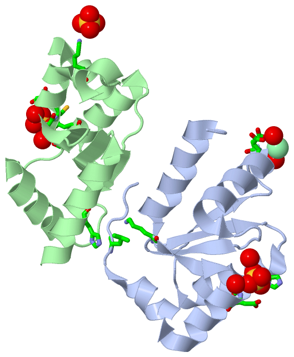
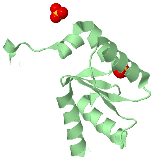
 Visualization

(-) Interactive Views

Asymmetric Unit
  Complete Structure
    Jena3D(integrated viewing of ligand, site, SAP, PROSITE, SCOP information)
    WebMol | AstexViewer[tm]@PDBe
(Java Applets, require no local installation except for Java; loading may be slow)
    STRAP
(Java WebStart application, automatic local installation, requires Java; full application with system access!)
    RasMol
(require local installation)
    Molscript (VRML)
(requires installation of a VRML viewer; select preferred view via VRML and generate a mono or stereo PDF format file)
 
  Ligands, Modified Residues, Ions
    CL  [ RasMol | Jena3D ]  +environment [ RasMol | Jena3D ]
    SO4  [ RasMol | Jena3D ]  +environment [ RasMol | Jena3D ]
 
  Sites
    AC1  [ RasMol ]  +environment [ RasMol ]
    AC2  [ RasMol ]  +environment [ RasMol ]
    AC3  [ RasMol ]  +environment [ RasMol ]
    AC4  [ RasMol ]  +environment [ RasMol ]
 
  Cis Peptide Bonds
    Val A:231 - Pro A:232   [ RasMol ]  
    Val B:231 - Pro B:232   [ RasMol ]  
 
Biological Units
  Complete Structure
    Biological Unit 1  [ Jena3D ]
    Biological Unit 2  [ Jena3D ]

(-) Still Images

Jmol
  protein: cartoon or spacefill or dots and stick; nucleic acid: cartoon and stick; ligands: spacefill; active site: stick
Molscript
  protein, nucleic acid: cartoon; ligands: spacefill; active site: ball and stick

 Databases and Analysis Tools

(-) Databases

Access by PDB/NDB ID
  3h8q
    Family and Domain InformationProDom | SYSTERS
    General Structural InformationGlycoscienceDB | MMDB | NDB | OCA | PDB | PDBe | PDBj | PDBsum | PDBWiki | PQS | PROTEOPEDIA
    Orientation in MembranesOPM
    Protein SurfaceSURFACE
    Secondary StructureDSSP (structure derived) | HSSP (homology derived)
    Structural GenomicsGeneCensus
    Structural NeighboursCE | VAST
    Structure ClassificationCATH | Dali | SCOP
    Validation and Original DataBMRB Data View | BMRB Restraints Grid | EDS | PROCHECK | RECOORD | WHAT_CHECK
 
Access by UniProt ID/Accession number
  TRXR3_HUMAN | Q86VQ6
    Comparative Protein Structure ModelsModBase
    Genomic InformationEnsembl
    Protein-protein InteractionDIP
    Sequence, Family and Domain InformationInterPro | Pfam | SMART | UniProtKB/SwissProt
 
Access by Enzyme Classificator   (EC Number)
  1.8.1.9
    General Enzyme InformationBRENDA | EC-PDB | Enzyme | IntEnz
    PathwayKEGG | MetaCyc
 
Access by Disease Identifier   (MIM ID)
  (no 'MIM ID' available)
    Disease InformationOMIM
 
Access by GenAge ID
  (no 'GenAge ID' available)
    Age Related InformationGenAge

(-) Analysis Tools

Access by PDB/NDB ID
    Domain InformationXDom
    Interatomic Contacts of Structural UnitsCSU
    Ligand-protein ContactsLPC
    Protein CavitiescastP
    Sequence and Secondary StructurePDBCartoon
    Structure AlignmentSTRAP(Java WebStart application, automatic local installation, requires Java; full application with system access!)
    Structure and Sequence BrowserSTING
 
Access by UniProt ID/Accession number
  TRXR3_HUMAN | Q86VQ6
    Protein Disorder PredictionDisEMBL | FoldIndex | GLOBPLOT (for more information see DisProt)

 Related Entries

(-) Entries Sharing at Least One Protein Chain (UniProt ID)

(no "Entries Sharing at Least One Protein Chain" available for 3H8Q)

(-) Related Entries Specified in the PDB File

(no "Related Entries Specified in the PDB File" available for 3H8Q)