|
|
|
|
 Description
|
|
Compounds
|
||||||||||||||||||||||||||||||||
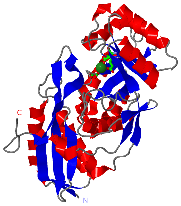
Chains, Units
Summary Information (see also Sequences/Alignments below) |
Ligands, Modified Residues, Ions (1, 1)
Asymmetric/Biological Unit (1, 1)
|
Sites (1, 1)
Asymmetric Unit (1, 1)
|
SS Bonds (0, 0)| (no "SS Bond" information available for 3H70) |
Cis Peptide Bonds (0, 0)| (no "Cis Peptide Bond" information available for 3H70) |
SAPs(SNPs)/Variants (0, 0)| (no "SAP(SNP)/Variant" information available for 3H70) |
PROSITE Motifs (0, 0)| (no "PROSITE Motif" information available for 3H70) |
Exons (0, 0)| (no "Exon" information available for 3H70) |
Sequences/Alignments
Asymmetric/Biological UnitChain A from PDB Type:PROTEIN Length:342 aligned with Q53635_STAAU | Q53635 from UniProtKB/TrEMBL Length:333 Alignment length:342 1 333 | 9 19 29 39 49 59 69 79 89 99 109 119 129 139 149 159 169 179 189 199 209 219 229 239 249 259 269 279 289 299 309 319 329 | - Q53635_STAAU - -MKLTALHFYKYSEPFKSQIVTPKVTLTHRDCLFIELIDDKGNAYFGECNAFQTDWYDHETIASVKHVIEQWFEDNRNKSFETYEAALKLVDSLENTPAARATIVMALYQMFHVLPSFSVAYGATASGLSNKQLESLKATKPTRIKLKWTPQIMHQIRVLRELDFHFQLVIDANESLDRQDFTQLQLLAREQVLYIEEPFKDISMLDEVADGTIPPIALDEKATSLLDIINLIELYNVKVVVLKPFRLGGIDKVQTAIDTLKSHGAKVVIGGMYEYGLSRYFTAMLARKGDYPGDVTPAGYYFEQDVVAHSGILKEGRLEFRPPLVDITQLQPY-------- - SCOP domains ------------------------------------------------------------------------------------------------------------------------------------------------------------------------------------------------------------------------------------------------------------------------------------------------------------------------------------------------------ SCOP domains CATH domains 3h70A01 A:0-116,A:322-333 Enolase-like, N-terminal domain 3h70A02 A:117-321 Enolase superfamily 3h70A01 -------- CATH domains Pfam domains ------------------------------------------------------------------------------------------------------------------------------------------------------------------------------------------------------------------------------------------------------------------------------------------------------------------------------------------------------ Pfam domains SAPs(SNPs) ------------------------------------------------------------------------------------------------------------------------------------------------------------------------------------------------------------------------------------------------------------------------------------------------------------------------------------------------------ SAPs(SNPs) PROSITE ------------------------------------------------------------------------------------------------------------------------------------------------------------------------------------------------------------------------------------------------------------------------------------------------------------------------------------------------------ PROSITE Transcript ------------------------------------------------------------------------------------------------------------------------------------------------------------------------------------------------------------------------------------------------------------------------------------------------------------------------------------------------------ Transcript 3h70 A 0 SLKLTALHFYKYSEPFKSQIVTPKVTLTHRDCLFIELIDDKGNAYFGECNAFQTDWYDHETIASVKHVIEQWFEDNRNKSFETYEAALKLVDSLENTPAARATIVMALYQMFHVLPSFSVAYGATASGLSNKQLESLKATKPTRIKLKWTPQIMHQIRVLRELDFHFQLVIDANESLDRQDFTQLQLLAREQVLYIEEPFKDISMLDEVADGTIPPIALDEKATSLLDIINLIELYNVKVVVLKPFRLGGIDKVQTAIDTLKSHGAKVVIGGMYEYGLSRYFTAMLARKGDYPGDVTPAGYYFEQDVVAHSGILKEGRLEFRPPLVDITQLQPYEGHHHHHH 341 9 19 29 39 49 59 69 79 89 99 109 119 129 139 149 159 169 179 189 199 209 219 229 239 249 259 269 279 289 299 309 319 329 339
|
||||||||||||||||||||
SCOP Domains (0, 0)| (no "SCOP Domain" information available for 3H70) |
CATH Domains (2, 2)
Asymmetric/Biological Unit

|
Pfam Domains (0, 0)| (no "Pfam Domain" information available for 3H70) |
Gene Ontology (7, 7)|
Asymmetric/Biological Unit(hide GO term definitions) Chain A (Q53635_STAAU | Q53635)
|
||||||||||||||||||||||||||||||||||||||||||||||||||||||
Interactive Views
|
||||||||||||||||||||||||||||||||||||||||||||||||||||||||||||||||||||||||||||||||||||||||||||||||||||||||||||||||||||||
Still Images
|
||||||||||||||||
Databases
|
||||||||||||||||||||||||||||||||||||||||||||||||||||||||||||||||||||||||||||||||||||||||||||||||||||||||||||||||||||||||||||||||||||||||||||||||||||||||||||||||
Analysis Tools
|
|||||||||||||||||||||||||||||||||||||||||||||||||||||||||||||
Entries Sharing at Least One Protein Chain (UniProt ID)
Related Entries Specified in the PDB File
|
|