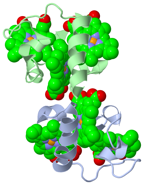
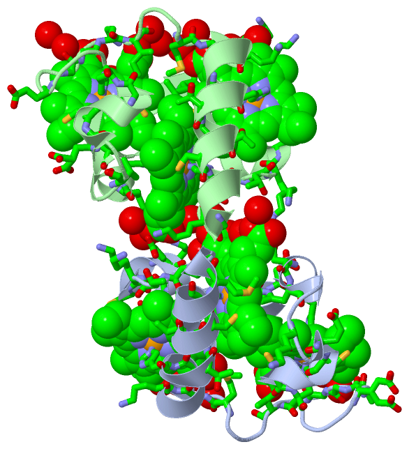
Asymmetric Unit (6, 6)
| No. | Name | Evidence | Residues | Description |
|---|
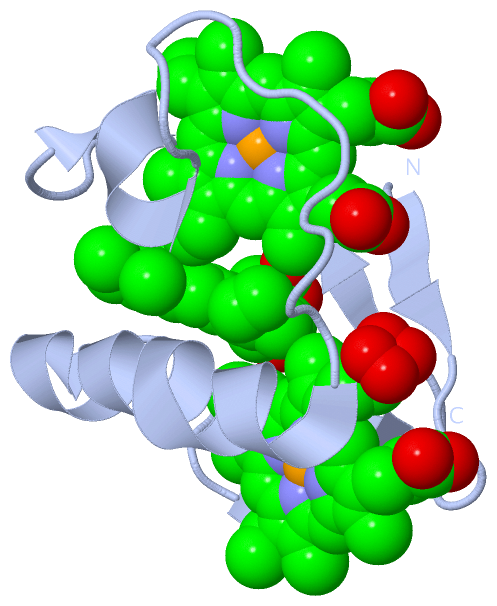
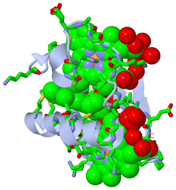
| 1 | AC1 | SOFTWARE | LYS A:9 , PHE A:15 , HIS A:17 , HIS A:20 , GLU A:26 , CYS A:27 , CYS A:30 , HIS A:31 , GLU A:33 , ALA A:34 , GLY A:35 , GLY A:36 , LYS A:37 , ILE A:38 , LYS A:43 , HEM A:73 , HEM A:74 , HOH A:84 , HOH A:109 , HOH A:112 , HOH A:133 , HOH A:142 | BINDING SITE FOR RESIDUE HEM A 72 |
| 2 | AC2 | SOFTWARE | VAL A:13 , THR A:14 , ASP A:16 , LYS A:19 , HIS A:20 , THR A:49A , CYS A:51 , CYS A:54 , HIS A:55 , MET A:58 , PRO A:62 , HEM A:72 , HOH A:77 , HOH A:78 , HOH A:116 , HOH A:127 , HOH A:161 , HOH A:172 , GLY B:59 , LYS B:60 , GLY B:61 , HOH B:100 , HOH B:181 | BINDING SITE FOR RESIDUE HEM A 73 |
| 3 | AC3 | SOFTWARE | LEU A:6 , GLU A:7 , ALA A:8 , LYS A:9 , ASN A:10 , LYS A:37 , MET A:41 , GLY A:42 , LYS A:43 , ALA A:46 , HIS A:47 , THR A:52 , PRO A:62 , THR A:63 , LYS A:64 , CYS A:65 , CYS A:68 , HIS A:69 , HEM A:72 , HOH A:87 , HOH A:113 | BINDING SITE FOR RESIDUE HEM A 74 |
| 4 | AC4 | SOFTWARE | PHE B:15 , HIS B:17 , HIS B:20 , VAL B:23 , GLU B:26 , CYS B:27 , CYS B:30 , HIS B:31 , GLU B:33 , GLY B:35 , GLY B:36 , LYS B:37 , ILE B:38 , MET B:41 , LYS B:43 , HEM B:73 , HEM B:74 , HOH B:80 , HOH B:92 , HOH B:107 , HOH B:131 , HOH B:168 | BINDING SITE FOR RESIDUE HEM B 72 |
| 5 | AC5 | SOFTWARE | GLU A:67 , LYS A:70 , THR B:14 , PHE B:15 , LYS B:19 , HIS B:20 , VAL B:23 , THR B:49A , CYS B:51 , CYS B:54 , HIS B:55 , LYS B:60 , PRO B:62 , HEM B:72 , HOH B:109 , HOH B:112 , HOH B:126 , HOH B:179 | BINDING SITE FOR RESIDUE HEM B 73 |
| 6 | AC6 | SOFTWARE | LYS A:24 , LEU B:6 , GLU B:7 , ALA B:8 , LYS B:9 , ASN B:10 , HIS B:17 , GLY B:42 , LYS B:43 , ALA B:46 , HIS B:47 , THR B:52 , PRO B:62 , THR B:63 , LYS B:64 , CYS B:65 , CYS B:68 , HIS B:69 , HEM B:72 , HOH B:102 , HOH B:138 , HOH B:193 , HOH B:197 | BINDING SITE FOR RESIDUE HEM B 74 |
|
 Description