|
|
|
|
 Description
|
|
Compounds
|
||||||||||||||||||||||||||||||||||||||||||||||||
Chains, Units
Summary Information (see also Sequences/Alignments below) |
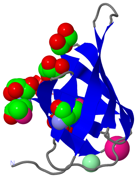
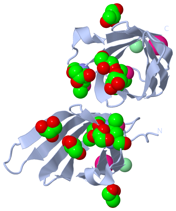
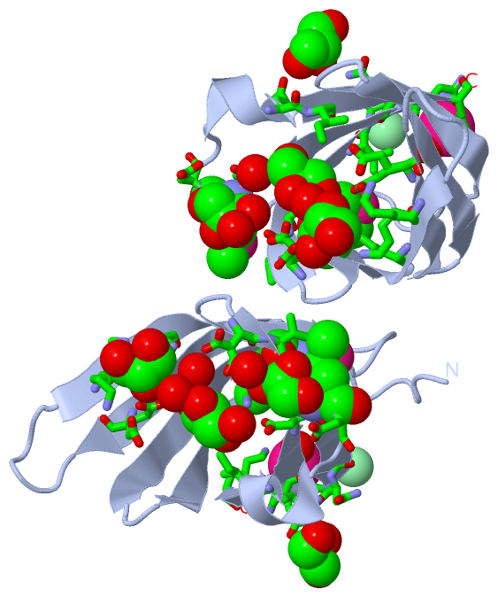
Ligands, Modified Residues, Ions (4, 8)| Asymmetric Unit (4, 8) Biological Unit 1 (2, 12) |
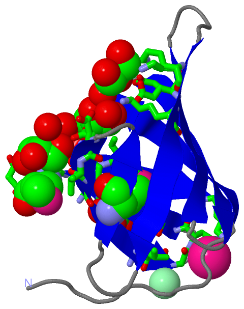
Sites (6, 6)
Asymmetric Unit (6, 6)
|
SS Bonds (0, 0)| (no "SS Bond" information available for 3GE2) |
Cis Peptide Bonds (0, 0)| (no "Cis Peptide Bond" information available for 3GE2) |
SAPs(SNPs)/Variants (0, 0)| (no "SAP(SNP)/Variant" information available for 3GE2) |
PROSITE Motifs (0, 0)| (no "PROSITE Motif" information available for 3GE2) |
Exons (0, 0)| (no "Exon" information available for 3GE2) |
Sequences/Alignments
Asymmetric UnitChain A from PDB Type:PROTEIN Length:89 aligned with A0A0H2UNA1_S | A0A0H2UNA1 from UniProtKB/TrEMBL Length:152 Alignment length:89 73 83 93 103 113 123 133 143 A0A0H2UNA1_S 64 QPVAQPTDIDGTYTGQDDGDRITLVVTGTTGTWTELESDGDQKVKQVTLDSANQRMIIGDDVKIYTVNGNQIVVDDMDRDPSDQIVLTK 152 SCOP domains ----------------------------------------------------------------------------------------- SCOP domains CATH domains 3ge2A00 A:65-153 [code=2.40.128.50, no name defined] CATH domains Pfam domains ----------------------------------------------------------------------------------------- Pfam domains SAPs(SNPs) ----------------------------------------------------------------------------------------- SAPs(SNPs) PROSITE ----------------------------------------------------------------------------------------- PROSITE Transcript ----------------------------------------------------------------------------------------- Transcript 3ge2 A 65 QPVAQPTDIDGTYTGQDDGDRITLVVTGTTGTWTELESDGDQKVKQVTFDSANQRmIIGDDVKIYTVNGNQIVVDDmDRDPSDQIVLTK 153 74 84 94 104 114 | 124 134 |144 120-MSE 141-MSE Chain A from PDB Type:PROTEIN Length:89 aligned with A5M522_STREE | A5M522 from UniProtKB/TrEMBL Length:152 Alignment length:89 73 83 93 103 113 123 133 143 A5M522_STREE 64 QPVAQPTDIDGTYTGQDDGDRITLVVTGTTGTWTELESDGDQKVKQVTFDSANQRMIIGDDVKIYTVNGNQIVVDDMDRDPSDQIVLTK 152 SCOP domains ----------------------------------------------------------------------------------------- SCOP domains CATH domains 3ge2A00 A:65-153 [code=2.40.128.50, no name defined] CATH domains Pfam domains ----------------------------------------------------------------------------------------- Pfam domains SAPs(SNPs) ----------------------------------------------------------------------------------------- SAPs(SNPs) PROSITE ----------------------------------------------------------------------------------------- PROSITE Transcript ----------------------------------------------------------------------------------------- Transcript 3ge2 A 65 QPVAQPTDIDGTYTGQDDGDRITLVVTGTTGTWTELESDGDQKVKQVTFDSANQRmIIGDDVKIYTVNGNQIVVDDmDRDPSDQIVLTK 153 74 84 94 104 114 | 124 134 |144 120-MSE 141-MSE
|
||||||||||||||||||||
SCOP Domains (0, 0)| (no "SCOP Domain" information available for 3GE2) |
CATH Domains (1, 1)
Asymmetric Unit
 |
Pfam Domains (0, 0)| (no "Pfam Domain" information available for 3GE2) |
Gene Ontology (0, 0)|
Asymmetric Unit(hide GO term definitions)
(no "Gene Ontology" information available for 3GE2)
|
Interactive Views
|
||||||||||||||||||||||||||||||||||||||||||||||||||||||||||||||||||||||||||||||||||||||||||||||||||||||||||||||||||||||||||||||||||||||||||||||||||||||||||||||||||||||||||||||||||||||||||||||||
Still Images
|
||||||||||||||||
Databases
|
||||||||||||||||||||||||||||||||||||||||||||||||||||||||||||||||||||||||||||||||||||||||||||||||||||||||||||||||||||||||||||||||||||||||||||||||||||||||||||||||||||||||||||||||||||||||||
Analysis Tools
|
||||||||||||||||||||||||||||||||||||||||||||||||||||||||||||||||||||||||
Entries Sharing at Least One Protein Chain (UniProt ID)
Related Entries Specified in the PDB File
|
|