Show PDB file:   
         Plain Text   HTML   (compressed file size)
QuickSearch:   
by PDB,NDB,UniProt,PROSITE Code or Search Term(s)  
(-)Asym./Biol. Unit
(-)Asym./Biol. Unit - sites
collapse expand < >
Image Asym./Biol. Unit
Asym./Biol. Unit  (Jmol Viewer)
Image Asym./Biol. Unit - sites
Asym./Biol. Unit - sites  (Jmol Viewer)

(-) Description

Title :  PDE7A CATALYTIC DOMAIN IN COMPLEX WITH 3-(2,6-DIFLUOROPHENYL)-2-(METHYLTHIO)QUINAZOLIN-4(3H)-ONE
 
Authors :  T. Castano, H. Wang
Date :  02 Feb 09  (Deposition) - 28 Apr 09  (Release) - 26 May 09  (Revision)
Method :  X-RAY DIFFRACTION
Resolution :  2.40
Chains :  Asym./Biol. Unit :  A
Keywords :  Pde7, Crystal, Inhibitor Complex, Alternative Splicing, Camp, Hydrolase, Phosphoprotein (Keyword Search: [Gene Ontology, PubMed, Web (Google))
 
Reference :  T. Castano, H. Wang, N. E. Campillo, S. Ballester, C. Gonzalez-Garcia, J. Hernandez, C. Perez, J. Cuenca, A. Perez-Castillo, A. Martinez, O. Huertas, J. L. Gelpi, F. J. Luque, H. Ke, C. Gil
Synthesis, Structural Analysis, And Biological Evaluation Of Thioxoquinazoline Derivatives As Phosphodiesterase 7 Inhibitors
Chemmedchem V. 4 866 2009
PubMed-ID: 19350606  |  Reference-DOI: 10.1002/CMDC.200900043
(for further references see the PDB file header)

(-) Compounds

Molecule 1 - HIGH AFFINITY CAMP-SPECIFIC 3',5'-CYCLIC PHOSPHODIESTERASE 7A
    ChainsA
    EC Number3.1.4.17
    EngineeredYES
    Expression SystemESCHERICHIA COLI
    Expression System PlasmidPET32
    Expression System StrainBL21
    Expression System Taxid562
    Expression System Vector TypePLASMID
    FragmentCATALYTIC DOMAIN RESIDUES 139-456
    GenePDE7A, PDE7A1
    Organism CommonHUMAN
    Organism ScientificHOMO SAPIENS
    Organism Taxid9606
    SynonymHCP1, TM22

 Structural Features

(-) Chains, Units

  1
Asymmetric/Biological Unit A

Summary Information (see also Sequences/Alignments below)
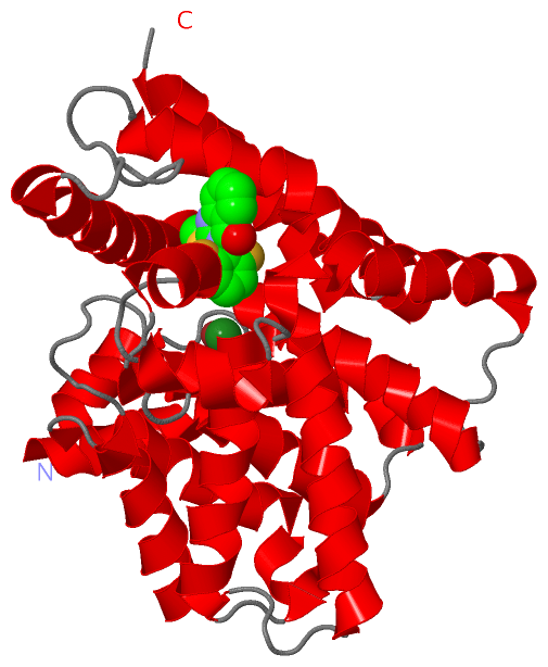
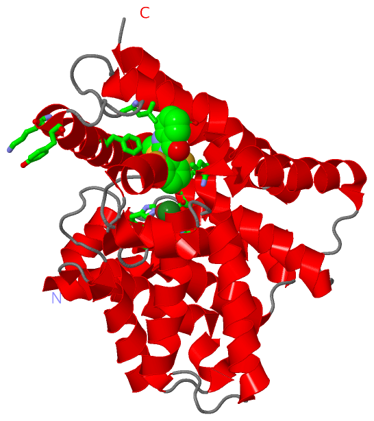
(-) Ligands, Modified Residues, Ions  (3, 3)

Asymmetric/Biological Unit (3, 3)
No.NameCountTypeFull Name
1MG1Ligand/IonMAGNESIUM ION
2TC81Ligand/Ion3-(2,6-DIFLUOROPHENYL)-2-(METHYLTHIO)QUINAZOLIN-4(3H)-ONE
3ZN1Ligand/IonZINC ION

(-) Sites  (3, 3)

Asymmetric Unit (3, 3)
No.NameEvidenceResiduesDescription
1AC1SOFTWAREILE A:323 , ILE A:363 , PHE A:384 , LYS A:393 , TYR A:394 , LEU A:401 , PHE A:416BINDING SITE FOR RESIDUE TC8 A 3
2AC2SOFTWAREHIS A:216 , HIS A:252 , ASP A:253 , ASP A:362BINDING SITE FOR RESIDUE ZN A 1
3AC3SOFTWAREASP A:253 , GLU A:282BINDING SITE FOR RESIDUE MG A 2

(-) SS Bonds  (0, 0)

(no "SS Bond" information available for 3G3N)

(-) Cis Peptide Bonds  (0, 0)

(no "Cis Peptide Bond" information available for 3G3N)

 Sequence-Structure Mapping

(-) SAPs(SNPs)/Variants  (0, 0)

(no "SAP(SNP)/Variant" information available for 3G3N)

(-) PROSITE Motifs  (1, 1)

Asymmetric/Biological Unit (1, 1)
 PROSITEUniProtKBPDB
No.IDACDescriptionIDLocationCountLocation
1PDEASE_IPS00126 3'5'-cyclic nucleotide phosphodiesterases signature.PDE7A_HUMAN252-263  1A:252-263

(-) Exons   (0, 0)

(no "Exon" information available for 3G3N)

(-) Sequences/Alignments

Asymmetric/Biological Unit
   Reformat: Number of residues per line =  ('0' or empty: single-line sequence representation)
  Number of residues per labelling interval =   
  UniProt sequence: complete  aligned part    
   Show mapping: SCOP domains CATH domains Pfam domains Secondary structure (by author)
SAPs(SNPs) PROSITE motifs Exons
(details for a mapped element are shown in a popup box when the mouse pointer rests over it)
Chain A from PDB  Type:PROTEIN  Length:318
 aligned with PDE7A_HUMAN | Q13946 from UniProtKB/Swiss-Prot  Length:482

    Alignment length:318
                                   148       158       168       178       188       198       208       218       228       238       248       258       268       278       288       298       308       318       328       338       348       358       368       378       388       398       408       418       428       438       448        
          PDE7A_HUMAN   139 DYNGQAKCMLEKVGNWNFDIFLFDRLTNGNSLVSLTFHLFSLHGLIEYFHLDMMKLRRFLVMIQEDYHSQNPYHNAVHAADVTQAMHCYLKEPKLANSVTPWDILLSLIAAATHDLDHPGVNQPFLIKTNHYLATLYKNTSVLENHHWRSAVGLLRESGLFSHLPLESRQQMETQIGALILATDISRQNEYLSLFRSHLDRGDLCLEDTRHRHLVLQMALKCADICNPCRTWELSKQWSEKVTEEFFHQGDIEKKYHLGVSPLCDRHTESIANIQIGFMTYLVEPLFTEWARFSNTRLSQTMLGHVGLNKASWKGLQR 456
               SCOP domains d3g3na_ A: automated matches                                                                                                                                                                                                                                                                                                   SCOP domains
               CATH domains ------------------------------------------------------------------------------------------------------------------------------------------------------------------------------------------------------------------------------------------------------------------------------------------------------------------------------ CATH domains
               Pfam domains ------------------------------------------------------------------------------------------------------------------------------------------------------------------------------------------------------------------------------------------------------------------------------------------------------------------------------ Pfam domains
         Sec.struct. author hhhhhhhhhhhhh.....hhhhhhhhh..hhhhhhhhhhhhhhhhhhhh..hhhhhhhhhhhhhhh........hhhhhhhhhhhhhhhhhhhhhhh..hhhhhhhhhhhhhhh.......hhhhhhhh.hhhhhhh...hhhhhhhhhhhhhhhhhhh.....hhhhhhhhhhhhhhhhhhhhhhhhhhhhhhhhhhhhhh.....hhhhhhhhhhhhhhhhhhhhhhhhhhhhhhhhhhhhhhhhhhhhhhhhh.............hhhhhhhhhhhhhhhhhhhhhhhhh.hhhhhhhhhhhhhhhhhhhhh.. Sec.struct. author
                 SAPs(SNPs) ------------------------------------------------------------------------------------------------------------------------------------------------------------------------------------------------------------------------------------------------------------------------------------------------------------------------------ SAPs(SNPs)
                    PROSITE -----------------------------------------------------------------------------------------------------------------PDEASE_I    ------------------------------------------------------------------------------------------------------------------------------------------------------------------------------------------------- PROSITE
                 Transcript ------------------------------------------------------------------------------------------------------------------------------------------------------------------------------------------------------------------------------------------------------------------------------------------------------------------------------ Transcript
                 3g3n A 139 DYNGQAKCMLEKVGNWNFDIFLFDRLTNGNSLVSLTFHLFSLHGLIEYFHLDMMKLRRFLVMIQEDYHSQNPYHNAVHAADVTQAMHCYLKEPKLANSVTPWDILLSLIAAATHDLDHPGVNQPFLIKTNHYLATLYKNTSVLENHHWRSAVGLLRESGLFSHLPLESRQQMETQIGALILATDISRQNEYLSLFRSHLDRGDLCLEDTRHRHLVLQMALKCADICNPCRTWELSKQWSEKVTEEFFHQGDIEKKYHLGVSPLCDRHTESIANIQIGFMTYLVEPLFTEWARFSNTRLSQTMLGHVGLNKASWKGLQR 456
                                   148       158       168       178       188       198       208       218       228       238       248       258       268       278       288       298       308       318       328       338       348       358       368       378       388       398       408       418       428       438       448        

   Legend:   → Mismatch (orange background)
  - → Gap (green background, '-', border residues have a numbering label)
    → Modified Residue (blue background, lower-case, 'x' indicates undefined single-letter code, labelled with number + name)
  x → Chemical Group (purple background, 'x', labelled with number + name, e.g. ACE or NH2)
  extra numbering lines below/above indicate numbering irregularities and modified residue names etc., number ends below/above '|'

 Classification and Annotation

(-) SCOP Domains  (1, 1)

Asymmetric/Biological Unit

(-) CATH Domains  (0, 0)

(no "CATH Domain" information available for 3G3N)

(-) Pfam Domains  (0, 0)

(no "Pfam Domain" information available for 3G3N)

(-) Gene Ontology  (11, 11)

Asymmetric/Biological Unit(hide GO term definitions)
Chain A   (PDE7A_HUMAN | Q13946)
molecular function
    GO:0004115    3',5'-cyclic-AMP phosphodiesterase activity    Catalysis of the reaction: adenosine 3',5'-cyclic phosphate + H2O = adenosine 5'-phosphate.
    GO:0004114    3',5'-cyclic-nucleotide phosphodiesterase activity    Catalysis of the reaction: nucleoside 3',5'-cyclic phosphate + H2O = nucleoside 5'-phosphate.
    GO:0004112    cyclic-nucleotide phosphodiesterase activity    Catalysis of the reaction: a nucleoside cyclic phosphate + H2O = a nucleoside phosphate.
    GO:0016787    hydrolase activity    Catalysis of the hydrolysis of various bonds, e.g. C-O, C-N, C-C, phosphoric anhydride bonds, etc. Hydrolase is the systematic name for any enzyme of EC class 3.
    GO:0046872    metal ion binding    Interacting selectively and non-covalently with any metal ion.
    GO:0008081    phosphoric diester hydrolase activity    Catalysis of the hydrolysis of a phosphodiester to give a phosphomonoester and a free hydroxyl group.
biological process
    GO:0006198    cAMP catabolic process    The chemical reactions and pathways resulting in the breakdown of the nucleotide cAMP (cyclic AMP, adenosine 3',5'-cyclophosphate).
    GO:0019933    cAMP-mediated signaling    Any intracellular signal transduction in which the signal is passed on within the cell via cyclic AMP (cAMP). Includes production of cAMP, and downstream effectors that further transmit the signal within the cell.
    GO:0007165    signal transduction    The cellular process in which a signal is conveyed to trigger a change in the activity or state of a cell. Signal transduction begins with reception of a signal (e.g. a ligand binding to a receptor or receptor activation by a stimulus such as light), or for signal transduction in the absence of ligand, signal-withdrawal or the activity of a constitutively active receptor. Signal transduction ends with regulation of a downstream cellular process, e.g. regulation of transcription or regulation of a metabolic process. Signal transduction covers signaling from receptors located on the surface of the cell and signaling via molecules located within the cell. For signaling between cells, signal transduction is restricted to events at and within the receiving cell.
cellular component
    GO:0005737    cytoplasm    All of the contents of a cell excluding the plasma membrane and nucleus, but including other subcellular structures.
    GO:0005829    cytosol    The part of the cytoplasm that does not contain organelles but which does contain other particulate matter, such as protein complexes.

 Visualization

(-) Interactive Views

Asymmetric/Biological Unit
  Complete Structure
    Jena3D(integrated viewing of ligand, site, SAP, PROSITE, SCOP information)
    WebMol | AstexViewer[tm]@PDBe
(Java Applets, require no local installation except for Java; loading may be slow)
    STRAP
(Java WebStart application, automatic local installation, requires Java; full application with system access!)
    RasMol
(require local installation)
    Molscript (VRML)
(requires installation of a VRML viewer; select preferred view via VRML and generate a mono or stereo PDF format file)
 
  Ligands, Modified Residues, Ions
    MG  [ RasMol | Jena3D ]  +environment [ RasMol | Jena3D ]
    TC8  [ RasMol | Jena3D ]  +environment [ RasMol | Jena3D ]
    ZN  [ RasMol | Jena3D ]  +environment [ RasMol | Jena3D ]
 
  Sites
    AC1  [ RasMol ]  +environment [ RasMol ]
    AC2  [ RasMol ]  +environment [ RasMol ]
    AC3  [ RasMol ]  +environment [ RasMol ]
 
  Cis Peptide Bonds
(no "Cis Peptide Bonds" information available for 3g3n)
 

(-) Still Images

Jmol
  protein: cartoon or spacefill or dots and stick; nucleic acid: cartoon and stick; ligands: spacefill; active site: stick
Molscript
  protein, nucleic acid: cartoon; ligands: spacefill; active site: ball and stick

 Databases and Analysis Tools

(-) Databases

Access by PDB/NDB ID
  3g3n
    Family and Domain InformationProDom | SYSTERS
    General Structural InformationGlycoscienceDB | MMDB | NDB | OCA | PDB | PDBe | PDBj | PDBsum | PDBWiki | PQS | PROTEOPEDIA
    Orientation in MembranesOPM
    Protein SurfaceSURFACE
    Secondary StructureDSSP (structure derived) | HSSP (homology derived)
    Structural GenomicsGeneCensus
    Structural NeighboursCE | VAST
    Structure ClassificationCATH | Dali | SCOP
    Validation and Original DataBMRB Data View | BMRB Restraints Grid | EDS | PROCHECK | RECOORD | WHAT_CHECK
 
Access by UniProt ID/Accession number
  PDE7A_HUMAN | Q13946
    Comparative Protein Structure ModelsModBase
    Genomic InformationEnsembl
    Protein-protein InteractionDIP
    Sequence, Family and Domain InformationInterPro | Pfam | SMART | UniProtKB/SwissProt
 
Access by Enzyme Classificator   (EC Number)
  3.1.4.17
    General Enzyme InformationBRENDA | EC-PDB | Enzyme | IntEnz
    PathwayKEGG | MetaCyc
 
Access by Disease Identifier   (MIM ID)
  (no 'MIM ID' available)
    Disease InformationOMIM
 
Access by GenAge ID
  (no 'GenAge ID' available)
    Age Related InformationGenAge

(-) Analysis Tools

Access by PDB/NDB ID
    Domain InformationXDom
    Interatomic Contacts of Structural UnitsCSU
    Ligand-protein ContactsLPC
    Protein CavitiescastP
    Sequence and Secondary StructurePDBCartoon
    Structure AlignmentSTRAP(Java WebStart application, automatic local installation, requires Java; full application with system access!)
    Structure and Sequence BrowserSTING
 
Access by UniProt ID/Accession number
  PDE7A_HUMAN | Q13946
    Protein Disorder PredictionDisEMBL | FoldIndex | GLOBPLOT (for more information see DisProt)

 Related Entries

(-) Entries Sharing at Least One Protein Chain (UniProt ID)

UniProtKB/Swiss-Prot
        PDE7A_HUMAN | Q139461zkl 4pm0 4y2b

(-) Related Entries Specified in the PDB File

(no "Related Entries Specified in the PDB File" available for 3G3N)