Show PDB file:   
         Plain Text   HTML   (compressed file size)
QuickSearch:   
by PDB,NDB,UniProt,PROSITE Code or Search Term(s)  
(-)Asym./Biol. Unit
(-)Asym./Biol. Unit - sites
collapse expand < >
Image Asym./Biol. Unit
Asym./Biol. Unit  (Jmol Viewer)
Image Asym./Biol. Unit - sites
Asym./Biol. Unit - sites  (Jmol Viewer)

(-) Description

Title :  LOW PH STRUCTURE OF THE RIESKE PROTEIN FROM THERMUS THERMOPHILUS AT 2.1 A
 
Authors :  L. M. Hunsicker-Wang, A. B. Taylor
Date :  02 Jan 09  (Deposition) - 20 Oct 09  (Release) - 13 Jul 11  (Revision)
Method :  X-RAY DIFFRACTION
Resolution :  2.10
Chains :  Asym./Biol. Unit :  A,B
Keywords :  Rieske Protein, Pr, [2Fe-2S], Electron Transport (Keyword Search: [Gene Ontology, PubMed, Web (Google))
 
Reference :  M. E. Konkle, S. K. Muellner, A. L. Schwander, M. M. Dicus, R. Pokhrel, R. D. Britt, A. B. Taylor, L. M. Hunsicker-Wang
Effects Of Ph On The Rieske Protein From Thermus Thermophilus: A Spectroscopic And Structural Analysis
Biochemistry V. 48 9848 2009
PubMed-ID: 19772300  |  Reference-DOI: 10.1021/BI901126U

(-) Compounds

Molecule 1 - QUINOL-CYTOCHROME C REDUCTASE, RIESKE IRON-SULFUR SUBUNIT
    ChainsA, B
    EngineeredYES
    Expression SystemESCHERICHIA COLI
    Expression System PlasmidPET17B
    Expression System StrainBL21(DE3)
    Expression System Taxid562
    Expression System Vector TypePLASMID
    GeneTTHA1931
    Organism ScientificTHERMUS THERMOPHILUS
    Organism Taxid300852
    StrainHB8

 Structural Features

(-) Chains, Units

  12
Asymmetric/Biological Unit AB

Summary Information (see also Sequences/Alignments below)

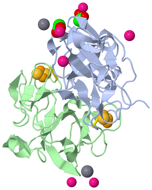
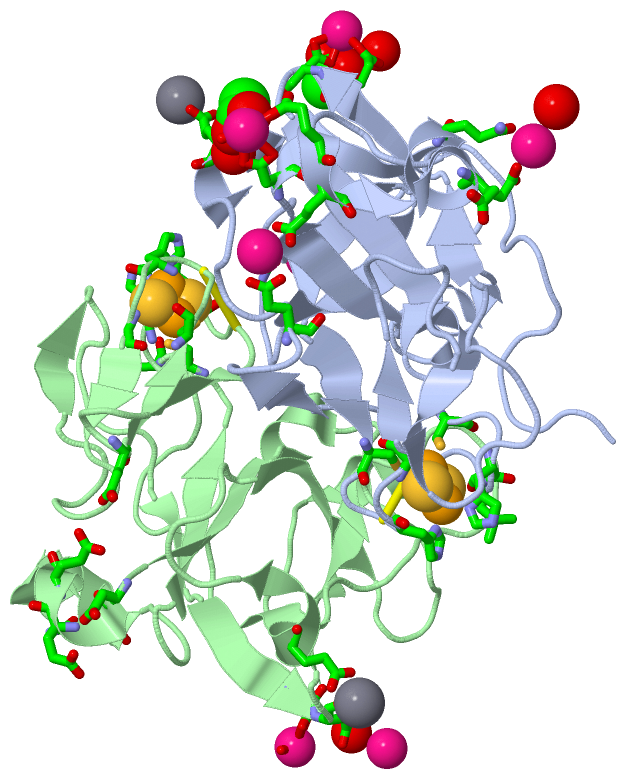
(-) Ligands, Modified Residues, Ions  (4, 12)

Asymmetric/Biological Unit (4, 12)
No.NameCountTypeFull Name
1ACT2Ligand/IonACETATE ION
2CA2Ligand/IonCALCIUM ION
3FES2Ligand/IonFE2/S2 (INORGANIC) CLUSTER
4PR6Ligand/IonPRASEODYMIUM ION

(-) Sites  (12, 12)

Asymmetric Unit (12, 12)
No.NameEvidenceResiduesDescription
01AC1SOFTWARECYS A:132 , HIS A:134 , LEU A:135 , CYS A:151 , HIS A:154 , GLY A:156BINDING SITE FOR RESIDUE FES A 204
02AC2SOFTWAREGLU A:66 , HOH A:203 , HOH A:227 , GLU B:116 , GLN B:119BINDING SITE FOR RESIDUE PR A 205
03AC3SOFTWAREASP A:109 , GLU A:111 , HOH A:210 , ACT A:213 , GLU B:112 , GLU B:147BINDING SITE FOR RESIDUE PR A 206
04AC4SOFTWAREGLU A:116 , GLN A:119 , HOH A:216 , HOH A:228BINDING SITE FOR RESIDUE PR A 207
05AC5SOFTWAREGLU A:112 , GLU A:147 , HOH A:211 , HOH A:233 , ASP B:109 , GLU B:111BINDING SITE FOR RESIDUE PR A 208
06AC6SOFTWAREGLU A:183 , ASP A:184 , ACT A:214 , ASP B:184 , HOH B:239BINDING SITE FOR RESIDUE PR A 209
07AC7SOFTWAREGLU A:73BINDING SITE FOR RESIDUE CA A 212
08AC8SOFTWAREHOH A:20 , ASP A:109 , GLU A:111 , PR A:206 , HOH A:210 , GLU B:147BINDING SITE FOR RESIDUE ACT A 213
09AC9SOFTWAREASP A:184 , PR A:209 , PR B:210BINDING SITE FOR RESIDUE ACT A 214
10BC1SOFTWARECYS B:132 , HIS B:134 , LEU B:135 , CYS B:151 , HIS B:154 , GLY B:156BINDING SITE FOR RESIDUE FES B 204
11BC2SOFTWAREGLU A:183 , ACT A:214 , HOH A:231 , GLU B:183 , GLN B:200 , HOH B:240BINDING SITE FOR RESIDUE PR B 210
12BC3SOFTWAREGLU B:73BINDING SITE FOR RESIDUE CA B 211

(-) SS Bonds  (2, 2)

Asymmetric/Biological Unit
No.Residues
1A:137 -A:153
2B:137 -B:153

(-) Cis Peptide Bonds  (0, 0)

(no "Cis Peptide Bond" information available for 3FOU)

 Sequence-Structure Mapping

(-) SAPs(SNPs)/Variants  (0, 0)

(no "SAP(SNP)/Variant" information available for 3FOU)

(-) PROSITE Motifs  (0, 0)

(no "PROSITE Motif" information available for 3FOU)

(-) Exons   (0, 0)

(no "Exon" information available for 3FOU)

(-) Sequences/Alignments

Asymmetric/Biological Unit
   Reformat: Number of residues per line =  ('0' or empty: single-line sequence representation)
  Number of residues per labelling interval =   
  UniProt sequence: complete  aligned part    
   Show mapping: SCOP domains CATH domains Pfam domains Secondary structure (by author)
SAPs(SNPs) PROSITE motifs Exons
(details for a mapped element are shown in a popup box when the mouse pointer rests over it)
Chain A from PDB  Type:PROTEIN  Length:156
 aligned with Q5SGZ9_THET8 | Q5SGZ9 from UniProtKB/TrEMBL  Length:210

    Alignment length:156
                                    55        65        75        85        95       105       115       125       135       145       155       165       175       185       195      
         Q5SGZ9_THET8    46 TPEKEPLKPGDILVYAQGGGEPKPIRLEELKPGDPFVLAYPMDPKTKVVKSGEAKNTLLVARFDPEELAPEVAQHAAEGVVAYSAVCTHLGCIVSQWVADEEAALCPCHGGVYDLRHGAQVIAGPPPRPVPQLPVRVEDGVLVAAGEFLGPVGVQA 201
               SCOP domains d3foua_ A: automated matches                                                                                                                                 SCOP domains
               CATH domains 3fouA00 A:46-201 'Rieske'-like iron-sulphur domains                                                                                                          CATH domains
               Pfam domains ------------------------------------------------------------------------------------------------------------------------------------------------------------ Pfam domains
         Sec.struct. author hhhhh......eeee..........hhhhh......eeeeee..........hhhh.eeeee.hhhhhhhhhhh.....eeeee...........eee....eee......ee.......eee........ee.eeee..eeee............ Sec.struct. author
                 SAPs(SNPs) ------------------------------------------------------------------------------------------------------------------------------------------------------------ SAPs(SNPs)
                    PROSITE ------------------------------------------------------------------------------------------------------------------------------------------------------------ PROSITE
                 Transcript ------------------------------------------------------------------------------------------------------------------------------------------------------------ Transcript
                 3fou A  46 TPEKEPLKPGDILVYAQGGGEPKPIRLEELKPGDPFVLAYPMDPKTKVVKSGEAKNTLLVARFDPEELAPEVAQHAAEGVVAYSAVCTHLGCIVSQWVADEEAALCPCHGGVYDLRHGAQVIAGPPPRPVPQLPVRVEDGVLVAAGEFLGPVGVQA 201
                                    55        65        75        85        95       105       115       125       135       145       155       165       175       185       195      

Chain B from PDB  Type:PROTEIN  Length:156
 aligned with Q5SGZ9_THET8 | Q5SGZ9 from UniProtKB/TrEMBL  Length:210

    Alignment length:156
                                    55        65        75        85        95       105       115       125       135       145       155       165       175       185       195      
         Q5SGZ9_THET8    46 TPEKEPLKPGDILVYAQGGGEPKPIRLEELKPGDPFVLAYPMDPKTKVVKSGEAKNTLLVARFDPEELAPEVAQHAAEGVVAYSAVCTHLGCIVSQWVADEEAALCPCHGGVYDLRHGAQVIAGPPPRPVPQLPVRVEDGVLVAAGEFLGPVGVQA 201
               SCOP domains d3foub_ B: automated matches                                                                                                                                 SCOP domains
               CATH domains 3fouB00 B:46-201 'Rieske'-like iron-sulphur domains                                                                                                          CATH domains
               Pfam domains ------------------------------------------------------------------------------------------------------------------------------------------------------------ Pfam domains
         Sec.struct. author hhhhh......eeee..........hhhhh......eeeeee..........hhhh.eeeee.hhhhhhhhhhh.....eeeee...........eee....eee......ee.......eee........ee.eeee..eeee............ Sec.struct. author
                 SAPs(SNPs) ------------------------------------------------------------------------------------------------------------------------------------------------------------ SAPs(SNPs)
                    PROSITE ------------------------------------------------------------------------------------------------------------------------------------------------------------ PROSITE
                 Transcript ------------------------------------------------------------------------------------------------------------------------------------------------------------ Transcript
                 3fou B  46 TPEKEPLKPGDILVYAQGGGEPKPIRLEELKPGDPFVLAYPMDPKTKVVKSGEAKNTLLVARFDPEELAPEVAQHAAEGVVAYSAVCTHLGCIVSQWVADEEAALCPCHGGVYDLRHGAQVIAGPPPRPVPQLPVRVEDGVLVAAGEFLGPVGVQA 201
                                    55        65        75        85        95       105       115       125       135       145       155       165       175       185       195      

   Legend:   → Mismatch (orange background)
  - → Gap (green background, '-', border residues have a numbering label)
    → Modified Residue (blue background, lower-case, 'x' indicates undefined single-letter code, labelled with number + name)
  x → Chemical Group (purple background, 'x', labelled with number + name, e.g. ACE or NH2)
  extra numbering lines below/above indicate numbering irregularities and modified residue names etc., number ends below/above '|'

 Classification and Annotation

(-) SCOP Domains  (1, 2)

Asymmetric/Biological Unit

(-) CATH Domains  (1, 2)

Asymmetric/Biological Unit
(-)
Class: Mainly Beta (13760)

(-) Pfam Domains  (0, 0)

(no "Pfam Domain" information available for 3FOU)

(-) Gene Ontology  (8, 8)

Asymmetric/Biological Unit(hide GO term definitions)
Chain A,B   (Q5SGZ9_THET8 | Q5SGZ9)
molecular function
    GO:0051537    2 iron, 2 sulfur cluster binding    Interacting selectively and non-covalently with a 2 iron, 2 sulfur (2Fe-2S) cluster; this cluster consists of two iron atoms, with two inorganic sulfur atoms found between the irons and acting as bridging ligands.
    GO:0051536    iron-sulfur cluster binding    Interacting selectively and non-covalently with an iron-sulfur cluster, a combination of iron and sulfur atoms.
    GO:0046872    metal ion binding    Interacting selectively and non-covalently with any metal ion.
    GO:0016491    oxidoreductase activity    Catalysis of an oxidation-reduction (redox) reaction, a reversible chemical reaction in which the oxidation state of an atom or atoms within a molecule is altered. One substrate acts as a hydrogen or electron donor and becomes oxidized, while the other acts as hydrogen or electron acceptor and becomes reduced.
    GO:0016679    oxidoreductase activity, acting on diphenols and related substances as donors    Catalysis of an oxidation-reduction (redox) reaction in which a diphenol or related substance acts as a hydrogen or electron donor and reduces a hydrogen or electron acceptor.
biological process
    GO:0055114    oxidation-reduction process    A metabolic process that results in the removal or addition of one or more electrons to or from a substance, with or without the concomitant removal or addition of a proton or protons.
cellular component
    GO:0016021    integral component of membrane    The component of a membrane consisting of the gene products and protein complexes having at least some part of their peptide sequence embedded in the hydrophobic region of the membrane.
    GO:0016020    membrane    A lipid bilayer along with all the proteins and protein complexes embedded in it an attached to it.

 Visualization

(-) Interactive Views

Asymmetric/Biological Unit
  Complete Structure
    Jena3D(integrated viewing of ligand, site, SAP, PROSITE, SCOP information)
    WebMol | AstexViewer[tm]@PDBe
(Java Applets, require no local installation except for Java; loading may be slow)
    STRAP
(Java WebStart application, automatic local installation, requires Java; full application with system access!)
    RasMol
(require local installation)
    Molscript (VRML)
(requires installation of a VRML viewer; select preferred view via VRML and generate a mono or stereo PDF format file)
 
  Ligands, Modified Residues, Ions
    ACT  [ RasMol | Jena3D ]  +environment [ RasMol | Jena3D ]
    CA  [ RasMol | Jena3D ]  +environment [ RasMol | Jena3D ]
    FES  [ RasMol | Jena3D ]  +environment [ RasMol | Jena3D ]
    PR  [ RasMol | Jena3D ]  +environment [ RasMol | Jena3D ]
 
  Sites
    AC1  [ RasMol ]  +environment [ RasMol ]
    AC2  [ RasMol ]  +environment [ RasMol ]
    AC3  [ RasMol ]  +environment [ RasMol ]
    AC4  [ RasMol ]  +environment [ RasMol ]
    AC5  [ RasMol ]  +environment [ RasMol ]
    AC6  [ RasMol ]  +environment [ RasMol ]
    AC7  [ RasMol ]  +environment [ RasMol ]
    AC8  [ RasMol ]  +environment [ RasMol ]
    AC9  [ RasMol ]  +environment [ RasMol ]
    BC1  [ RasMol ]  +environment [ RasMol ]
    BC2  [ RasMol ]  +environment [ RasMol ]
    BC3  [ RasMol ]  +environment [ RasMol ]
 
  Cis Peptide Bonds
(no "Cis Peptide Bonds" information available for 3fou)
 

(-) Still Images

Jmol
  protein: cartoon or spacefill or dots and stick; nucleic acid: cartoon and stick; ligands: spacefill; active site: stick
Molscript
  protein, nucleic acid: cartoon; ligands: spacefill; active site: ball and stick

 Databases and Analysis Tools

(-) Databases

Access by PDB/NDB ID
  3fou
    Family and Domain InformationProDom | SYSTERS
    General Structural InformationGlycoscienceDB | MMDB | NDB | OCA | PDB | PDBe | PDBj | PDBsum | PDBWiki | PQS | PROTEOPEDIA
    Orientation in MembranesOPM
    Protein SurfaceSURFACE
    Secondary StructureDSSP (structure derived) | HSSP (homology derived)
    Structural GenomicsGeneCensus
    Structural NeighboursCE | VAST
    Structure ClassificationCATH | Dali | SCOP
    Validation and Original DataBMRB Data View | BMRB Restraints Grid | EDS | PROCHECK | RECOORD | WHAT_CHECK
 
Access by UniProt ID/Accession number
  Q5SGZ9_THET8 | Q5SGZ9
    Comparative Protein Structure ModelsModBase
    Genomic InformationEnsembl
    Protein-protein InteractionDIP
    Sequence, Family and Domain InformationInterPro | Pfam | SMART | UniProtKB/TrEMBL
 
Access by Enzyme Classificator   (EC Number)
  (no 'Enzyme Classificator' available)
    General Enzyme InformationBRENDA | EC-PDB | Enzyme | IntEnz
    PathwayKEGG | MetaCyc
 
Access by Disease Identifier   (MIM ID)
  (no 'MIM ID' available)
    Disease InformationOMIM
 
Access by GenAge ID
  (no 'GenAge ID' available)
    Age Related InformationGenAge

(-) Analysis Tools

Access by PDB/NDB ID
    Domain InformationXDom
    Interatomic Contacts of Structural UnitsCSU
    Ligand-protein ContactsLPC
    Protein CavitiescastP
    Sequence and Secondary StructurePDBCartoon
    Structure AlignmentSTRAP(Java WebStart application, automatic local installation, requires Java; full application with system access!)
    Structure and Sequence BrowserSTING
 
Access by UniProt ID/Accession number
  Q5SGZ9_THET8 | Q5SGZ9
    Protein Disorder PredictionDisEMBL | FoldIndex | GLOBPLOT (for more information see DisProt)

 Related Entries

(-) Entries Sharing at Least One Protein Chain (UniProt ID)

UniProtKB/TrEMBL
        Q5SGZ9_THET8 | Q5SGZ91nyk

(-) Related Entries Specified in the PDB File

1nyk HIGH PH STRUCTURE OF THE RIESKE PROTEIN FROM THERMUS THERMOPHILUS AT 1.3A