|
|
|
|
 Description
|
|
Compounds
|
||||||||||||||||||||||||
Chains, Units
Summary Information (see also Sequences/Alignments below) |
Ligands, Modified Residues, Ions (2, 5)| Asymmetric/Biological Unit (2, 5) |
Sites (5, 5)
Asymmetric Unit (5, 5)
|
SS Bonds (7, 7)
Asymmetric/Biological Unit
|
||||||||||||||||||||||||||||||||
Cis Peptide Bonds (1, 1)
Asymmetric/Biological Unit
|
||||||||
SAPs(SNPs)/Variants (0, 0)| (no "SAP(SNP)/Variant" information available for 3FO7) |
PROSITE Motifs (0, 0)| (no "PROSITE Motif" information available for 3FO7) |
Exons (0, 0)| (no "Exon" information available for 3FO7) |
Sequences/Alignments
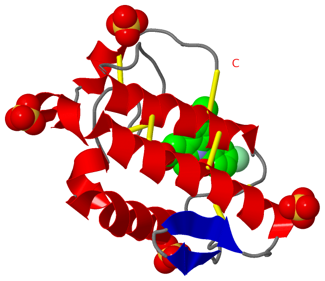
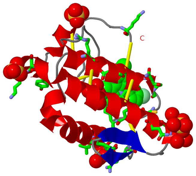
Asymmetric/Biological UnitChain A from PDB Type:PROTEIN Length:121 aligned with D0VX11_9SAUR | D0VX11 from UniProtKB/TrEMBL Length:121 Alignment length:121 10 20 30 40 50 60 70 80 90 100 110 120 D0VX11_9SAUR 1 SLLEFGKMILEETGKLAIPSYSSYGCYCGWGGKGTPKDATDRCCFVHDCCYGNLPDCNPKSDRYKYKRVNGAIVCEKGTSCENRICECDKAAAICFRQNLNTYSKKYMLYPDFLCKGELKC 121 SCOP domains d3fo7a_ A: Snake phospholipase A2 SCOP domains CATH domains 3fo7A00 A:1-133 Phospholipase A2 CATH domains Pfam domains ------------------------------------------------------------------------------------------------------------------------- Pfam domains SAPs(SNPs) ------------------------------------------------------------------------------------------------------------------------- SAPs(SNPs) PROSITE ------------------------------------------------------------------------------------------------------------------------- PROSITE Transcript ------------------------------------------------------------------------------------------------------------------------- Transcript 3fo7 A 1 SLLEFGKMILEETGKLAIPSYSSYGCYCGWGGKGTPKDATDRCCFVHDCCYGNLPDCNPKSDRYKYKRVNGAIVCEKGTSCENRICECDKAAAICFRQNLNTYSKKYMLYPDFLCKGELKC 133 10 || 21 31 41 51 ||||69 79 ||90 100 110 120 || 131| 14| 56||| 86| 122| 131| 16 59|| 88 124 133 61| 67
|
||||||||||||||||||||
SCOP Domains (1, 1)
Asymmetric/Biological Unit
 |
CATH Domains (1, 1)
Asymmetric/Biological Unit
|
Pfam Domains (0, 0)| (no "Pfam Domain" information available for 3FO7) |
Gene Ontology (4, 4)|
Asymmetric/Biological Unit(hide GO term definitions) Chain A (D0VX11_9SAUR | D0VX11)
|
||||||||||||||||||||||||||||||||||||||||||
Interactive Views
|
||||||||||||||||||||||||||||||||||||||||||||||||||||||||||||||||||||||||||||||||||||||||||||||||||||||||||||||||||||||||||||||||||||||||||||||||||||||||||
Still Images
|
||||||||||||||||
Databases
|
||||||||||||||||||||||||||||||||||||||||||||||||||||||||||||||||||||||||||||||||||||||||||||||||||||||||||||||||||||||||||||||||||||||||||||||||||||||||||||||||
Analysis Tools
|
|||||||||||||||||||||||||||||||||||||||||||||||||||||||||||||
Entries Sharing at Least One Protein Chain (UniProt ID)
Related Entries Specified in the PDB File
|
|