Show PDB file:   
         Plain Text   HTML   (compressed file size)
QuickSearch:   
by PDB,NDB,UniProt,PROSITE Code or Search Term(s)  
(-)Asym./Biol. Unit
(-)Asym./Biol. Unit - sites
collapse expand < >
Image Asym./Biol. Unit
Asym./Biol. Unit  (Jmol Viewer)
Image Asym./Biol. Unit - sites
Asym./Biol. Unit - sites  (Jmol Viewer)

(-) Description

Title :  CRYSTAL STRUCTURE OF MINER1: THE REDOX-ACTIVE 2FE-2S PROTEIN CAUSATIVE IN WOLFRAM SYNDROME 2
 
Authors :  A. R. Conlan, H. L. Axelrod, A. E. Cohen, E. C. Abresch, D. Yee, J. Zuris, R. Nechushtai, P. A. Jennings, M. L. Paddock
Date :  26 Dec 08  (Deposition) - 18 Aug 09  (Release) - 13 Jul 11  (Revision)
Method :  X-RAY DIFFRACTION
Resolution :  2.10
Chains :  Asym./Biol. Unit :  A,B
Keywords :  Diabetes, Membrane Bound, Thiazolidinedione, Oxidative Stress, Cdgsh, Endoplasmic Reticulum, Iron, Iron-Sulfur, Membrane, Metal-Binding, Transmembrane, Zinc-Finger, Metal Binding Protein (Keyword Search: [Gene Ontology, PubMed, Web (Google))
 
Reference :  A. R. Conlan, H. L. Axelrod, A. E. Cohen, E. C. Abresch, J. Zuris, D. Yee, R. Nechushtai, P. A. Jennings, M. L. Paddock
Crystal Structure Of Miner1: The Redox-Active 2Fe-2S Protei Causative In Wolfram Syndrome 2.
J. Mol. Biol. V. 392 143 2009
PubMed-ID: 19580816  |  Reference-DOI: 10.1016/J.JMB.2009.06.079

(-) Compounds

Molecule 1 - CDGSH IRON SULFUR DOMAIN-CONTAINING PROTEIN 2
    ChainsA, B
    EngineeredYES
    Expression SystemESCHERICHIA COLI
    Expression System PlasmidPET28A(+)
    Expression System Taxid562
    Expression System Vector TypePLASMID
    FragmentC-TERMINAL WATER-SOLUBLE DOMAIN: UNP RESIDUES 57-135
    GeneCDGSH2, CISD2, ERIS, ZCD2
    MutationYES
    Organism CommonHUMAN
    Organism ScientificHOMO SAPIENS
    Organism Taxid9606
    SynonymENDOPLASMIC RETICULUM INTERMEMBRANE SMALL PROTEIN, MITONEET- RELATED 1 PROTEIN, MINER1

 Structural Features

(-) Chains, Units

  12
Asymmetric/Biological Unit AB

Summary Information (see also Sequences/Alignments below)

(-) Ligands, Modified Residues, Ions  (1, 2)

Asymmetric/Biological Unit (1, 2)
No.NameCountTypeFull Name
1FES2Ligand/IonFE2/S2 (INORGANIC) CLUSTER

(-) Sites  (2, 2)

Asymmetric Unit (2, 2)
No.NameEvidenceResiduesDescription
1AC1SOFTWARECYS A:99 , ARG A:100 , CYS A:101 , CYS A:110 , ASP A:111 , GLY A:112 , SER A:113 , HIS A:114BINDING SITE FOR RESIDUE FES A 200
2AC2SOFTWARECYS B:99 , ARG B:100 , CYS B:101 , CYS B:110 , ASP B:111 , SER B:113 , HIS B:114BINDING SITE FOR RESIDUE FES B 200

(-) SS Bonds  (0, 0)

(no "SS Bond" information available for 3FNV)

(-) Cis Peptide Bonds  (2, 2)

Asymmetric/Biological Unit
No.Residues
1Phe A:107 -Pro A:108
2Phe B:107 -Pro B:108

 Sequence-Structure Mapping

(-) SAPs(SNPs)/Variants  (0, 0)

(no "SAP(SNP)/Variant" information available for 3FNV)

(-) PROSITE Motifs  (0, 0)

(no "PROSITE Motif" information available for 3FNV)

(-) Exons   (0, 0)

(no "Exon" information available for 3FNV)

(-) Sequences/Alignments

Asymmetric/Biological Unit
   Reformat: Number of residues per line =  ('0' or empty: single-line sequence representation)
  Number of residues per labelling interval =   
  UniProt sequence: complete  aligned part    
   Show mapping: SCOP domains CATH domains Pfam domains Secondary structure (by author)
SAPs(SNPs) PROSITE motifs Exons
(details for a mapped element are shown in a popup box when the mouse pointer rests over it)
Chain A from PDB  Type:PROTEIN  Length:67
 aligned with CISD2_HUMAN | Q8N5K1 from UniProtKB/Swiss-Prot  Length:135

    Alignment length:67
                                    77        87        97       107       117       127       
          CISD2_HUMAN    68 DSLINLKIQKENPKVVNEINIEDLCLTKAAYCRCWRSKTFPACDGSHNKHNELTGDNVGPLILKKKE 134
               SCOP domains ------------------------------------------------------------------- SCOP domains
               CATH domains ------------------------------------------------------------------- CATH domains
               Pfam domains ------------------------------------------------------------------- Pfam domains
         Sec.struct. author ..............eeeeee.hhh...eeee.............hhhhhhhhhhh..eeeeeee... Sec.struct. author
                 SAPs(SNPs) ------------------------------------------------------------------- SAPs(SNPs)
                    PROSITE ------------------------------------------------------------------- PROSITE
                 Transcript ------------------------------------------------------------------- Transcript
                 3fnv A  68 DSLINLKIQKENPKVVNEINIEDLSLTKAAYCRCWRSKTFPACDGSHNKHNELTGDNVGPLILKKKE 134
                                    77        87        97       107       117       127       

Chain B from PDB  Type:PROTEIN  Length:68
 aligned with CISD2_HUMAN | Q8N5K1 from UniProtKB/Swiss-Prot  Length:135

    Alignment length:68
                                    77        87        97       107       117       127        
          CISD2_HUMAN    68 DSLINLKIQKENPKVVNEINIEDLCLTKAAYCRCWRSKTFPACDGSHNKHNELTGDNVGPLILKKKEV 135
               SCOP domains -------------------------------------------------------------------- SCOP domains
               CATH domains -------------------------------------------------------------------- CATH domains
               Pfam domains -------------------------------------------------------------------- Pfam domains
         Sec.struct. author ..............eeeeee.hhh...eeee.............hhhhhhhhhhh..eeeeeee.... Sec.struct. author
                 SAPs(SNPs) -------------------------------------------------------------------- SAPs(SNPs)
                    PROSITE -------------------------------------------------------------------- PROSITE
                 Transcript -------------------------------------------------------------------- Transcript
                 3fnv B  68 DSLINLKIQKENPKVVNEINIEDLSLTKAAYCRCWRSKTFPACDGSHNKHNELTGDNVGPLILKKKEV 135
                                    77        87        97       107       117       127        

   Legend:   → Mismatch (orange background)
  - → Gap (green background, '-', border residues have a numbering label)
    → Modified Residue (blue background, lower-case, 'x' indicates undefined single-letter code, labelled with number + name)
  x → Chemical Group (purple background, 'x', labelled with number + name, e.g. ACE or NH2)
  extra numbering lines below/above indicate numbering irregularities and modified residue names etc., number ends below/above '|'

 Classification and Annotation

(-) SCOP Domains  (0, 0)

(no "SCOP Domain" information available for 3FNV)

(-) CATH Domains  (0, 0)

(no "CATH Domain" information available for 3FNV)

(-) Pfam Domains  (0, 0)

(no "Pfam Domain" information available for 3FNV)

(-) Gene Ontology  (17, 17)

Asymmetric/Biological Unit(hide GO term definitions)
Chain A,B   (CISD2_HUMAN | Q8N5K1)
molecular function
    GO:0051537    2 iron, 2 sulfur cluster binding    Interacting selectively and non-covalently with a 2 iron, 2 sulfur (2Fe-2S) cluster; this cluster consists of two iron atoms, with two inorganic sulfur atoms found between the irons and acting as bridging ligands.
    GO:0051536    iron-sulfur cluster binding    Interacting selectively and non-covalently with an iron-sulfur cluster, a combination of iron and sulfur atoms.
    GO:0046872    metal ion binding    Interacting selectively and non-covalently with any metal ion.
    GO:0005515    protein binding    Interacting selectively and non-covalently with any protein or protein complex (a complex of two or more proteins that may include other nonprotein molecules).
    GO:0042803    protein homodimerization activity    Interacting selectively and non-covalently with an identical protein to form a homodimer.
biological process
    GO:0006914    autophagy    The process in which cells digest parts of their own cytoplasm; allows for both recycling of macromolecular constituents under conditions of cellular stress and remodeling the intracellular structure for cell differentiation.
    GO:0000422    autophagy of mitochondrion    The autophagic process in which mitochondria are delivered to the vacuole and degraded in response to changing cellular conditions.
    GO:0010259    multicellular organism aging    An aging process that has as participant a whole multicellular organism. Multicellular organism aging includes loss of functions such as resistance to disease, homeostasis, and fertility, as well as wear and tear. Multicellular organisms aging includes processes like cellular senescence and organ senescence, but is more inclusive. May precede death (GO:0016265) of an organism and may succeed developmental maturation (GO:0021700).
    GO:0010506    regulation of autophagy    Any process that modulates the frequency, rate or extent of autophagy. Autophagy is the process in which cells digest parts of their own cytoplasm.
cellular component
    GO:0005783    endoplasmic reticulum    The irregular network of unit membranes, visible only by electron microscopy, that occurs in the cytoplasm of many eukaryotic cells. The membranes form a complex meshwork of tubular channels, which are often expanded into slitlike cavities called cisternae. The ER takes two forms, rough (or granular), with ribosomes adhering to the outer surface, and smooth (with no ribosomes attached).
    GO:0005789    endoplasmic reticulum membrane    The lipid bilayer surrounding the endoplasmic reticulum.
    GO:0016021    integral component of membrane    The component of a membrane consisting of the gene products and protein complexes having at least some part of their peptide sequence embedded in the hydrophobic region of the membrane.
    GO:0043231    intracellular membrane-bounded organelle    Organized structure of distinctive morphology and function, bounded by a single or double lipid bilayer membrane and occurring within the cell. Includes the nucleus, mitochondria, plastids, vacuoles, and vesicles. Excludes the plasma membrane.
    GO:0016020    membrane    A lipid bilayer along with all the proteins and protein complexes embedded in it an attached to it.
    GO:0005741    mitochondrial outer membrane    The outer, i.e. cytoplasm-facing, lipid bilayer of the mitochondrial envelope.
    GO:0005739    mitochondrion    A semiautonomous, self replicating organelle that occurs in varying numbers, shapes, and sizes in the cytoplasm of virtually all eukaryotic cells. It is notably the site of tissue respiration.
    GO:0043234    protein complex    A stable macromolecular complex composed (only) of two or more polypeptide subunits along with any covalently attached molecules (such as lipid anchors or oligosaccharide) or non-protein prosthetic groups (such as nucleotides or metal ions). Prosthetic group in this context refers to a tightly bound cofactor. The component polypeptide subunits may be identical.
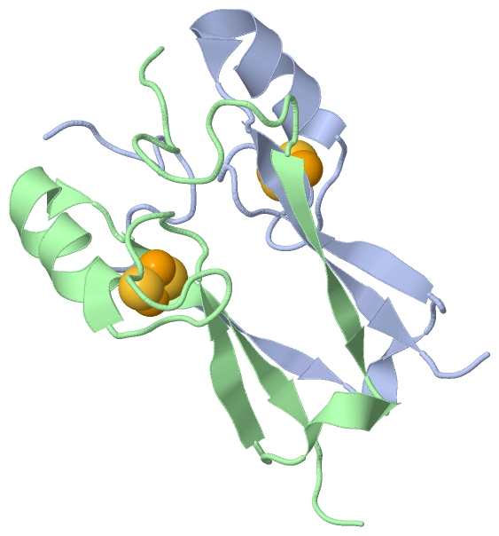
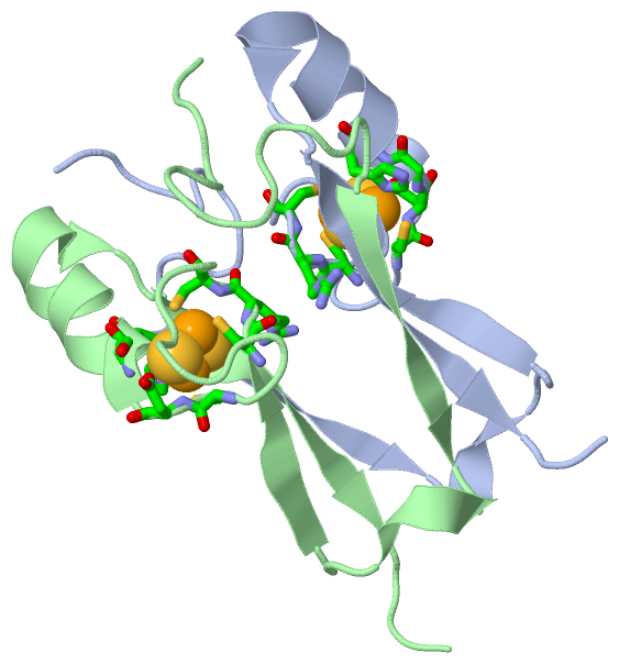
 Visualization

(-) Interactive Views

Asymmetric/Biological Unit
  Complete Structure
    Jena3D(integrated viewing of ligand, site, SAP, PROSITE, SCOP information)
    WebMol | AstexViewer[tm]@PDBe
(Java Applets, require no local installation except for Java; loading may be slow)
    STRAP
(Java WebStart application, automatic local installation, requires Java; full application with system access!)
    RasMol
(require local installation)
    Molscript (VRML)
(requires installation of a VRML viewer; select preferred view via VRML and generate a mono or stereo PDF format file)
 
  Ligands, Modified Residues, Ions
    FES  [ RasMol | Jena3D ]  +environment [ RasMol | Jena3D ]
 
  Sites
    AC1  [ RasMol ]  +environment [ RasMol ]
    AC2  [ RasMol ]  +environment [ RasMol ]
 
  Cis Peptide Bonds
    Phe A:107 - Pro A:108   [ RasMol ]  
    Phe B:107 - Pro B:108   [ RasMol ]  
 

(-) Still Images

Jmol
  protein: cartoon or spacefill or dots and stick; nucleic acid: cartoon and stick; ligands: spacefill; active site: stick
Molscript
  protein, nucleic acid: cartoon; ligands: spacefill; active site: ball and stick

 Databases and Analysis Tools

(-) Databases

Access by PDB/NDB ID
  3fnv
    Family and Domain InformationProDom | SYSTERS
    General Structural InformationGlycoscienceDB | MMDB | NDB | OCA | PDB | PDBe | PDBj | PDBsum | PDBWiki | PQS | PROTEOPEDIA
    Orientation in MembranesOPM
    Protein SurfaceSURFACE
    Secondary StructureDSSP (structure derived) | HSSP (homology derived)
    Structural GenomicsGeneCensus
    Structural NeighboursCE | VAST
    Structure ClassificationCATH | Dali | SCOP
    Validation and Original DataBMRB Data View | BMRB Restraints Grid | EDS | PROCHECK | RECOORD | WHAT_CHECK
 
Access by UniProt ID/Accession number
  CISD2_HUMAN | Q8N5K1
    Comparative Protein Structure ModelsModBase
    Genomic InformationEnsembl
    Protein-protein InteractionDIP
    Sequence, Family and Domain InformationInterPro | Pfam | SMART | UniProtKB/SwissProt
 
Access by Enzyme Classificator   (EC Number)
  (no 'Enzyme Classificator' available)
    General Enzyme InformationBRENDA | EC-PDB | Enzyme | IntEnz
    PathwayKEGG | MetaCyc
 
Access by Disease Identifier   (MIM ID)
  (no 'MIM ID' available)
    Disease InformationOMIM
 
Access by GenAge ID
  (no 'GenAge ID' available)
    Age Related InformationGenAge

(-) Analysis Tools

Access by PDB/NDB ID
    Domain InformationXDom
    Interatomic Contacts of Structural UnitsCSU
    Ligand-protein ContactsLPC
    Protein CavitiescastP
    Sequence and Secondary StructurePDBCartoon
    Structure AlignmentSTRAP(Java WebStart application, automatic local installation, requires Java; full application with system access!)
    Structure and Sequence BrowserSTING
 
Access by UniProt ID/Accession number
  CISD2_HUMAN | Q8N5K1
    Protein Disorder PredictionDisEMBL | FoldIndex | GLOBPLOT (for more information see DisProt)

 Related Entries

(-) Entries Sharing at Least One Protein Chain (UniProt ID)

UniProtKB/Swiss-Prot
        CISD2_HUMAN | Q8N5K14oo7 4ooa

(-) Related Entries Specified in the PDB File

2qh7 MITONEET IS A UNIQUELY FOLDED 2FE-2S OUTER MITOCHONDRIAL MEMBRANE PROTEIN STABILIZED BY PIOGLITAZONE