Show PDB file:   
         Plain Text   HTML   (compressed file size)
QuickSearch:   
by PDB,NDB,UniProt,PROSITE Code or Search Term(s)  
(-)Asym./Biol. Unit
(-)Asym./Biol. Unit - sites
collapse expand < >
Image Asym./Biol. Unit
Asym./Biol. Unit  (Jmol Viewer)
Image Asym./Biol. Unit - sites
Asym./Biol. Unit - sites  (Jmol Viewer)

(-) Description

Title :  CRYSTAL STRUCTURE OF D235A MUTANT OF HUMAN PYRIDOXAL KINASE
 
Authors :  M. K. Safo, A. K. Gandhi, F. N. Musayev, M. Ghatge, M. L. Di Salvo, V. Schi
Date :  10 Dec 08  (Deposition) - 23 Dec 08  (Release) - 13 Jul 11  (Revision)
Method :  X-RAY DIFFRACTION
Resolution :  2.50
Chains :  Asym./Biol. Unit :  A,B
Keywords :  Beta Sheet With Alpha Helix, Atp Complex, Metal Ion, Transferase, Atp-Binding, Kinase, Metal-Binding, Nucleotide-Binding (Keyword Search: [Gene Ontology, PubMed, Web (Google))
 
Reference :  A. K. Gandhi, M. S. Ghatge, F. N. Musayev, A. Sease, S. O. Aboagye, M. L. Di Salvo, V. Schirch, M. K. Safo
Kinetic And Structural Studies Of The Role Of The Active Site Residue Asp235 Of Human Pyridoxal Kinase.
Biochem. Biophys. Res. Commun. V. 381 12 2009
PubMed-ID: 19351586  |  Reference-DOI: 10.1016/J.BBRC.2009.01.170

(-) Compounds

Molecule 1 - PYRIDOXAL KINASE
    ChainsA, B
    EC Number2.7.1.35
    EngineeredYES
    Expression SystemESCHERICHIA COLI
    Expression System PlasmidPET28
    Expression System StrainRPSETTA(DE3)PLYSS COMPETENT CELLS
    Expression System Taxid562
    Expression System Vector TypePLASMID
    GeneC21ORF124, C21ORF97, PDXK, PKH, PNK
    MutationYES
    Organism CommonHUMAN
    Organism ScientificHOMO SAPIENS
    Organism Taxid9606
    SynonymPYRIDOXINE KINASE

 Structural Features

(-) Chains, Units

  12
Asymmetric/Biological Unit AB

Summary Information (see also Sequences/Alignments below)

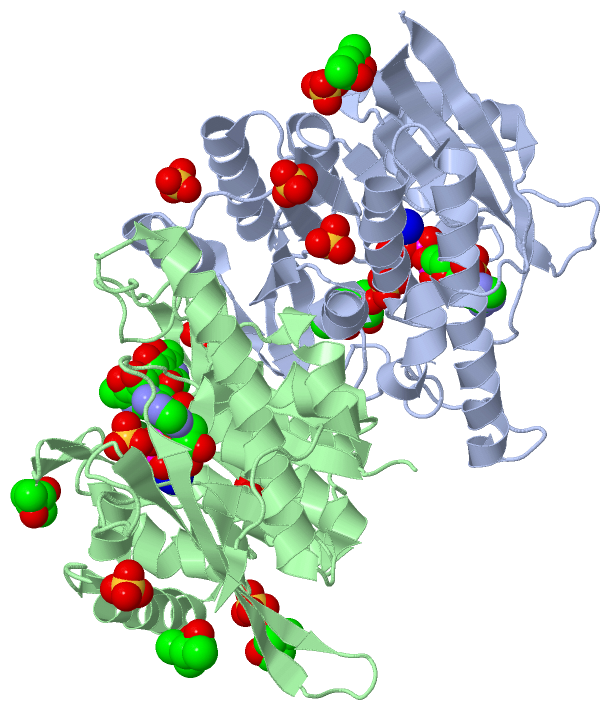
(-) Ligands, Modified Residues, Ions  (7, 24)

Asymmetric/Biological Unit (7, 24)
No.NameCountTypeFull Name
1ATP2Ligand/IonADENOSINE-5'-TRIPHOSPHATE
2MG2Ligand/IonMAGNESIUM ION
3MPD4Ligand/Ion(4S)-2-METHYL-2,4-PENTANEDIOL
4NA2Ligand/IonSODIUM ION
5PLP1Ligand/IonPYRIDOXAL-5'-PHOSPHATE
6PXL2Ligand/Ion3-HYDROXY-5-(HYDROXYMETHYL)-2-METHYLISONICOTINALDEHYDE
7SO411Ligand/IonSULFATE ION

(-) Sites  (24, 24)

Asymmetric Unit (24, 24)
No.NameEvidenceResiduesDescription
01AC1SOFTWAREASP A:118 , SO4 A:315 , HOH A:389 , HOH A:390 , ATP A:407BINDING SITE FOR RESIDUE MG A 400
02AC2SOFTWAREASP A:113 , THR A:148 , PRO A:149 , ASN A:150 , THR A:186 , HOH A:392 , ATP A:407BINDING SITE FOR RESIDUE NA A 402
03AC3SOFTWAREASP A:113 , ASP A:118 , TYR A:127 , ASN A:150 , GLU A:153 , THR A:186 , SER A:187 , ARG A:224 , LYS A:225 , VAL A:226 , ALA A:228 , THR A:233 , LEU A:267 , SO4 A:314 , SO4 A:315 , HOH A:349 , HOH A:389 , HOH A:390 , HOH A:392 , MG A:400 , NA A:402BINDING SITE FOR RESIDUE ATP A 407
04AC4SOFTWARESER A:12 , HIS A:46 , THR A:47 , VAL A:231 , SO4 A:314 , HOH A:342 , HOH A:367BINDING SITE FOR RESIDUE PXL A 313
05AC5SOFTWAREVAL A:231 , GLY A:232 , GLY A:234 , ALA A:235 , PXL A:313 , HOH A:342 , ATP A:407BINDING SITE FOR RESIDUE SO4 A 314
06AC6SOFTWARELEU A:199 , HOH A:390 , MG A:400 , ATP A:407BINDING SITE FOR RESIDUE SO4 A 315
07AC7SOFTWAREARG A:70 , MET A:93 , GLU A:100BINDING SITE FOR RESIDUE SO4 A 316
08AC8SOFTWAREGLU A:3 , GLU A:4 , CYS A:5 , ASP A:78 , HIS A:246 , HOH A:383BINDING SITE FOR RESIDUE SO4 A 317
09AC9SOFTWAREGLY A:32 , LYS A:247BINDING SITE FOR RESIDUE SO4 A 318
10BC1SOFTWAREASP A:145 , GLY A:179 , ASP A:181 , MPD A:321 , HOH A:334BINDING SITE FOR RESIDUE SO4 A 319
11BC2SOFTWAREVAL A:56 , GLU A:61 , HOH A:340 , GLU B:61 , GLU B:280BINDING SITE FOR RESIDUE SO4 A 320
12BC3SOFTWAREASP A:181 , SO4 A:319BINDING SITE FOR RESIDUE MPD A 321
13BC4SOFTWAREASP B:118 , SO4 B:315 , HOH B:344 , HOH B:383 , ATP B:409BINDING SITE FOR RESIDUE MG B 404
14BC5SOFTWAREASP B:113 , THR B:148 , PRO B:149 , ASN B:150 , GLU B:153 , THR B:186 , HOH B:385 , ATP B:409BINDING SITE FOR RESIDUE NA B 406
15BC6SOFTWAREASP B:113 , ASP B:118 , TYR B:127 , ASN B:150 , GLU B:153 , THR B:186 , SER B:187 , LEU B:199 , ARG B:224 , LYS B:225 , VAL B:226 , ALA B:228 , THR B:233 , LEU B:263 , LEU B:267 , PLP B:314 , SO4 B:315 , HOH B:344 , HOH B:353 , MG B:404 , NA B:406BINDING SITE FOR RESIDUE ATP B 409
16BC7SOFTWARESER B:12 , VAL B:19 , PHE B:43 , ASN B:45 , HIS B:46 , THR B:47 , GLY B:48 , VAL B:231 , PLP B:314BINDING SITE FOR RESIDUE PXL B 313
17BC8SOFTWAREVAL B:231 , GLY B:232 , THR B:233 , GLY B:234 , PXL B:313 , HOH B:384 , ATP B:409BINDING SITE FOR RESIDUE PLP B 314
18BC9SOFTWAREHOH B:359 , MG B:404 , ATP B:409BINDING SITE FOR RESIDUE SO4 B 315
19CC1SOFTWAREGLN B:63 , ARG B:70 , GLU B:100BINDING SITE FOR RESIDUE SO4 B 316
20CC2SOFTWAREALA B:144 , ASP B:145 , GLY B:179 , ASP B:181BINDING SITE FOR RESIDUE SO4 B 317
21CC3SOFTWAREGLN B:165 , HOH B:351BINDING SITE FOR RESIDUE SO4 B 318
22CC4SOFTWAREARG A:170 , GLY B:124 , GLU B:130BINDING SITE FOR RESIDUE MPD B 319
23CC5SOFTWAREASP B:173 , SER B:177 , LEU B:312BINDING SITE FOR RESIDUE MPD B 320
24CC6SOFTWAREASP B:181 , ARG B:206 , ARG B:208BINDING SITE FOR RESIDUE MPD B 321

(-) SS Bonds  (0, 0)

(no "SS Bond" information available for 3FHX)

(-) Cis Peptide Bonds  (1, 1)

Asymmetric/Biological Unit
No.Residues
1Glu B:3 -Glu B:4

 Sequence-Structure Mapping

(-) SAPs(SNPs)/Variants  (0, 0)

(no "SAP(SNP)/Variant" information available for 3FHX)

(-) PROSITE Motifs  (0, 0)

(no "PROSITE Motif" information available for 3FHX)

(-) Exons   (11, 22)

Asymmetric/Biological Unit (11, 22)
 ENSEMBLUniProtKBPDB
No.Transcript IDExonExon IDGenome LocationLengthIDLocationLengthCountLocationLength
1.1cENST000002915651cENSE00001861574chr21:45138993-45139262270PDXK_HUMAN1-29292A:3-29
B:3-29
27
27
1.6ENST000002915656ENSE00001050589chr21:45153950-4515400455PDXK_HUMAN30-48192A:30-48
B:30-48
19
19
1.9eENST000002915659eENSE00001050594chr21:45161548-45161652105PDXK_HUMAN48-83362A:48-83
B:48-83
36
36
1.10bENST0000029156510bENSE00001050590chr21:45163605-4516368884PDXK_HUMAN83-111292A:83-111
B:83-111
29
29
1.12bENST0000029156512bENSE00001050584chr21:45165960-4516600647PDXK_HUMAN111-126162A:111-126
B:111-126
16
16
1.14ENST0000029156514ENSE00001050578chr21:45168876-4516896186PDXK_HUMAN127-155292A:127-155
B:127-155
29
29
1.15ENST0000029156515ENSE00001050582chr21:45170381-4517042646PDXK_HUMAN155-170162A:155-170
B:155-170
16
16
1.16ENST0000029156516ENSE00001050593chr21:45172399-45172510112PDXK_HUMAN171-208382A:171-208
B:171-208
38
38
1.17cENST0000029156517cENSE00001050587chr21:45173464-45173600137PDXK_HUMAN208-253462A:208-253 (gaps)
B:208-253
46
46
1.18bENST0000029156518bENSE00001050583chr21:45175579-4517564567PDXK_HUMAN254-276232A:254-276
B:254-276
23
23
1.19eENST0000029156519eENSE00001431057chr21:45175832-451821886357PDXK_HUMAN276-312372A:276-312
B:276-312
37
37

(-) Sequences/Alignments

Asymmetric/Biological Unit
   Reformat: Number of residues per line =  ('0' or empty: single-line sequence representation)
  Number of residues per labelling interval =   
  UniProt sequence: complete  aligned part    
   Show mapping: SCOP domains CATH domains Pfam domains Secondary structure (by author)
SAPs(SNPs) PROSITE motifs Exons
(details for a mapped element are shown in a popup box when the mouse pointer rests over it)
Chain A from PDB  Type:PROTEIN  Length:308
 aligned with PDXK_HUMAN | O00764 from UniProtKB/Swiss-Prot  Length:312

    Alignment length:310
                                    12        22        32        42        52        62        72        82        92       102       112       122       132       142       152       162       172       182       192       202       212       222       232       242       252       262       272       282       292       302       312
           PDXK_HUMAN     3 EECRVLSIQSHVIRGYVGNRAATFPLQVLGFEIDAVNSVQFSNHTGYAHWKGQVLNSDELQELYEGLRLNNMNKYDYVLTGYTRDKSFLAMVVDIVQELKQQNPRLVYVCDPVLGDKWDGEGSMYVPEDLLPVYKEKVVPLADIITPNQFEAELLSGRKIHSQEEALRVMDMLHSMGPDTVVITSSDLPSPQGSNYLIVLGSQRRRNPAGSVVMERIRMDIRKVDAVFVGTGDLFAAMLLAWTHKHPNNLKVACEKTVSTLHHVLQRTIQCAKAQAGEGVRPSPMQLELRMVQSKRDIEDPEIVVQATVL 312
               SCOP domains d3fhxa_ A: automated matches                                                                                                                                                                                                                                                                                           SCOP domains
               CATH domains 3fhxA00 A:3-312  [code=3.40.1190.20, no name defined]                                                                                                                                                                                                                                                                  CATH domains
               Pfam domains ---------------------------------------------------------------------------------------------------------------------------------------------------------------------------------------------------------------------------------------------------------------------------------------------------------------------- Pfam domains
         Sec.struct. author ...eeeeeeeee....hhhhhhhhhhhhh..eeeeeeeeee..........eee.hhhhhhhhhhhhhhh......eeee....hhhhhhhhhhhhhhhhhhh...eeee....eeee..eeee....hhhhhhhhh......ee..hhhhhhhhhh....hhhhhhhhhhhhhhhh..eeee............eeeeeeeeeee..--.eeeeeeeeeee.......hhhhhhhhhhhhhhhhh..hhhhhhhhhhhhhhhhhhhhhhhhhhhh..............hhhhhhhhhh.......ee. Sec.struct. author
                 SAPs(SNPs) ---------------------------------------------------------------------------------------------------------------------------------------------------------------------------------------------------------------------------------------------------------------------------------------------------------------------- SAPs(SNPs)
                    PROSITE ---------------------------------------------------------------------------------------------------------------------------------------------------------------------------------------------------------------------------------------------------------------------------------------------------------------------- PROSITE
           Transcript 1 (1) Exon 1.1c  PDB: A:3-29     Exon 1.6           ----------------------------------Exon 1.10b  PDB: A:83-111    ---------------Exon 1.14  PDB: A:127-155    ---------------Exon 1.16  PDB: A:171-208             ---------------------------------------------Exon 1.18b             ------------------------------------ Transcript 1 (1)
           Transcript 1 (2) ---------------------------------------------Exon 1.9e  PDB: A:48-83             ---------------------------Exon 1.12b      ----------------------------Exon 1.15       -------------------------------------Exon 1.17c  PDB: A:208-253 (gaps)             ----------------------Exon 1.19e  PDB: A:276-312            Transcript 1 (2)
                 3fhx A   3 EECRVLSIQSHVIRGYVGNRAATFPLQVLGFEIDAVNSVQFSNHTGYAHWKGQVLNSDELQELYEGLRLNNMNKYDYVLTGYTRDKSFLAMVVDIVQELKQQNPRLVYVCDPVLGDKWDGEGSMYVPEDLLPVYKEKVVPLADIITPNQFEAELLSGRKIHSQEEALRVMDMLHSMGPDTVVITSSDLPSPQGSNYLIVLGSQRRRNP--SVVMERIRMDIRKVDAVFVGTGALFAAMLLAWTHKHPNNLKVACEKTVSTLHHVLQRTIQCAKAQAGEGVRPSPMQLELRMVQSKRDIEDPEIVVQATVL 312
                                    12        22        32        42        52        62        72        82        92       102       112       122       132       142       152       162       172       182       192       202       | -|      222       232       242       252       262       272       282       292       302       312
                                                                                                                                                                                                                                         210  |                                                                                                   
                                                                                                                                                                                                                                            213                                                                                                   

Chain B from PDB  Type:PROTEIN  Length:310
 aligned with PDXK_HUMAN | O00764 from UniProtKB/Swiss-Prot  Length:312

    Alignment length:310
                                    12        22        32        42        52        62        72        82        92       102       112       122       132       142       152       162       172       182       192       202       212       222       232       242       252       262       272       282       292       302       312
           PDXK_HUMAN     3 EECRVLSIQSHVIRGYVGNRAATFPLQVLGFEIDAVNSVQFSNHTGYAHWKGQVLNSDELQELYEGLRLNNMNKYDYVLTGYTRDKSFLAMVVDIVQELKQQNPRLVYVCDPVLGDKWDGEGSMYVPEDLLPVYKEKVVPLADIITPNQFEAELLSGRKIHSQEEALRVMDMLHSMGPDTVVITSSDLPSPQGSNYLIVLGSQRRRNPAGSVVMERIRMDIRKVDAVFVGTGDLFAAMLLAWTHKHPNNLKVACEKTVSTLHHVLQRTIQCAKAQAGEGVRPSPMQLELRMVQSKRDIEDPEIVVQATVL 312
               SCOP domains d3fhxb_ B: automated matches                                                                                                                                                                                                                                                                                           SCOP domains
               CATH domains 3fhxB00 B:3-312  [code=3.40.1190.20, no name defined]                                                                                                                                                                                                                                                                  CATH domains
               Pfam domains ---------------------------------------------------------------------------------------------------------------------------------------------------------------------------------------------------------------------------------------------------------------------------------------------------------------------- Pfam domains
         Sec.struct. author ...eeeeeeeee....hhhhhhhhhhhhh..eeeeeeeeee..........eee.hhhhhhhhhhhhhhh......eeee....hhhhhhhhhhhhhhhhhhh...eeee....eeee..eeee....hhhhhhhhhhhhhh.ee..hhhhhhhhhh....hhhhhhhhhhhhhhhh..eeee............eeeeeeeeeee.....eeeeeeeeeee.......hhhhhhhhhhhhhhhhh..hhhhhhhhhhhhhhhhhhhhhhhhhhhhh.............hhhhhhhhhh.......ee. Sec.struct. author
                 SAPs(SNPs) ---------------------------------------------------------------------------------------------------------------------------------------------------------------------------------------------------------------------------------------------------------------------------------------------------------------------- SAPs(SNPs)
                    PROSITE ---------------------------------------------------------------------------------------------------------------------------------------------------------------------------------------------------------------------------------------------------------------------------------------------------------------------- PROSITE
           Transcript 1 (1) Exon 1.1c  PDB: B:3-29     Exon 1.6           ----------------------------------Exon 1.10b  PDB: B:83-111    ---------------Exon 1.14  PDB: B:127-155    ---------------Exon 1.16  PDB: B:171-208             ---------------------------------------------Exon 1.18b             ------------------------------------ Transcript 1 (1)
           Transcript 1 (2) ---------------------------------------------Exon 1.9e  PDB: B:48-83             ---------------------------Exon 1.12b      ----------------------------Exon 1.15       -------------------------------------Exon 1.17c  PDB: B:208-253 UniProt: 208-253   ----------------------Exon 1.19e  PDB: B:276-312            Transcript 1 (2)
                 3fhx B   3 EECRVLSIQSHVIRGYVGNRAATFPLQVLGFEIDAVNSVQFSNHTGYAHWKGQVLNSDELQELYEGLRLNNMNKYDYVLTGYTRDKSFLAMVVDIVQELKQQNPRLVYVCDPVLGDKWDGEGSMYVPEDLLPVYKEKVVPLADIITPNQFEAELLSGRKIHSQEEALRVMDMLHSMGPDTVVITSSDLPSPQGSNYLIVLGSQRRRNPAGSVVMERIRMDIRKVDAVFVGTGALFAAMLLAWTHKHPNNLKVACEKTVSTLHHVLQRTIQCAKAQAGEGVRPSPMQLELRMVQSKRDIEDPEIVVQATVL 312
                                    12        22        32        42        52        62        72        82        92       102       112       122       132       142       152       162       172       182       192       202       212       222       232       242       252       262       272       282       292       302       312

   Legend:   → Mismatch (orange background)
  - → Gap (green background, '-', border residues have a numbering label)
    → Modified Residue (blue background, lower-case, 'x' indicates undefined single-letter code, labelled with number + name)
  x → Chemical Group (purple background, 'x', labelled with number + name, e.g. ACE or NH2)
  extra numbering lines below/above indicate numbering irregularities and modified residue names etc., number ends below/above '|'

 Classification and Annotation

(-) SCOP Domains  (1, 2)

Asymmetric/Biological Unit

(-) CATH Domains  (1, 2)

Asymmetric/Biological Unit
(-)
Class: Alpha Beta (26913)

(-) Pfam Domains  (0, 0)

(no "Pfam Domain" information available for 3FHX)

(-) Gene Ontology  (23, 23)

Asymmetric/Biological Unit(hide GO term definitions)
Chain A,B   (PDXK_HUMAN | O00764)
molecular function
    GO:0005524    ATP binding    Interacting selectively and non-covalently with ATP, adenosine 5'-triphosphate, a universally important coenzyme and enzyme regulator.
    GO:0016301    kinase activity    Catalysis of the transfer of a phosphate group, usually from ATP, to a substrate molecule.
    GO:0031403    lithium ion binding    Interacting selectively and non-covalently with lithium ions (Li+).
    GO:0000287    magnesium ion binding    Interacting selectively and non-covalently with magnesium (Mg) ions.
    GO:0046872    metal ion binding    Interacting selectively and non-covalently with any metal ion.
    GO:0000166    nucleotide binding    Interacting selectively and non-covalently with a nucleotide, any compound consisting of a nucleoside that is esterified with (ortho)phosphate or an oligophosphate at any hydroxyl group on the ribose or deoxyribose.
    GO:0030955    potassium ion binding    Interacting selectively and non-covalently with potassium (K+) ions.
    GO:0042803    protein homodimerization activity    Interacting selectively and non-covalently with an identical protein to form a homodimer.
    GO:0008478    pyridoxal kinase activity    Catalysis of the reaction: ATP + pyridoxal = ADP + pyridoxal 5'-phosphate.
    GO:0030170    pyridoxal phosphate binding    Interacting selectively and non-covalently with pyridoxal 5' phosphate, 3-hydroxy-5-(hydroxymethyl)-2-methyl4-pyridine carboxaldehyde 5' phosphate, the biologically active form of vitamin B6.
    GO:0031402    sodium ion binding    Interacting selectively and non-covalently with sodium ions (Na+).
    GO:0016740    transferase activity    Catalysis of the transfer of a group, e.g. a methyl group, glycosyl group, acyl group, phosphorus-containing, or other groups, from one compound (generally regarded as the donor) to another compound (generally regarded as the acceptor). Transferase is the systematic name for any enzyme of EC class 2.
    GO:0008270    zinc ion binding    Interacting selectively and non-covalently with zinc (Zn) ions.
biological process
    GO:0008283    cell proliferation    The multiplication or reproduction of cells, resulting in the expansion of a cell population.
    GO:0016310    phosphorylation    The process of introducing a phosphate group into a molecule, usually with the formation of a phosphoric ester, a phosphoric anhydride or a phosphoric amide.
    GO:0009443    pyridoxal 5'-phosphate salvage    Any process that generates pyridoxal 5'-phosphate, the active form of vitamin B6, from derivatives of it without de novo synthesis.
    GO:0042823    pyridoxal phosphate biosynthetic process    The chemical reactions and pathways resulting in the formation of pyridoxal phosphate, pyridoxal phosphorylated at the hydroxymethyl group of C-5, the active form of vitamin B6.
    GO:0042816    vitamin B6 metabolic process    The chemical reactions and pathways involving any of the vitamin B6 compounds: pyridoxal, pyridoxamine and pyridoxine and the active form, pyridoxal phosphate.
cellular component
    GO:0005737    cytoplasm    All of the contents of a cell excluding the plasma membrane and nucleus, but including other subcellular structures.
    GO:0005829    cytosol    The part of the cytoplasm that does not contain organelles but which does contain other particulate matter, such as protein complexes.
    GO:0070062    extracellular exosome    A vesicle that is released into the extracellular region by fusion of the limiting endosomal membrane of a multivesicular body with the plasma membrane. Extracellular exosomes, also simply called exosomes, have a diameter of about 40-100 nm.
    GO:0005654    nucleoplasm    That part of the nuclear content other than the chromosomes or the nucleolus.
    GO:0005634    nucleus    A membrane-bounded organelle of eukaryotic cells in which chromosomes are housed and replicated. In most cells, the nucleus contains all of the cell's chromosomes except the organellar chromosomes, and is the site of RNA synthesis and processing. In some species, or in specialized cell types, RNA metabolism or DNA replication may be absent.

 Visualization

(-) Interactive Views

Asymmetric/Biological Unit
  Complete Structure
    Jena3D(integrated viewing of ligand, site, SAP, PROSITE, SCOP information)
    WebMol | AstexViewer[tm]@PDBe
(Java Applets, require no local installation except for Java; loading may be slow)
    STRAP
(Java WebStart application, automatic local installation, requires Java; full application with system access!)
    RasMol
(require local installation)
    Molscript (VRML)
(requires installation of a VRML viewer; select preferred view via VRML and generate a mono or stereo PDF format file)
 
  Ligands, Modified Residues, Ions
    ATP  [ RasMol | Jena3D ]  +environment [ RasMol | Jena3D ]
    MG  [ RasMol | Jena3D ]  +environment [ RasMol | Jena3D ]
    MPD  [ RasMol | Jena3D ]  +environment [ RasMol | Jena3D ]
    NA  [ RasMol | Jena3D ]  +environment [ RasMol | Jena3D ]
    PLP  [ RasMol | Jena3D ]  +environment [ RasMol | Jena3D ]
    PXL  [ RasMol | Jena3D ]  +environment [ RasMol | Jena3D ]
    SO4  [ RasMol | Jena3D ]  +environment [ RasMol | Jena3D ]
 
  Sites
    AC1  [ RasMol ]  +environment [ RasMol ]
    AC2  [ RasMol ]  +environment [ RasMol ]
    AC3  [ RasMol ]  +environment [ RasMol ]
    AC4  [ RasMol ]  +environment [ RasMol ]
    AC5  [ RasMol ]  +environment [ RasMol ]
    AC6  [ RasMol ]  +environment [ RasMol ]
    AC7  [ RasMol ]  +environment [ RasMol ]
    AC8  [ RasMol ]  +environment [ RasMol ]
    AC9  [ RasMol ]  +environment [ RasMol ]
    BC1  [ RasMol ]  +environment [ RasMol ]
    BC2  [ RasMol ]  +environment [ RasMol ]
    BC3  [ RasMol ]  +environment [ RasMol ]
    BC4  [ RasMol ]  +environment [ RasMol ]
    BC5  [ RasMol ]  +environment [ RasMol ]
    BC6  [ RasMol ]  +environment [ RasMol ]
    BC7  [ RasMol ]  +environment [ RasMol ]
    BC8  [ RasMol ]  +environment [ RasMol ]
    BC9  [ RasMol ]  +environment [ RasMol ]
    CC1  [ RasMol ]  +environment [ RasMol ]
    CC2  [ RasMol ]  +environment [ RasMol ]
    CC3  [ RasMol ]  +environment [ RasMol ]
    CC4  [ RasMol ]  +environment [ RasMol ]
    CC5  [ RasMol ]  +environment [ RasMol ]
    CC6  [ RasMol ]  +environment [ RasMol ]
 
  Cis Peptide Bonds
    Glu B:3 - Glu B:4   [ RasMol ]  
 

(-) Still Images

Jmol
  protein: cartoon or spacefill or dots and stick; nucleic acid: cartoon and stick; ligands: spacefill; active site: stick
Molscript
  protein, nucleic acid: cartoon; ligands: spacefill; active site: ball and stick

 Databases and Analysis Tools

(-) Databases

Access by PDB/NDB ID
  3fhx
    Family and Domain InformationProDom | SYSTERS
    General Structural InformationGlycoscienceDB | MMDB | NDB | OCA | PDB | PDBe | PDBj | PDBsum | PDBWiki | PQS | PROTEOPEDIA
    Orientation in MembranesOPM
    Protein SurfaceSURFACE
    Secondary StructureDSSP (structure derived) | HSSP (homology derived)
    Structural GenomicsGeneCensus
    Structural NeighboursCE | VAST
    Structure ClassificationCATH | Dali | SCOP
    Validation and Original DataBMRB Data View | BMRB Restraints Grid | EDS | PROCHECK | RECOORD | WHAT_CHECK
 
Access by UniProt ID/Accession number
  PDXK_HUMAN | O00764
    Comparative Protein Structure ModelsModBase
    Genomic InformationEnsembl
    Protein-protein InteractionDIP
    Sequence, Family and Domain InformationInterPro | Pfam | SMART | UniProtKB/SwissProt
 
Access by Enzyme Classificator   (EC Number)
  2.7.1.35
    General Enzyme InformationBRENDA | EC-PDB | Enzyme | IntEnz
    PathwayKEGG | MetaCyc
 
Access by Disease Identifier   (MIM ID)
  (no 'MIM ID' available)
    Disease InformationOMIM
 
Access by GenAge ID
  (no 'GenAge ID' available)
    Age Related InformationGenAge

(-) Analysis Tools

Access by PDB/NDB ID
    Domain InformationXDom
    Interatomic Contacts of Structural UnitsCSU
    Ligand-protein ContactsLPC
    Protein CavitiescastP
    Sequence and Secondary StructurePDBCartoon
    Structure AlignmentSTRAP(Java WebStart application, automatic local installation, requires Java; full application with system access!)
    Structure and Sequence BrowserSTING
 
Access by UniProt ID/Accession number
  PDXK_HUMAN | O00764
    Protein Disorder PredictionDisEMBL | FoldIndex | GLOBPLOT (for more information see DisProt)

 Related Entries

(-) Entries Sharing at Least One Protein Chain (UniProt ID)

UniProtKB/Swiss-Prot
        PDXK_HUMAN | O007642ajp 2f7k 2yxt 2yxu 3fhy 3keu 4en4 4eoh

(-) Related Entries Specified in the PDB File

3fhy CRYSTAL STRUCTURE OF D235N MUTANT OF HUMAN PYRIDOXAL KINASE