| molecular function |
|---|
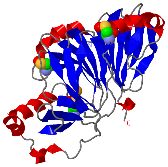
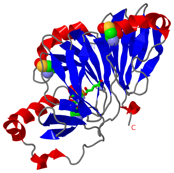
| | GO:0000334 | | 3-hydroxyanthranilate 3,4-dioxygenase activity | | Catalysis of the reaction: 3-hydroxyanthranilate + O(2) = cis,cis-2-amino-3-(3-oxoprop-1-enyl)but-2-enedioate + H(+). |
| | GO:0051213 | | dioxygenase activity | | Catalysis of an oxidation-reduction (redox) reaction in which both atoms of oxygen from one molecule of O2 are incorporated into the (reduced) product(s) of the reaction. The two atoms of oxygen may be distributed between two different products. |
| | GO:0008198 | | ferrous iron binding | | Interacting selectively and non-covalently with ferrous iron, Fe(II). |
| | GO:0005506 | | iron ion binding | | Interacting selectively and non-covalently with iron (Fe) ions. |
| | GO:0046872 | | metal ion binding | | Interacting selectively and non-covalently with any metal ion. |
| | GO:0016491 | | oxidoreductase activity | | Catalysis of an oxidation-reduction (redox) reaction, a reversible chemical reaction in which the oxidation state of an atom or atoms within a molecule is altered. One substrate acts as a hydrogen or electron donor and becomes oxidized, while the other acts as hydrogen or electron acceptor and becomes reduced. |
| biological process |
|---|
| | GO:0034354 | | 'de novo' NAD biosynthetic process from tryptophan | | The chemical reactions and pathways resulting in the formation of nicotinamide adenine dinucleotide (NAD), beginning with the synthesis of tryptophan from simpler precursors; biosynthesis may be of either the oxidized form, NAD, or the reduced form, NADH. |
| | GO:0009435 | | NAD biosynthetic process | | The chemical reactions and pathways resulting in the formation of nicotinamide adenine dinucleotide, a coenzyme present in most living cells and derived from the B vitamin nicotinic acid; biosynthesis may be of either the oxidized form, NAD, or the reduced form, NADH. |
| | GO:0043420 | | anthranilate metabolic process | | The chemical reactions and pathways involving anthranilate (2-aminobenzoate). |
| | GO:0070050 | | neuron cellular homeostasis | | The cellular homeostatic process that preserves a neuron in a stable, differentiated functional and structural state. |
| | GO:0055114 | | oxidation-reduction process | | A metabolic process that results in the removal or addition of one or more electrons to or from a substance, with or without the concomitant removal or addition of a proton or protons. |
| | GO:0019363 | | pyridine nucleotide biosynthetic process | | The chemical reactions and pathways resulting in the formation of a pyridine nucleotide, a nucleotide characterized by a pyridine derivative as a nitrogen base. |
| | GO:0019805 | | quinolinate biosynthetic process | | The chemical reactions and pathways resulting in the formation of quinolinate, the anion of quinolinic acid, also known as 2,3-pyridinedicarboxylic acid. |
| | GO:0046874 | | quinolinate metabolic process | | The chemical reactions and pathways involving quinolinate, the anion of quinolinic acid, also known as 2,3-pyridinedicarboxylic acid. |
| | GO:0046686 | | response to cadmium ion | | Any process that results in a change in state or activity of a cell or an organism (in terms of movement, secretion, enzyme production, gene expression, etc.) as a result of a cadmium (Cd) ion stimulus. |
| | GO:0010043 | | response to zinc ion | | Any process that results in a change in state or activity of a cell or an organism (in terms of movement, secretion, enzyme production, gene expression, etc.) as a result of a zinc ion stimulus. |
| | GO:0006569 | | tryptophan catabolic process | | The chemical reactions and pathways resulting in the breakdown of tryptophan, the chiral amino acid 2-amino-3-(1H-indol-3-yl)propanoic acid. |
| cellular component |
|---|
| | GO:0005737 | | cytoplasm | | All of the contents of a cell excluding the plasma membrane and nucleus, but including other subcellular structures. |
| | GO:0005829 | | cytosol | | The part of the cytoplasm that does not contain organelles but which does contain other particulate matter, such as protein complexes. |
| | GO:0070062 | | extracellular exosome | | A vesicle that is released into the extracellular region by fusion of the limiting endosomal membrane of a multivesicular body with the plasma membrane. Extracellular exosomes, also simply called exosomes, have a diameter of about 40-100 nm. |
 Description