|
|
|
|
 Description
|
|
Compounds
|
||||||||||||||||||||||||||||||||||||||||||||||||||||||||||||||||
Chains, Units
Summary Information (see also Sequences/Alignments below) |
Ligands, Modified Residues, Ions (1, 4)
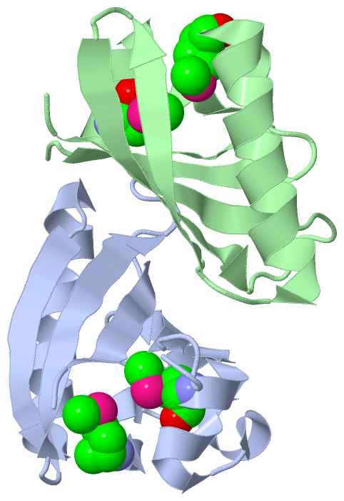
Asymmetric Unit (1, 4)
|
Sites (0, 0)| (no "Site" information available for 3FC7) |
SS Bonds (0, 0)| (no "SS Bond" information available for 3FC7) |
Cis Peptide Bonds (0, 0)| (no "Cis Peptide Bond" information available for 3FC7) |
SAPs(SNPs)/Variants (0, 0)| (no "SAP(SNP)/Variant" information available for 3FC7) |
PROSITE Motifs (0, 0)| (no "PROSITE Motif" information available for 3FC7) |
Exons (0, 0)| (no "Exon" information available for 3FC7) |
Sequences/Alignments
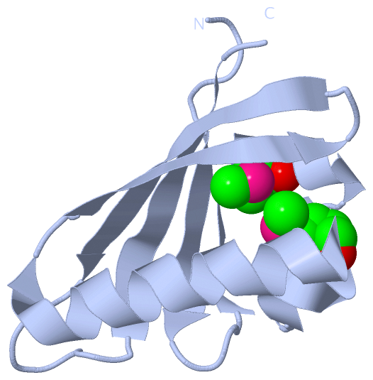
Asymmetric UnitChain A from PDB Type:PROTEIN Length:100 aligned with Q5V4P0_HALMA | Q5V4P0 from UniProtKB/TrEMBL Length:461 Alignment length:100 151 161 171 181 191 201 211 221 231 241 Q5V4P0_HALMA 142 SDSPDGIVHLTTNGTILSVNPSMAGRLGADPDTLVGQQLSAVMDSEAANQRLEAGKSAVENGTATRSEDAVGGRHYHNQYIPVDSHRKSDTFQLVSRDIT 241 SCOP domains ---------------------------------------------------------------------------------------------------- SCOP domains CATH domains 3fc7A00 A:142-241 [code=3.30.450.20, no name defined] CATH domains Pfam domains ---------------------------------------------------------------------------------------------------- Pfam domains SAPs(SNPs) ---------------------------------------------------------------------------------------------------- SAPs(SNPs) PROSITE ---------------------------------------------------------------------------------------------------- PROSITE Transcript ---------------------------------------------------------------------------------------------------- Transcript 3fc7 A 142 SDSPDGIVHLTTNGTILSVNPSmAGRLGADPDTLVGQQLSAVmDSEAANQRLEAGKSAVENGTATRSEDAVGGRHYHNQYIPVDSHRKSDTFQLVSRDIT 241 151 161 | 171 181 | 191 201 211 221 231 241 164-MSE 184-MSE Chain B from PDB Type:PROTEIN Length:98 aligned with Q5V4P0_HALMA | Q5V4P0 from UniProtKB/TrEMBL Length:461 Alignment length:98 153 163 173 183 193 203 213 223 233 Q5V4P0_HALMA 144 SPDGIVHLTTNGTILSVNPSMAGRLGADPDTLVGQQLSAVMDSEAANQRLEAGKSAVENGTATRSEDAVGGRHYHNQYIPVDSHRKSDTFQLVSRDIT 241 SCOP domains -------------------------------------------------------------------------------------------------- SCOP domains CATH domains 3fc7B00 B:144-241 [code=3.30.450.20, no name defined] CATH domains Pfam domains -------------------------------------------------------------------------------------------------- Pfam domains SAPs(SNPs) -------------------------------------------------------------------------------------------------- SAPs(SNPs) PROSITE -------------------------------------------------------------------------------------------------- PROSITE Transcript -------------------------------------------------------------------------------------------------- Transcript 3fc7 B 144 SPDGIVHLTTNGTILSVNPSmAGRLGADPDTLVGQQLSAVmDSEAANQRLEAGKSAVENGTATRSEDAVGGRHYHNQYIPVDSHRKSDTFQLVSRDIT 241 153 163| 173 183| 193 203 213 223 233 164-MSE 184-MSE
|
||||||||||||||||||||
SCOP Domains (0, 0)| (no "SCOP Domain" information available for 3FC7) |
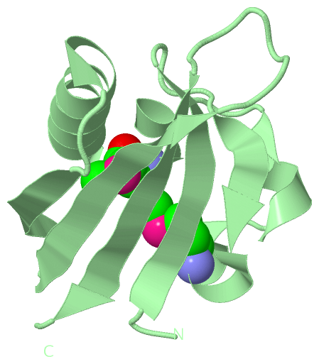
CATH Domains (1, 2)
Asymmetric Unit
 |
Pfam Domains (0, 0)| (no "Pfam Domain" information available for 3FC7) |
Gene Ontology (5, 5)|
Asymmetric Unit(hide GO term definitions) Chain A,B (Q5V4P0_HALMA | Q5V4P0)
|
||||||||||||||||||||||||||||||||||||||||||||||||
Interactive Views
|
||||||||||||||||||||||||||||||||||||||||||||||||||||||||||||||||||||||||||||||||||||||||||||||||||||||||||||||||||||||||||||||||||||||||||||
Still Images
|
||||||||||||||||
Databases
|
||||||||||||||||||||||||||||||||||||||||||||||||||||||||||||||||||||||||||||||||||||||||||||||||||||||||||||||||||||||||||||||||||||||||||||||||||||||||||||||||
Analysis Tools
|
|||||||||||||||||||||||||||||||||||||||||||||||||||||||||||||
Entries Sharing at Least One Protein Chain (UniProt ID)
Related Entries Specified in the PDB File
|
|