|
|
|
|
 Description
|
|
Compounds
|
||||||||||||||||||||
Chains, Units
Summary Information (see also Sequences/Alignments below) |
Ligands, Modified Residues, Ions (0, 0)| (no "Ligand,Modified Residues,Ions" information available for 3F7M) |
Sites (0, 0)| (no "Site" information available for 3F7M) |
SS Bonds (2, 2)
Asymmetric/Biological Unit
|
||||||||||||
Cis Peptide Bonds (1, 1)
Asymmetric/Biological Unit
|
||||||||
SAPs(SNPs)/Variants (0, 0)| (no "SAP(SNP)/Variant" information available for 3F7M) |
PROSITE Motifs (3, 3)
Asymmetric/Biological Unit (3, 3)
|
||||||||||||||||||||||||||||||||||||||||
Exons (0, 0)| (no "Exon" information available for 3F7M) |
Sequences/Alignments
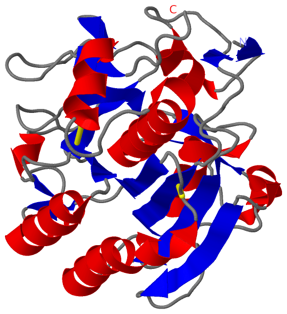
Asymmetric/Biological UnitChain A from PDB Type:PROTEIN Length:279 aligned with ALP_LECPS | Q68GV9 from UniProtKB/Swiss-Prot Length:382 Alignment length:279 113 123 133 143 153 163 173 183 193 203 213 223 233 243 253 263 273 283 293 303 313 323 333 343 353 363 373 ALP_LECPS 104 ITQQQGATWGLTRISHRARGSTAYAYDTSAGAGACVYVIDTGVEDTHPDFEGRAKQIKSYASTARDGHGHGTHCAGTIGSKTWGVAKKVSIFGVKVLDDSGSGSLSNIVAGMDFVASDRQSRNCPRRTVASMSLGGGYSAALNQAAARLQSSGVFVAVAAGNDNRDAANTSPASEPTVCTVGATDSNDVRSTFSNYGRVVDIFAPGTSITSTWIGGRTNTISGTSMATPHIAGLAAYLFGLEGGSAGAMCGRIQTLSTKNVLTSIPSGTVNYLAFNGAT 382 SCOP domains d3f7ma_ A: automated matches SCOP domains CATH domains 3f7mA00 A:104-382 [code=3.40.50.200, no name defined] CATH domains Pfam domains --------------------------------------------------------------------------------------------------------------------------------------------------------------------------------------------------------------------------------------------------------------------------------------- Pfam domains SAPs(SNPs) --------------------------------------------------------------------------------------------------------------------------------------------------------------------------------------------------------------------------------------------------------------------------------------- SAPs(SNPs) PROSITE -----------------------------------SUBTILASE_AS----------------------SUBTILASE_H----------------------------------------------------------------------------------------------------------------------------------------------SUBTILASE_S---------------------------------------------- PROSITE Transcript --------------------------------------------------------------------------------------------------------------------------------------------------------------------------------------------------------------------------------------------------------------------------------------- Transcript 3f7m A 104 ITQQQGATWGLTRISHRARGSTAYAYDTSAGAGACVYVIDTGVEDTHPDFEGRAKQIKSYASTARDGHGHGTHCAGTIGSKTWGVAKKVSIFGVKVLDDSGSGSLSNIIAGMDFVASDRQSRNCPRRTVASMSLGGGYSAALNQAAARLQSSGVFVAVAAGNDNRDAANTSPASEPTVCTVGATDSNDVRSTFSNYGRVVDIFAPGTSITSTWIGGRTNTISGTSMATPHIAGLAAYLFGLEGGSAGAMCGRIQTLSTKNVLTSIPSGTVNYLAFNGAT 382 113 123 133 143 153 163 173 183 193 203 213 223 233 243 253 263 273 283 293 303 313 323 333 343 353 363 373
|
||||||||||||||||||||
SCOP Domains (1, 1)
Asymmetric/Biological Unit
 |
CATH Domains (1, 1)
Asymmetric/Biological Unit
|
Pfam Domains (0, 0)| (no "Pfam Domain" information available for 3F7M) |
Gene Ontology (6, 6)|
Asymmetric/Biological Unit(hide GO term definitions) Chain A (ALP_LECPS | Q68GV9)
|
||||||||||||||||||||||||||||||||||||||||||||||||||||||
Interactive Views
|
|||||||||||||||||||||||||||||||||||||||||||||||||||||||||||||||||||||||||||||||||||||||||||||||||||||||||||||||||||||
Still Images
|
||||||||||||||||
Databases
|
||||||||||||||||||||||||||||||||||||||||||||||||||||||||||||||||||||||||||||||||||||||||||||||||||||||||||||||||||||||||||||||||||||||||||||||||||||||||||||||||
Analysis Tools
|
|||||||||||||||||||||||||||||||||||||||||||||||||||||||||||||
Entries Sharing at Least One Protein Chain (UniProt ID)
Related Entries Specified in the PDB File
|
|