|
|
|
|
 Description
|
|
Compounds
|
||||||||||||||||||||||||||||||||||||||||||||||||||||||||||||
Chains, Units
Summary Information (see also Sequences/Alignments below) |
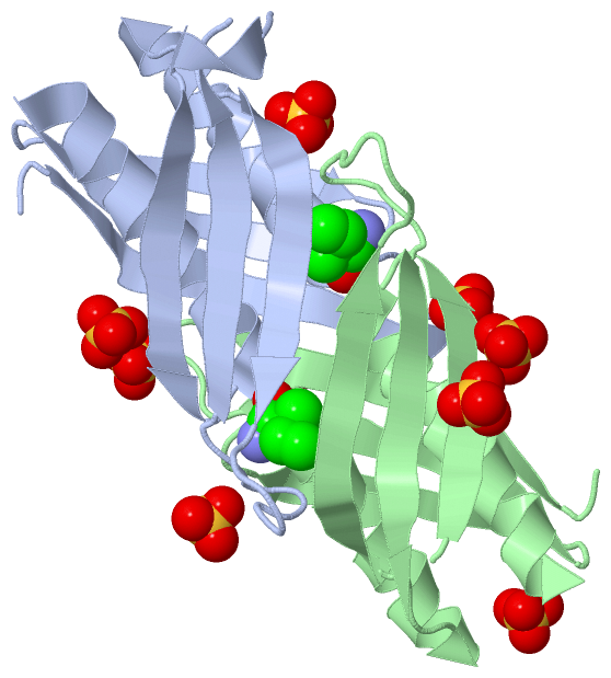
Ligands, Modified Residues, Ions (3, 12)| Asymmetric/Biological Unit (3, 12) |
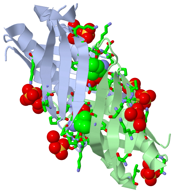
Sites (12, 12)
Asymmetric Unit (12, 12)
|
SS Bonds (0, 0)| (no "SS Bond" information available for 3F6G) |
Cis Peptide Bonds (0, 0)| (no "Cis Peptide Bond" information available for 3F6G) |
SAPs(SNPs)/Variants (0, 0)| (no "SAP(SNP)/Variant" information available for 3F6G) |
PROSITE Motifs (0, 0)| (no "PROSITE Motif" information available for 3F6G) |
Exons (0, 0)| (no "Exon" information available for 3F6G) |
Sequences/Alignments
Asymmetric/Biological UnitChain A from PDB Type:PROTEIN Length:121 aligned with Q8F3Q1_LEPIN | Q8F3Q1 from UniProtKB/TrEMBL Length:516 Alignment length:123 399 409 419 429 439 449 459 469 479 489 499 509 Q8F3Q1_LEPIN 390 KVLTIKSCNIHSGIGIRPHAQIELEYQGKIHKEISEGDGGYDAFMNALTKITNRLGISIPKLIDYEVRIPPGGKTDALVETRITWNKSLDLEEDQTFKTMGVHPDQTVAAVHATEKMLNQILQ 512 SCOP domains --------------------------------------------------------------------------------------------------------------------------- SCOP domains CATH domains --------------------------------------------------------------------------------------------------------------------------- CATH domains Pfam domains --------------------------------------------------------------------------------------------------------------------------- Pfam domains SAPs(SNPs) --------------------------------------------------------------------------------------------------------------------------- SAPs(SNPs) PROSITE --------------------------------------------------------------------------------------------------------------------------- PROSITE Transcript --------------------------------------------------------------------------------------------------------------------------- Transcript 3f6g A 390 KVLTIKSCNIHSGIGIRPHAQIELEYQGKIHKEISEGDGGYDAFMNALTKITNRLGISIPKLIDYEVRIPPGGKTDALVETRITWNKS--LEEDQTFKTMGVHPDQTVAAVHATEKMLNQILQ 512 399 409 419 429 439 449 459 469 | -| 489 499 509 477 | 480 Chain B from PDB Type:PROTEIN Length:115 aligned with Q8F3Q1_LEPIN | Q8F3Q1 from UniProtKB/TrEMBL Length:516 Alignment length:123 399 409 419 429 439 449 459 469 479 489 499 509 Q8F3Q1_LEPIN 390 KVLTIKSCNIHSGIGIRPHAQIELEYQGKIHKEISEGDGGYDAFMNALTKITNRLGISIPKLIDYEVRIPPGGKTDALVETRITWNKSLDLEEDQTFKTMGVHPDQTVAAVHATEKMLNQILQ 512 SCOP domains --------------------------------------------------------------------------------------------------------------------------- SCOP domains CATH domains --------------------------------------------------------------------------------------------------------------------------- CATH domains Pfam domains --------------------------------------------------------------------------------------------------------------------------- Pfam domains SAPs(SNPs) --------------------------------------------------------------------------------------------------------------------------- SAPs(SNPs) PROSITE --------------------------------------------------------------------------------------------------------------------------- PROSITE Transcript --------------------------------------------------------------------------------------------------------------------------- Transcript 3f6g B 390 KVLTIKSCNIHSGIGIRPHAQIELEYQGKIHKEISEGDGGYDAFMNALTKITNRLGISIPKLIDYEVRIPPGGKTDALVETRITWNK--------TFKTMGVHPDQTVAAVHATEKMLNQILQ 512 399 409 419 429 439 449 459 469 | - | 489 499 509 476 485
|
||||||||||||||||||||
SCOP Domains (0, 0)| (no "SCOP Domain" information available for 3F6G) |
CATH Domains (0, 0)| (no "CATH Domain" information available for 3F6G) |
Pfam Domains (0, 0)| (no "Pfam Domain" information available for 3F6G) |
Gene Ontology (9, 9)|
Asymmetric/Biological Unit(hide GO term definitions) Chain A,B (Q8F3Q1_LEPIN | Q8F3Q1)
|
||||||||||||||||||||||||||||||||||||||||||||||||||||||||||||||||||
Interactive Views
|
|||||||||||||||||||||||||||||||||||||||||||||||||||||||||||||||||||||||||||||||||||||||||||||||||||||||||||||||||||||||||||||||||||||||||||||||||||||||||||||||||||||||||||||||||||||||||||||||||||||||||||||||||
Still Images
|
||||||||||||||||
Databases
|
||||||||||||||||||||||||||||||||||||||||||||||||||||||||||||||||||||||||||||||||||||||||||||||||||||||||||||||||||||||||||||||||||||||||||||||||||||||||||||||||
Analysis Tools
|
|||||||||||||||||||||||||||||||||||||||||||||||||||||||||||||
Entries Sharing at Least One Protein Chain (UniProt ID)
Related Entries Specified in the PDB File
|
|