Show PDB file:   
         Plain Text   HTML   (compressed file size)
QuickSearch:   
by PDB,NDB,UniProt,PROSITE Code or Search Term(s)  
(-)Asymmetric Unit
(-)Biological Unit 1
(-)Biological Unit 2
collapse expand < >
Image Asymmetric Unit
Asymmetric Unit  (Jmol Viewer)
Image Biological Unit 1
Biological Unit 1  (Jmol Viewer)
Image Biological Unit 2
Biological Unit 2  (Jmol Viewer)

(-) Description

Title :  CRYSTAL STRUCTURE OF CONSERVED PROTEIN BF1870 OF UNKNOWN FUNCTION FROM BACTEROIDES FRAGILIS
 
Authors :  Y. Kim, C. Tesar, G. Cobb, A. Joachimiak, Midwest Center For Structu Genomics (Mcsg)
Date :  15 Oct 08  (Deposition) - 28 Oct 08  (Release) - 13 Jul 11  (Revision)
Method :  X-RAY DIFFRACTION
Resolution :  2.00
Chains :  Asym. Unit :  A,B
Biol. Unit 1:  A  (1x)
Biol. Unit 2:  B  (1x)
Keywords :  Alpha-Beta Fold, Structural Genomics, Psi-2, Protein Structure Initiative, Midwest Center For Structural Genomics, Mcsg, Unknown Function (Keyword Search: [Gene Ontology, PubMed, Web (Google))
 
Reference :  Y. Kim, C. Tesar, G. Cobb, A. Joachimiak
Crystal Structure Of Conserved Protein Bf1870 Of Unknown Function From Bacteroides Fragilis
To Be Published
PubMed: search

(-) Compounds

Molecule 1 - UNCHARACTERIZED CONSERVED PROTEIN BF1870
    ChainsA, B
    EngineeredYES
    Expression SystemESCHERICHIA COLI
    Expression System PlasmidPMCSG19
    Expression System StrainBL21DE3
    Expression System Taxid562
    Expression System Vector TypePLASMID
    FragmentRESIDUES 189-327
    GeneBF1870
    Organism ScientificBACTEROIDES FRAGILIS
    Organism Taxid272559
    StrainNCTC 9343

 Structural Features

(-) Chains, Units

  12
Asymmetric Unit AB
Biological Unit 1 (1x)A 
Biological Unit 2 (1x) B

Summary Information (see also Sequences/Alignments below)

(-) Ligands, Modified Residues, Ions  (1, 12)

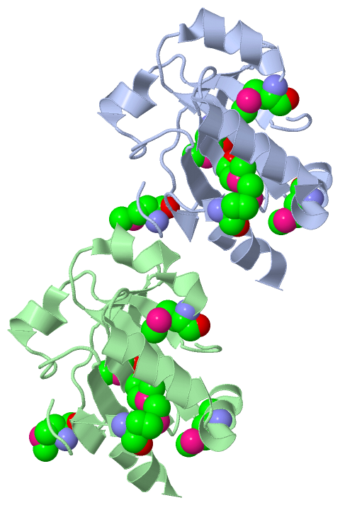
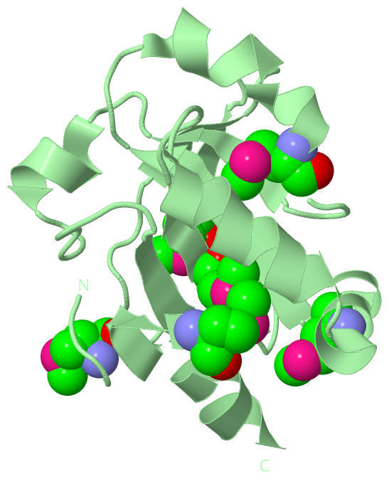
Asymmetric Unit (1, 12)
No.NameCountTypeFull Name
1MSE12Mod. Amino AcidSELENOMETHIONINE
Biological Unit 1 (1, 6)
No.NameCountTypeFull Name
1MSE6Mod. Amino AcidSELENOMETHIONINE
Biological Unit 2 (1, 6)
No.NameCountTypeFull Name
1MSE6Mod. Amino AcidSELENOMETHIONINE

(-) Sites  (0, 0)

(no "Site" information available for 3EWL)

(-) SS Bonds  (0, 0)

(no "SS Bond" information available for 3EWL)

(-) Cis Peptide Bonds  (2, 2)

Asymmetric Unit
No.Residues
1Thr A:107 -Pro A:108
2Thr B:107 -Pro B:108

 Sequence-Structure Mapping

(-) SAPs(SNPs)/Variants  (0, 0)

(no "SAP(SNP)/Variant" information available for 3EWL)

(-) PROSITE Motifs  (0, 0)

(no "PROSITE Motif" information available for 3EWL)

(-) Exons   (0, 0)

(no "Exon" information available for 3EWL)

(-) Sequences/Alignments

Asymmetric Unit
   Reformat: Number of residues per line =  ('0' or empty: single-line sequence representation)
  Number of residues per labelling interval =   
  UniProt sequence: complete  aligned part    
   Show mapping: SCOP domains CATH domains Pfam domains Secondary structure (by author)
SAPs(SNPs) PROSITE motifs Exons
(details for a mapped element are shown in a popup box when the mouse pointer rests over it)
Chain A from PDB  Type:PROTEIN  Length:138
 aligned with Q5LE83_BACFN | Q5LE83 from UniProtKB/TrEMBL  Length:327

    Alignment length:138
                                   195       205       215       225       235       245       255       265       275       285       295       305       315        
         Q5LE83_BACFN   186 NRPGMKAADFTYVTVHGDNSRMSRLKAQYTMLFFYDPDCSNCRKFEKLFAEIPAFVEMVENGTLRVLAIYPDENREEWATKAVYMPQGWIVGWNKAGDIRTRQLYDIRATPTIYLLDGRKRVILKDTSMEQLIDYLAT 323
               SCOP domains d3ewla_ A: automated matches                                                                                                               SCOP domains
               CATH domains 3ewlA00 A:-2-135 Glutaredoxin                                                                                                              CATH domains
               Pfam domains ------------------------------------------------------------------------------------------------------------------------------------------ Pfam domains
         Sec.struct. author ..........eee.....eee.hhh...eeeeee....hhhhhhhhhhhhhhhhhhhhhhh..eeeeeee...hhhhhhhhhh......eeee...hhhhhh.........eeeee.....eee...hhhhhhhhhhh Sec.struct. author
                 SAPs(SNPs) ------------------------------------------------------------------------------------------------------------------------------------------ SAPs(SNPs)
                    PROSITE ------------------------------------------------------------------------------------------------------------------------------------------ PROSITE
                 Transcript ------------------------------------------------------------------------------------------------------------------------------------------ Transcript
                 3ewl A  -2 SNAGmKAADFTYVTVHGDNSRmSRLKAQYTmLFFYDPDCSNCRKFEKLFAEIPAFVEmVENGTLRVLAIYPDENREEWATKAVYmPQGWIVGWNKAGDIRTRQLYDIRATPTIYLLDGRKRVILKDTSmEQLIDYLAT 135
                                |    7        17 |      27|       37        47       |57        67        77    |   87        97       107       117       127        
                                |               19-MSE   28-MSE                     55-MSE                     82-MSE                                     126-MSE     
                                2-MSE                                                                                                                                 

Chain B from PDB  Type:PROTEIN  Length:137
 aligned with Q5LE83_BACFN | Q5LE83 from UniProtKB/TrEMBL  Length:327

    Alignment length:137
                                   195       205       215       225       235       245       255       265       275       285       295       305       315       
         Q5LE83_BACFN   186 NRPGMKAADFTYVTVHGDNSRMSRLKAQYTMLFFYDPDCSNCRKFEKLFAEIPAFVEMVENGTLRVLAIYPDENREEWATKAVYMPQGWIVGWNKAGDIRTRQLYDIRATPTIYLLDGRKRVILKDTSMEQLIDYLA 322
               SCOP domains d3ewlb_ B: automated matches                                                                                                              SCOP domains
               CATH domains 3ewlB00 B:-2-134 Glutaredoxin                                                                                                             CATH domains
               Pfam domains ----------------------------------------------------------------------------------------------------------------------------------------- Pfam domains
         Sec.struct. author ..........eee.....eee.hhh...eeeeee....hhhhhhhhhhhh.hhhhhhhhhh..eeeeeee...hhhhhhhhhhhh....eeee...hhhhhh.........eeeee.....eeeeeehhhhhhhhhh Sec.struct. author
                 SAPs(SNPs) ----------------------------------------------------------------------------------------------------------------------------------------- SAPs(SNPs)
                    PROSITE ----------------------------------------------------------------------------------------------------------------------------------------- PROSITE
                 Transcript ----------------------------------------------------------------------------------------------------------------------------------------- Transcript
                 3ewl B  -2 SNAGmKAADFTYVTVHGDNSRmSRLKAQYTmLFFYDPDCSNCRKFEKLFAEIPAFVEmVENGTLRVLAIYPDENREEWATKAVYmPQGWIVGWNKAGDIRTRQLYDIRATPTIYLLDGRKRVILKDTSmEQLIDYLA 134
                                |    7        17 |      27|       37        47       |57        67        77    |   87        97       107       117       127       
                                2-MSE           19-MSE   28-MSE                     55-MSE                     82-MSE                                     126-MSE    

   Legend:   → Mismatch (orange background)
  - → Gap (green background, '-', border residues have a numbering label)
    → Modified Residue (blue background, lower-case, 'x' indicates undefined single-letter code, labelled with number + name)
  x → Chemical Group (purple background, 'x', labelled with number + name, e.g. ACE or NH2)
  extra numbering lines below/above indicate numbering irregularities and modified residue names etc., number ends below/above '|'

 Classification and Annotation

(-) SCOP Domains  (1, 2)

Asymmetric Unit

(-) CATH Domains  (1, 2)

Asymmetric Unit
(-)
Class: Alpha Beta (26913)

(-) Pfam Domains  (0, 0)

(no "Pfam Domain" information available for 3EWL)

(-) Gene Ontology  (2, 2)

Asymmetric Unit(hide GO term definitions)
Chain A,B   (Q5LE83_BACFN | Q5LE83)
cellular component
    GO:0016021    integral component of membrane    The component of a membrane consisting of the gene products and protein complexes having at least some part of their peptide sequence embedded in the hydrophobic region of the membrane.
    GO:0016020    membrane    A lipid bilayer along with all the proteins and protein complexes embedded in it an attached to it.

 Visualization

(-) Interactive Views

Asymmetric Unit
  Complete Structure
    Jena3D(integrated viewing of ligand, site, SAP, PROSITE, SCOP information)
    WebMol | AstexViewer[tm]@PDBe
(Java Applets, require no local installation except for Java; loading may be slow)
    STRAP
(Java WebStart application, automatic local installation, requires Java; full application with system access!)
    RasMol
(require local installation)
    Molscript (VRML)
(requires installation of a VRML viewer; select preferred view via VRML and generate a mono or stereo PDF format file)
 
  Ligands, Modified Residues, Ions
    MSE  [ RasMol | Jena3D ]  +environment [ RasMol | Jena3D ]
 
  Sites
(no "Sites" information available for 3ewl)
 
  Cis Peptide Bonds
    Thr A:107 - Pro A:108   [ RasMol ]  
    Thr B:107 - Pro B:108   [ RasMol ]  
 
Biological Units
  Complete Structure
    Biological Unit 1  [ Jena3D ]
    Biological Unit 2  [ Jena3D ]

(-) Still Images

Jmol
  protein: cartoon or spacefill or dots and stick; nucleic acid: cartoon and stick; ligands: spacefill; active site: stick
Molscript
  protein, nucleic acid: cartoon; ligands: spacefill; active site: ball and stick

 Databases and Analysis Tools

(-) Databases

Access by PDB/NDB ID
  3ewl
    Family and Domain InformationProDom | SYSTERS
    General Structural InformationGlycoscienceDB | MMDB | NDB | OCA | PDB | PDBe | PDBj | PDBsum | PDBWiki | PQS | PROTEOPEDIA
    Orientation in MembranesOPM
    Protein SurfaceSURFACE
    Secondary StructureDSSP (structure derived) | HSSP (homology derived)
    Structural GenomicsGeneCensus
    Structural NeighboursCE | VAST
    Structure ClassificationCATH | Dali | SCOP
    Validation and Original DataBMRB Data View | BMRB Restraints Grid | EDS | PROCHECK | RECOORD | WHAT_CHECK
 
Access by UniProt ID/Accession number
  Q5LE83_BACFN | Q5LE83
    Comparative Protein Structure ModelsModBase
    Genomic InformationEnsembl
    Protein-protein InteractionDIP
    Sequence, Family and Domain InformationInterPro | Pfam | SMART | UniProtKB/TrEMBL
 
Access by Enzyme Classificator   (EC Number)
  (no 'Enzyme Classificator' available)
    General Enzyme InformationBRENDA | EC-PDB | Enzyme | IntEnz
    PathwayKEGG | MetaCyc
 
Access by Disease Identifier   (MIM ID)
  (no 'MIM ID' available)
    Disease InformationOMIM
 
Access by GenAge ID
  (no 'GenAge ID' available)
    Age Related InformationGenAge

(-) Analysis Tools

Access by PDB/NDB ID
    Domain InformationXDom
    Interatomic Contacts of Structural UnitsCSU
    Ligand-protein ContactsLPC
    Protein CavitiescastP
    Sequence and Secondary StructurePDBCartoon
    Structure AlignmentSTRAP(Java WebStart application, automatic local installation, requires Java; full application with system access!)
    Structure and Sequence BrowserSTING
 
Access by UniProt ID/Accession number
  Q5LE83_BACFN | Q5LE83
    Protein Disorder PredictionDisEMBL | FoldIndex | GLOBPLOT (for more information see DisProt)

 Related Entries

(-) Entries Sharing at Least One Protein Chain (UniProt ID)

(no "Entries Sharing at Least One Protein Chain" available for 3EWL)

(-) Related Entries Specified in the PDB File

(no "Related Entries Specified in the PDB File" available for 3EWL)