|
|
|
|
 Description
|
|
Compounds
|
||||||||||||||||||||
Chains, Units
Summary Information (see also Sequences/Alignments below) |
Ligands, Modified Residues, Ions (0, 0)| (no "Ligand,Modified Residues,Ions" information available for 3EIV) |
Sites (0, 0)| (no "Site" information available for 3EIV) |
SS Bonds (0, 0)| (no "SS Bond" information available for 3EIV) |
Cis Peptide Bonds (0, 0)| (no "Cis Peptide Bond" information available for 3EIV) |
SAPs(SNPs)/Variants (0, 0)| (no "SAP(SNP)/Variant" information available for 3EIV) |
PROSITE Motifs (1, 1)
Asymmetric Unit (1, 1)
|
||||||||||||||||||||||||||||||||||||||||||||||||||||||||||||||||||||||||||||||||||||||||||||||||
Exons (0, 0)| (no "Exon" information available for 3EIV) |
Sequences/Alignments
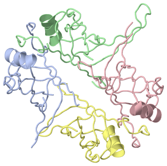
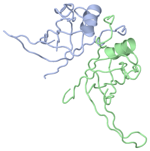
Asymmetric UnitChain A from PDB Type:PROTEIN Length:98 aligned with SSB2_STRCO | Q9X8U3 from UniProtKB/Swiss-Prot Length:199 Alignment length:117 13 23 33 43 53 63 73 83 93 103 113 SSB2_STRCO 4 ETVITVVGNLVDDPELRFTPSGAAVAKFRVASTPRTFDRQTNEWKDGESLFLTCSVWRQAAENVAESLQRGMRVIVQGRLKQRSYEDREGVKRTVYELDVDEVGASLRSATAKVTKT 120 SCOP domains d3eiva_ A: automated matches SCOP domains CATH domains 3eivA00 A:4-120 Nucleic acid-bindi ng proteins CATH domains Pfam domains --------------------------------------------------------------------------------------------------------------------- Pfam domains SAPs(SNPs) --------------------------------------------------------------------------------------------------------------------- SAPs(SNPs) PROSITE SSB PDB: - UniProt: 1-110 ---------- PROSITE Transcript --------------------------------------------------------------------------------------------------------------------- Transcript 3eiv A 4 ETVITVVGNLVDDPELRFTPSGAAVAKFRVASTP-----------DGESLFLTCSVWRQAAENVAESLQRGMRVIVQGRLKQRS--------RTVYELDVDEVGASLRSATAKVTKT 120 13 23 33 | - | 53 63 73 83 | - | 103 113 37 49 87 96 Chain B from PDB Type:PROTEIN Length:111 aligned with SSB2_STRCO | Q9X8U3 from UniProtKB/Swiss-Prot Length:199 Alignment length:120 10 20 30 40 50 60 70 80 90 100 110 120 SSB2_STRCO 1 MAGETVITVVGNLVDDPELRFTPSGAAVAKFRVASTPRTFDRQTNEWKDGESLFLTCSVWRQAAENVAESLQRGMRVIVQGRLKQRSYEDREGVKRTVYELDVDEVGASLRSATAKVTKT 120 SCOP domains d3eivb_ B: automated matches SCOP domains CATH domains 3eivB00 B:1-120 Nucleic acid-binding pr oteins CATH domains Pfam domains ------------------------------------------------------------------------------------------------------------------------ Pfam domains SAPs(SNPs) ------------------------------------------------------------------------------------------------------------------------ SAPs(SNPs) PROSITE SSB PDB: B:1-110 UniProt: 1-110 ---------- PROSITE Transcript ------------------------------------------------------------------------------------------------------------------------ Transcript 3eiv B 1 MAGETVITVVGNLVDDPELRFTPSGAAVAKFRVASTPRT---------DGESLFLTCSVWRQAAENVAESLQRGMRVIVQGRLKQRSYEDREGVKRTVYELDVDEVGASLRSATAKVTKT 120 10 20 30 |- 50 60 70 80 90 100 110 120 39 49 Chain C from PDB Type:PROTEIN Length:111 aligned with SSB2_STRCO | Q9X8U3 from UniProtKB/Swiss-Prot Length:199 Alignment length:119 11 21 31 41 51 61 71 81 91 101 111 SSB2_STRCO 2 AGETVITVVGNLVDDPELRFTPSGAAVAKFRVASTPRTFDRQTNEWKDGESLFLTCSVWRQAAENVAESLQRGMRVIVQGRLKQRSYEDREGVKRTVYELDVDEVGASLRSATAKVTKT 120 SCOP domains d3eivc_ C: automated matches SCOP domains CATH domains 3eivC00 C:2-120 Nucleic acid-binding proteins CATH domains Pfam domains ----------------------------------------------------------------------------------------------------------------------- Pfam domains SAPs(SNPs) ----------------------------------------------------------------------------------------------------------------------- SAPs(SNPs) PROSITE SSB PDB: - UniProt: 1-110 ---------- PROSITE Transcript ----------------------------------------------------------------------------------------------------------------------- Transcript 3eiv C 2 AGETVITVVGNLVDDPELRFTPSGAAVAKFRVASTPRTFDRQTNEWKDGESLFLTCSVWRQAAENVAESLQRGMRVIVQGRLKQRS--------RTVYELDVDEVGASLRSATAKVTKT 120 11 21 31 41 51 61 71 81 | - | 101 111 87 96 Chain D from PDB Type:PROTEIN Length:100 aligned with SSB2_STRCO | Q9X8U3 from UniProtKB/Swiss-Prot Length:199 Alignment length:119 11 21 31 41 51 61 71 81 91 101 111 SSB2_STRCO 2 AGETVITVVGNLVDDPELRFTPSGAAVAKFRVASTPRTFDRQTNEWKDGESLFLTCSVWRQAAENVAESLQRGMRVIVQGRLKQRSYEDREGVKRTVYELDVDEVGASLRSATAKVTKT 120 SCOP domains d3eivd_ D: automated matches SCOP domains CATH domains 3eivD00 D:2-120 Nucleic acid-binding proteins CATH domains Pfam domains ----------------------------------------------------------------------------------------------------------------------- Pfam domains SAPs(SNPs) ----------------------------------------------------------------------------------------------------------------------- SAPs(SNPs) PROSITE SSB PDB: - UniProt: 1-110 ---------- PROSITE Transcript ----------------------------------------------------------------------------------------------------------------------- Transcript 3eiv D 2 AGETVITVVGNLVDDPELRFTPSGAAVAKFRVASTPR----------DGESLFLTCSVWRQAAENVAESLQRGMRVIVQGRLKQRS---------TVYELDVDEVGASLRSATAKVTKT 120 11 21 31 | - |51 61 71 81 | - | 101 111 38 49 87 97
|
||||||||||||||||||||
SCOP Domains (1, 4)
Asymmetric Unit
 |
CATH Domains (1, 4)
Asymmetric Unit
|
Pfam Domains (0, 0)| (no "Pfam Domain" information available for 3EIV) |
Gene Ontology (3, 3)|
Asymmetric Unit(hide GO term definitions) Chain A,B,C,D (SSB2_STRCO | Q9X8U3)
|
||||||||||||||||||||||||||||||
Interactive Views
|
||||||||||||||||||||||||||||||||||||||||||||||||||||||||||||||||||||||||||||||||||||||||||||||||||||||||||||||||||||||||||||||||||||||||||||||||
Still Images
|
||||||||||||||||
Databases
|
||||||||||||||||||||||||||||||||||||||||||||||||||||||||||||||||||||||||||||||||||||||||||||||||||||||||||||||||||||||||||||||||||||||||||||||||||||||||||||||||
Analysis Tools
|
|||||||||||||||||||||||||||||||||||||||||||||||||||||||||||||
Entries Sharing at Least One Protein Chain (UniProt ID)
Related Entries Specified in the PDB File
|
|