|
|
|
|
 Description
|
|
Compounds
|
||||||||||||||||||||
Chains, Units
Summary Information (see also Sequences/Alignments below) |
Ligands, Modified Residues, Ions (0, 0)| (no "Ligand,Modified Residues,Ions" information available for 3E4H) |
Sites (0, 0)| (no "Site" information available for 3E4H) |
SS Bonds (3, 3)
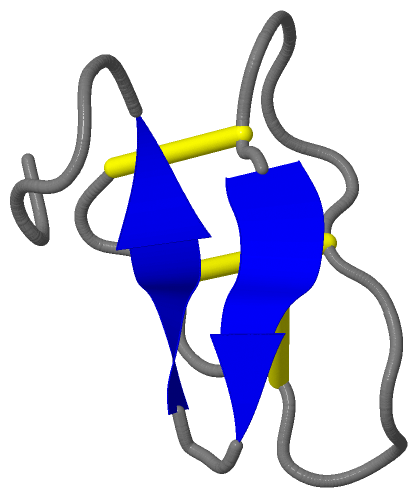
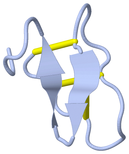
Asymmetric Unit
|
||||||||||||||||
Cis Peptide Bonds (1, 1)
Asymmetric Unit
|
||||||||
SAPs(SNPs)/Variants (0, 0)| (no "SAP(SNP)/Variant" information available for 3E4H) |
PROSITE Motifs (1, 1)
Asymmetric Unit (1, 1)
|
||||||||||||||||||||||||||||||||||||||||||||||||||||||||||||||||||||||||
Exons (0, 0)| (no "Exon" information available for 3E4H) |
Sequences/Alignments
Asymmetric UnitChain A from PDB Type:PROTEIN Length:29 aligned with VARF_VIOAR | P58451 from UniProtKB/Swiss-Prot Length:29 Alignment length:29 29 14 24 | VARF_VIOAR 5 CGETCTLGTCYTAGCSCSWPVCTRN---- - SCOP domains d3e4ha_ A: automated matches SCOP domains CATH domains ----------------------------- CATH domains Pfam domains ----------------------------- Pfam domains SAPs(SNPs) ----------------------------- SAPs(SNPs) PROSITE CYCLOTIDE_------------------- PROSITE Transcript ----------------------------- Transcript 3e4h A 1 CGETCTLGTCYTAGCSCSWPVCTRNGVPI 29 10 20
|
||||||||||||||||||||
SCOP Domains (1, 1)
Asymmetric Unit
 |
CATH Domains (0, 0)| (no "CATH Domain" information available for 3E4H) |
Pfam Domains (0, 0)| (no "Pfam Domain" information available for 3E4H) |
Gene Ontology (3, 3)|
Asymmetric Unit(hide GO term definitions) Chain A (VARF_VIOAR | P58451)
|
||||||||||||||||||||||||||||||||||||
Interactive Views
|
||||||||||||||||||||||||||||||||||||||||||||||||||||||||||||||||||||||||||||||||||||||||||||||||||||||||||||||||||||||||||||||||||||||||||||
Still Images
|
||||||||||||||||
Databases
|
||||||||||||||||||||||||||||||||||||||||||||||||||||||||||||||||||||||||||||||||||||||||||||||||||||||||||||||||||||||||||||||||||||||||||||||||||||||||||||||||
Analysis Tools
|
|||||||||||||||||||||||||||||||||||||||||||||||||||||||||||||
Entries Sharing at Least One Protein Chain (UniProt ID)
Related Entries Specified in the PDB File
|
|