Show PDB file:   
         Plain Text   HTML   (compressed file size)
QuickSearch:   
by PDB,NDB,UniProt,PROSITE Code or Search Term(s)  
(-)Asym./Biol. Unit
(-)Asym./Biol. Unit - sites
collapse expand < >
Image Asym./Biol. Unit
Asym./Biol. Unit  (Jmol Viewer)
Image Asym./Biol. Unit - sites
Asym./Biol. Unit - sites  (Jmol Viewer)

(-) Description

Title :  IRON RECONSTITUTED FERRIC BINDING PROTEIN FROM CAMPYLOBACTER JEJUNI
 
Authors :  M. E. P. Murphy, S. A. L. Tom-Yew, E. G. Bekker
Date :  01 Aug 08  (Deposition) - 11 Aug 09  (Release) - 11 Aug 09  (Revision)
Method :  X-RAY DIFFRACTION
Resolution :  1.60
Chains :  Asym./Biol. Unit :  X
Keywords :  Beta Sheet Surrounded By Alpha Helices, Anti-Parallel Beta Sheet Containing Hinge Region, Metal Binding Protein (Keyword Search: [Gene Ontology, PubMed, Web (Google))
 
Reference :  S. A. L. Tom-Yew, B. H. Shilton, E. G. Bekker, E. C. Gaynor, M. E. P. Murphy
Small Hinge Motion By The Anion-Independent Ferric Binding Protein From Campylobacter Jejuni
To Be Published
PubMed: search
(for further references see the PDB file header)

(-) Compounds

Molecule 1 - PUTATIVE IRON-UPTAKE ABC TRANSPORT SYSTEM, PERIPLASMIC IRON-BINDING PROTEIN
    ChainsX
    EngineeredYES
    Expression SystemESCHERICHIA COLI
    Expression System PlasmidPET28A
    Expression System StrainHMS174(DE3)
    Expression System Taxid562
    Expression System Vector TypePLASMID
    GeneCFBPA, CJ0175C
    Organism ScientificCAMPYLOBACTER JEJUNI
    Organism Taxid197
    StrainNCTC 11168

 Structural Features

(-) Chains, Units

  1
Asymmetric/Biological Unit X

Summary Information (see also Sequences/Alignments below)

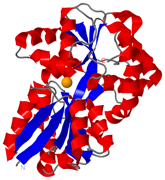
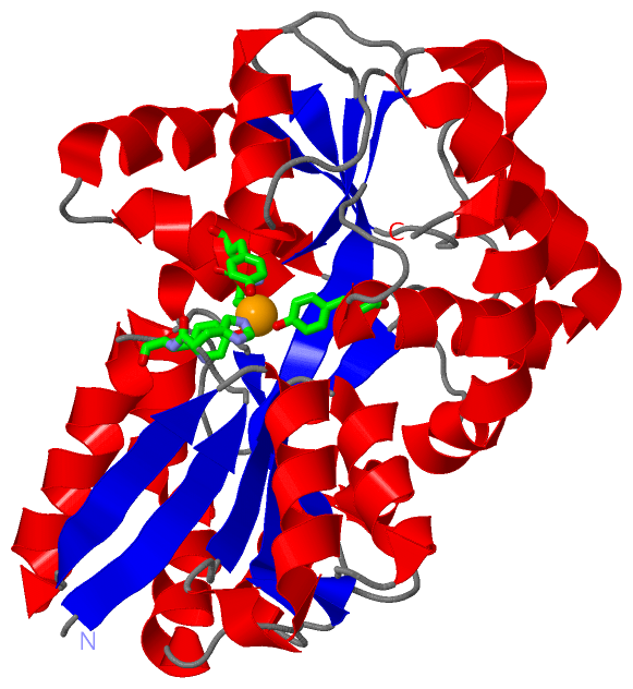
(-) Ligands, Modified Residues, Ions  (1, 1)

Asymmetric/Biological Unit (1, 1)
No.NameCountTypeFull Name
1FE1Ligand/IonFE (III) ION

(-) Sites  (1, 1)

Asymmetric Unit (1, 1)
No.NameEvidenceResiduesDescription
1AC1SOFTWAREHIS X:14 , TYR X:15 , TYR X:146 , TYR X:202 , TYR X:203BINDING SITE FOR RESIDUE FE X 326

(-) SS Bonds  (0, 0)

(no "SS Bond" information available for 3E13)

(-) Cis Peptide Bonds  (0, 0)

(no "Cis Peptide Bond" information available for 3E13)

 Sequence-Structure Mapping

(-) SAPs(SNPs)/Variants  (0, 0)

(no "SAP(SNP)/Variant" information available for 3E13)

(-) PROSITE Motifs  (0, 0)

(no "PROSITE Motif" information available for 3E13)

(-) Exons   (0, 0)

(no "Exon" information available for 3E13)

(-) Sequences/Alignments

Asymmetric/Biological Unit
   Reformat: Number of residues per line =  ('0' or empty: single-line sequence representation)
  Number of residues per labelling interval =   
  UniProt sequence: complete  aligned part    
   Show mapping: SCOP domains CATH domains Pfam domains Secondary structure (by author)
SAPs(SNPs) PROSITE motifs Exons
(details for a mapped element are shown in a popup box when the mouse pointer rests over it)
Chain X from PDB  Type:PROTEIN  Length:317
 aligned with Q0PBW4_CAMJE | Q0PBW4 from UniProtKB/TrEMBL  Length:334

    Alignment length:317
                                    27        37        47        57        67        77        87        97       107       117       127       137       147       157       167       177       187       197       207       217       227       237       247       257       267       277       287       297       307       317       327       
         Q0PBW4_CAMJE    18 SELNIYSARHYNADFEIIKKFEEKTGIKVNHTQAKASELIKRLSLEGSNSPADIFITADISNLTEAKNLGLLSPVSSKYLEEFIPAHLRDKDKEWFAITKRARIIAYNKNTNIDISKMKNYEDLAKAEFKGEIVMRSATAPYSKTLLASIIANDGNKEAKAWAKGVLENLATNPKGGDRDQARQVFAGEAKFAVMNTYYIGLLKNSKNPKDVEVGNSLGIIFPNQDNRGTHINISGIAMTKSSKNQDAAKKFMEFMLSPEIQKILTDSNYEFPIRNDVELSQTVKDFGTFKEDQIPVSKIAENIKEAVKIYDEVGFR 334
               SCOP domains d3e13x_ X: Ferric-binding protein FbpA                                                                                                                                                                                                                                                                                        SCOP domains
               CATH domains 3e13X01 X:5-105,X:238-284 Periplasmic binding protein-like II                                        ------------------------------------------------------------------------------------------------------------------------------------3e13X01 X:5-105,X:238-284                      ------------------------------------- CATH domains
               Pfam domains ----------------------------------------------------------------------------------------------------------------------------------------------------------------------------------------------------------------------------------------------------------------------------------------------------------------------------- Pfam domains
         Sec.struct. author .eeeeee....hhhhhhhhhhhhhhh.eeeeee.hhhhhhhhhhhhhhhh...eeee.hhhhhhhhhhh..ee...hhhhhhhhhhhhh......eeeeeeeeeeeee...........hhhhhhhhhhh..ee.....hhhhhhhhhhhhhhhhhhhhhhhhhhhhhhh......hhhhhhhhhhh....eeeeehhhhhhhhh..hhhhhhhhhheeee..........eeeeeeeee....hhhhhhhhhhhhhhhhhhhhhhhh...ee.......hhhhhhhh.......hhhhhhhhhhhhhhhhhhhh.. Sec.struct. author
                 SAPs(SNPs) ----------------------------------------------------------------------------------------------------------------------------------------------------------------------------------------------------------------------------------------------------------------------------------------------------------------------------- SAPs(SNPs)
                    PROSITE ----------------------------------------------------------------------------------------------------------------------------------------------------------------------------------------------------------------------------------------------------------------------------------------------------------------------------- PROSITE
                 Transcript ----------------------------------------------------------------------------------------------------------------------------------------------------------------------------------------------------------------------------------------------------------------------------------------------------------------------------- Transcript
                 3e13 X   5 SELNIYSARHYNADFEIIKKFEEKTGIKVNHTQAKASELIKRLSLEGSNSPADIFITADISNLTEAKNLGLLSPVSSKYLEEFIPAHLRDKDKEWFAITKRARIIAYNKNTNIDISKMKNYEDLAKAEFKGEIVMRSATAPYSKTLLASIIANDGNKEAKAWAKGVLENLATNPKGGDRDQARQVFAGEAKFAVMNTYYIGLLKNSKNPKDVEVGNSLGIIFPNQDNRGTHINISGIAMTKSSKNQDAAKKFMEFMLSPEIQKILTDSNYEFPIRNDVELSQTVKDFGTFKEDQIPVSKIAENIKEAVKIYDEVGFR 321
                                    14        24        34        44        54        64        74        84        94       104       114       124       134       144       154       164       174       184       194       204       214       224       234       244       254       264       274       284       294       304       314       

   Legend:   → Mismatch (orange background)
  - → Gap (green background, '-', border residues have a numbering label)
    → Modified Residue (blue background, lower-case, 'x' indicates undefined single-letter code, labelled with number + name)
  x → Chemical Group (purple background, 'x', labelled with number + name, e.g. ACE or NH2)
  extra numbering lines below/above indicate numbering irregularities and modified residue names etc., number ends below/above '|'

 Classification and Annotation

(-) SCOP Domains  (1, 1)

Asymmetric/Biological Unit

(-) CATH Domains  (1, 1)

Asymmetric/Biological Unit
(-)
Class: Alpha Beta (26913)

(-) Pfam Domains  (0, 0)

(no "Pfam Domain" information available for 3E13)

(-) Gene Ontology  (1, 1)

Asymmetric/Biological Unit(hide GO term definitions)
Chain X   (Q0PBW4_CAMJE | Q0PBW4)
molecular function
    GO:0046872    metal ion binding    Interacting selectively and non-covalently with any metal ion.

 Visualization

(-) Interactive Views

Asymmetric/Biological Unit
  Complete Structure
    Jena3D(integrated viewing of ligand, site, SAP, PROSITE, SCOP information)
    WebMol | AstexViewer[tm]@PDBe
(Java Applets, require no local installation except for Java; loading may be slow)
    STRAP
(Java WebStart application, automatic local installation, requires Java; full application with system access!)
    RasMol
(require local installation)
    Molscript (VRML)
(requires installation of a VRML viewer; select preferred view via VRML and generate a mono or stereo PDF format file)
 
  Ligands, Modified Residues, Ions
    FE  [ RasMol | Jena3D ]  +environment [ RasMol | Jena3D ]
 
  Sites
    AC1  [ RasMol ]  +environment [ RasMol ]
 
  Cis Peptide Bonds
(no "Cis Peptide Bonds" information available for 3e13)
 

(-) Still Images

Jmol
  protein: cartoon or spacefill or dots and stick; nucleic acid: cartoon and stick; ligands: spacefill; active site: stick
Molscript
  protein, nucleic acid: cartoon; ligands: spacefill; active site: ball and stick

 Databases and Analysis Tools

(-) Databases

Access by PDB/NDB ID
  3e13
    Family and Domain InformationProDom | SYSTERS
    General Structural InformationGlycoscienceDB | MMDB | NDB | OCA | PDB | PDBe | PDBj | PDBsum | PDBWiki | PQS | PROTEOPEDIA
    Orientation in MembranesOPM
    Protein SurfaceSURFACE
    Secondary StructureDSSP (structure derived) | HSSP (homology derived)
    Structural GenomicsGeneCensus
    Structural NeighboursCE | VAST
    Structure ClassificationCATH | Dali | SCOP
    Validation and Original DataBMRB Data View | BMRB Restraints Grid | EDS | PROCHECK | RECOORD | WHAT_CHECK
 
Access by UniProt ID/Accession number
  Q0PBW4_CAMJE | Q0PBW4
    Comparative Protein Structure ModelsModBase
    Genomic InformationEnsembl
    Protein-protein InteractionDIP
    Sequence, Family and Domain InformationInterPro | Pfam | SMART | UniProtKB/TrEMBL
 
Access by Enzyme Classificator   (EC Number)
  (no 'Enzyme Classificator' available)
    General Enzyme InformationBRENDA | EC-PDB | Enzyme | IntEnz
    PathwayKEGG | MetaCyc
 
Access by Disease Identifier   (MIM ID)
  (no 'MIM ID' available)
    Disease InformationOMIM
 
Access by GenAge ID
  (no 'GenAge ID' available)
    Age Related InformationGenAge

(-) Analysis Tools

Access by PDB/NDB ID
    Domain InformationXDom
    Interatomic Contacts of Structural UnitsCSU
    Ligand-protein ContactsLPC
    Protein CavitiescastP
    Sequence and Secondary StructurePDBCartoon
    Structure AlignmentSTRAP(Java WebStart application, automatic local installation, requires Java; full application with system access!)
    Structure and Sequence BrowserSTING
 
Access by UniProt ID/Accession number
  Q0PBW4_CAMJE | Q0PBW4
    Protein Disorder PredictionDisEMBL | FoldIndex | GLOBPLOT (for more information see DisProt)

 Related Entries

(-) Entries Sharing at Least One Protein Chain (UniProt ID)

UniProtKB/TrEMBL
        Q0PBW4_CAMJE | Q0PBW41y4t

(-) Related Entries Specified in the PDB File

1y4t RECOMBINANT HOLO FBPA PROTEIN FROM CAMPYLOBACTER JEJUNI, AS ISOLATED FROM E. COLI