|
|
|
|
 Description
|
|
Compounds
|
||||||||||||||||||||||||||||||||||||||||||||||||
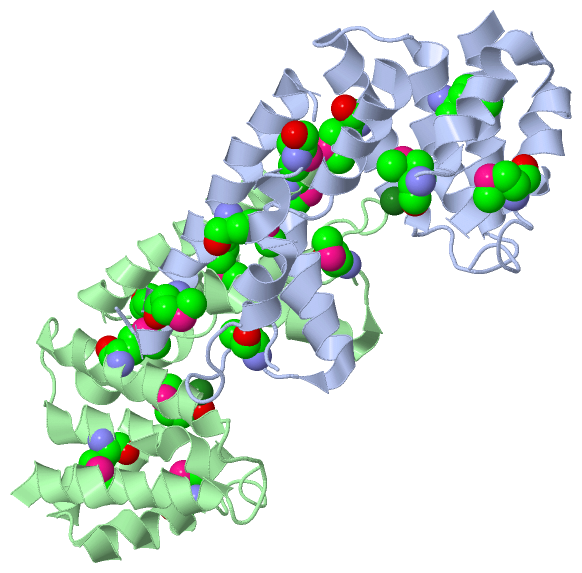
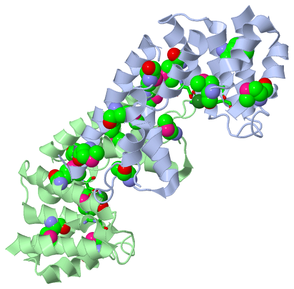
Chains, Units
Summary Information (see also Sequences/Alignments below) |
Ligands, Modified Residues, Ions (2, 18)| Asymmetric/Biological Unit (2, 18) |
Sites (2, 2)
Asymmetric Unit (2, 2)
|
SS Bonds (0, 0)| (no "SS Bond" information available for 3DJB) |
Cis Peptide Bonds (0, 0)| (no "Cis Peptide Bond" information available for 3DJB) |
SAPs(SNPs)/Variants (0, 0)| (no "SAP(SNP)/Variant" information available for 3DJB) |
PROSITE Motifs (0, 0)| (no "PROSITE Motif" information available for 3DJB) |
Exons (0, 0)| (no "Exon" information available for 3DJB) |
Sequences/Alignments
Asymmetric/Biological UnitChain A from PDB Type:PROTEIN Length:188 aligned with Q6HJG6_BACHK | Q6HJG6 from UniProtKB/TrEMBL Length:215 Alignment length:213 11 21 31 41 51 61 71 81 91 101 111 121 131 141 151 161 171 181 191 201 211 Q6HJG6_BACHK 2 TKQEKIEKTITFVKHILEKDASGHDWYHIERVHKLAISLSEQEGGNRFIIEMAALLHDVADEKLNESEEAGMKKVSDWLEELHVEEEESKHVLHIIANMSYKGGHGGKVESIEGKIVQDADRLDALGAIGIARTFAYGGAKGRLMYDPTIPPREVMTKDEYRKNNDPSLNHFYEKLLKLKDLMNTNAAKQEAEVRHRYMEQFIEQFMKEWNAQ 214 SCOP domains d3djba1 A:2-214 Uncharacterized protein BT9727_1981 SCOP domains CATH domains 3djbA01 A:2-99 HD-domain/PDEase-like - ------------------------------------------- ------------------------------------------------ CATH domains Pfam domains --------------------------------------------------------------------------------------------------------------------------------------------------------------------------------------------------------------------- Pfam domains SAPs(SNPs) --------------------------------------------------------------------------------------------------------------------------------------------------------------------------------------------------------------------- SAPs(SNPs) PROSITE --------------------------------------------------------------------------------------------------------------------------------------------------------------------------------------------------------------------- PROSITE Transcript --------------------------------------------------------------------------------------------------------------------------------------------------------------------------------------------------------------------- Transcript 3djb A 2 TKQEKIEKTITFVKHILEKDASGHDWYHIRRVHKmAISLSEQEGGNRFIIEmAALLHDVAD--LNESEEAGmKKVSDWLEELHVEEEESKHVLHIIANm-----------SIEGKLVQDADRLDALGAIGIARTFAYGGAKGRLmYDPTIPPR------------DPSLNHFYEKLLKLKDLmNTNAAKQEAEVRHRYmEQFIEQFmKEWNAQ 214 11 21 31 | 41 51 | 61| | 71 | 81 91 |- -| 121 131 141 | 151 | - | 171 181 | 191 201 |211 36-MSE 53-MSE 62 65 73-MSE 100-MSE 112 146-MSE 154 167 184-MSE 200-MSE 208-MSE Chain B from PDB Type:PROTEIN Length:188 aligned with Q6HJG6_BACHK | Q6HJG6 from UniProtKB/TrEMBL Length:215 Alignment length:213 11 21 31 41 51 61 71 81 91 101 111 121 131 141 151 161 171 181 191 201 211 Q6HJG6_BACHK 2 TKQEKIEKTITFVKHILEKDASGHDWYHIERVHKLAISLSEQEGGNRFIIEMAALLHDVADEKLNESEEAGMKKVSDWLEELHVEEEESKHVLHIIANMSYKGGHGGKVESIEGKIVQDADRLDALGAIGIARTFAYGGAKGRLMYDPTIPPREVMTKDEYRKNNDPSLNHFYEKLLKLKDLMNTNAAKQEAEVRHRYMEQFIEQFMKEWNAQ 214 SCOP domains d3djbb_ B: Uncharacterized protein BT9727_1981 SCOP domains CATH domains 3djbB01 B:2-99 HD-domain/PDEase-like - ------------------------------------------- ------------------------------------------------ CATH domains Pfam domains --------------------------------------------------------------------------------------------------------------------------------------------------------------------------------------------------------------------- Pfam domains SAPs(SNPs) --------------------------------------------------------------------------------------------------------------------------------------------------------------------------------------------------------------------- SAPs(SNPs) PROSITE --------------------------------------------------------------------------------------------------------------------------------------------------------------------------------------------------------------------- PROSITE Transcript --------------------------------------------------------------------------------------------------------------------------------------------------------------------------------------------------------------------- Transcript 3djb B 2 TKQEKIEKTITFVKHILEKDASGHDWYHIRRVHKmAISLSEQEGGNRFIIEmAALLHDVAD--LNESEEAGmKKVSDWLEELHVEEEESKHVLHIIANm-----------SIEGKLVQDADRLDALGAIGIARTFAYGGAKGRLmYDPTIPPR------------DPSLNHFYEKLLKLKDLmNTNAAKQEAEVRHRYmEQFIEQFmKEWNAQ 214 11 21 31 | 41 51 | 61| | 71 | 81 91 |- -| 121 131 141 | 151 | - | 171 181 | 191 201 |211 36-MSE 53-MSE 62 65 73-MSE 100-MSE 112 146-MSE 154 167 184-MSE 200-MSE 208-MSE
|
||||||||||||||||||||
SCOP Domains (1, 2)
Asymmetric/Biological Unit
 |
CATH Domains (1, 2)
Asymmetric/Biological Unit
|
Pfam Domains (0, 0)| (no "Pfam Domain" information available for 3DJB) |
Gene Ontology (2, 2)|
Asymmetric/Biological Unit(hide GO term definitions) Chain A,B (Q6HJG6_BACHK | Q6HJG6)
|
||||||||||||||||||
Interactive Views
|
||||||||||||||||||||||||||||||||||||||||||||||||||||||||||||||||||||||||||||||||||||||||||||||||||||||||||||||||||||||||||||||||||||
Still Images
|
||||||||||||||||
Databases
|
||||||||||||||||||||||||||||||||||||||||||||||||||||||||||||||||||||||||||||||||||||||||||||||||||||||||||||||||||||||||||||||||||||||||||||||||||||||||||||||||
Analysis Tools
|
|||||||||||||||||||||||||||||||||||||||||||||||||||||||||||||
Entries Sharing at Least One Protein Chain (UniProt ID)
Related Entries Specified in the PDB File
|
|