Show PDB file:   
         Plain Text   HTML   (compressed file size)
QuickSearch:   
by PDB,NDB,UniProt,PROSITE Code or Search Term(s)  
(-)Asymmetric Unit
(-)Asym. Unit - sites
(-)Biological Unit 1
(-)Biol. Unit 1 - sites
(-)Biological Unit 2
(-)Biological Unit 3
collapse expand < >
Image Asymmetric Unit
Asymmetric Unit  (Jmol Viewer)
Image Asym. Unit - sites
Asym. Unit - sites  (Jmol Viewer)
Image Biological Unit 1
Biological Unit 1  (Jmol Viewer)
Image Biol. Unit 1 - sites
Biol. Unit 1 - sites  (Jmol Viewer)
Image Biological Unit 2
Biological Unit 2  (Jmol Viewer)
Image Biological Unit 3
Biological Unit 3  (Jmol Viewer)

(-) Description

Title :  C-TERMINAL DOMAIN OF PROBABLE HEMOLYSIN FROM CHROMOBACTERIUM VIOLACEUM
 
Authors :  C. Chang, X. Xu, H. Cui, A. Savchenko, A. Edwards, A. Joachimiak, Midwes For Structural Genomics (Mcsg)
Date :  09 Jun 08  (Deposition) - 05 Aug 08  (Release) - 13 Jul 11  (Revision)
Method :  X-RAY DIFFRACTION
Resolution :  2.14
Chains :  Asym. Unit :  A,B,C,D,E,F
Biol. Unit 1:  A,B  (1x)
Biol. Unit 2:  C,D  (1x)
Biol. Unit 3:  E,F  (1x)
Keywords :  Hemolysin, Chromobacterium Violaceum, Structural Genomics, Psi-2, Protein Structure Initiative, Midwest Center For Structural Genomics, Mcsg, Membrane, Transmembrane, Membrane Protein (Keyword Search: [Gene Ontology, PubMed, Web (Google))
 
Reference :  C. Chang, Xiaohui Xu, H. Cui, A. Savchenko, A. Edwards, A. Joachimiak
Crystal Structure Of C-Terminal Domain Of Probable Hemolysi From Chromobacterium Violaceum
To Be Published
PubMed: search

(-) Compounds

Molecule 1 - PROBABLE HEMOLYSIN
    ChainsA, B, C, D, E, F
    EngineeredYES
    Expression SystemESCHERICHIA COLI
    Expression System PlasmidPET DERIVATIVES
    Expression System StrainBL21(DE3) DERIVATIVES
    Expression System Vector TypePLASMID
    FragmentRESIDUES 337-427
    GeneCV_0231
    Organism ScientificCHROMOBACTERIUM VIOLACEUM
    Organism Taxid536

 Structural Features

(-) Chains, Units

  123456
Asymmetric Unit ABCDEF
Biological Unit 1 (1x)AB    
Biological Unit 2 (1x)  CD  
Biological Unit 3 (1x)    EF

Summary Information (see also Sequences/Alignments below)

(-) Ligands, Modified Residues, Ions  (2, 27)

Asymmetric Unit (2, 27)
No.NameCountTypeFull Name
1CA9Ligand/IonCALCIUM ION
2MSE18Mod. Amino AcidSELENOMETHIONINE
Biological Unit 1 (1, 6)
No.NameCountTypeFull Name
1CA-1Ligand/IonCALCIUM ION
2MSE6Mod. Amino AcidSELENOMETHIONINE
Biological Unit 2 (1, 6)
No.NameCountTypeFull Name
1CA-1Ligand/IonCALCIUM ION
2MSE6Mod. Amino AcidSELENOMETHIONINE
Biological Unit 3 (1, 6)
No.NameCountTypeFull Name
1CA-1Ligand/IonCALCIUM ION
2MSE6Mod. Amino AcidSELENOMETHIONINE

(-) Sites  (9, 9)

Asymmetric Unit (9, 9)
No.NameEvidenceResiduesDescription
1AC1SOFTWAREASP E:412 , ASP E:414 , ASP F:419BINDING SITE FOR RESIDUE CA E 501
2AC2SOFTWAREASP E:412 , ASP E:414 , ASP E:419 , ASP F:412 , ASP F:414 , ASP F:419BINDING SITE FOR RESIDUE CA E 502
3AC3SOFTWAREASP E:419 , ASP F:412 , ASP F:414BINDING SITE FOR RESIDUE CA F 503
4AC4SOFTWAREASP A:412 , ASP A:414 , ASP B:419BINDING SITE FOR RESIDUE CA A 504
5AC5SOFTWAREASP A:412 , ASP A:414 , ASP A:419 , ASP B:412 , ASP B:414 , ASP B:419BINDING SITE FOR RESIDUE CA A 505
6AC6SOFTWAREASP A:419 , ASP B:412 , ASP B:414BINDING SITE FOR RESIDUE CA B 506
7AC7SOFTWAREASP C:412 , ASP C:414 , ASP C:419 , ASP D:412 , ASP D:414 , ASP D:419BINDING SITE FOR RESIDUE CA C 507
8AC8SOFTWAREASP C:412 , ASP C:414 , ASP D:419BINDING SITE FOR RESIDUE CA C 508
9AC9SOFTWAREASP C:419 , ASP D:412 , ASP D:414BINDING SITE FOR RESIDUE CA D 509

(-) SS Bonds  (0, 0)

(no "SS Bond" information available for 3DED)

(-) Cis Peptide Bonds  (1, 1)

Asymmetric Unit
No.Residues
1Gly C:338 -Glu C:339

 Sequence-Structure Mapping

(-) SAPs(SNPs)/Variants  (0, 0)

(no "SAP(SNP)/Variant" information available for 3DED)

(-) PROSITE Motifs  (0, 0)

(no "PROSITE Motif" information available for 3DED)

(-) Exons   (0, 0)

(no "Exon" information available for 3DED)

(-) Sequences/Alignments

Asymmetric Unit
   Reformat: Number of residues per line =  ('0' or empty: single-line sequence representation)
  Number of residues per labelling interval =   
  UniProt sequence: complete  aligned part    
   Show mapping: SCOP domains CATH domains Pfam domains Secondary structure (by author)
SAPs(SNPs) PROSITE motifs Exons
(details for a mapped element are shown in a popup box when the mouse pointer rests over it)
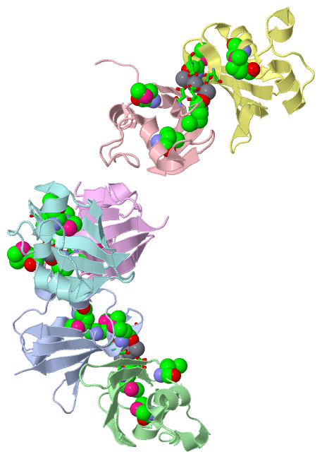
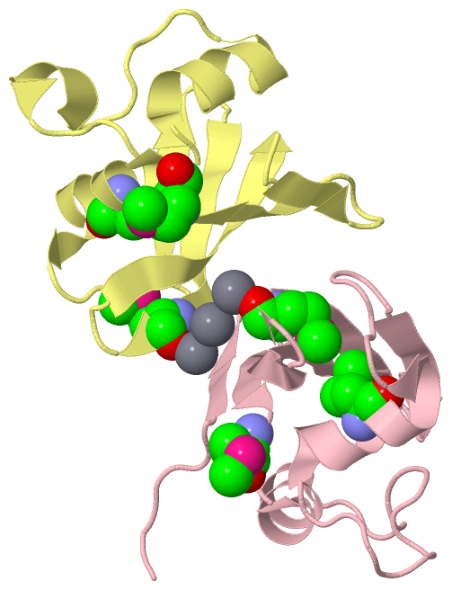
Chain A from PDB  Type:PROTEIN  Length:87
 aligned with Q7P1I2_CHRVO | Q7P1I2 from UniProtKB/TrEMBL  Length:436

    Alignment length:87
                                   350       360       370       380       390       400       410       420       
         Q7P1I2_CHRVO   341 DEIVQREDGSWLVDGMVSLDRFREFFELEAPLPGEAGGNIHTLAGVMLYQLGRVPSVTDRFEWNGFSFEVVDMDRTRVDKILVQRHH 427
               SCOP domains d3deda1 A:341-427 Probable hemolysin CV0231                                             SCOP domains
               CATH domains 3dedA00 A:341-427  [code=3.30.465.10, no name defined]                                  CATH domains
               Pfam domains --------------------------------------------------------------------------------------- Pfam domains
         Sec.struct. author ..eee.....eeee...hhhhhhhhh.......hhhhh...hhhhhhhhhhh.......eeee..eeeeeeeee..eeeeeeeee.. Sec.struct. author
                 SAPs(SNPs) --------------------------------------------------------------------------------------- SAPs(SNPs)
                    PROSITE --------------------------------------------------------------------------------------- PROSITE
                 Transcript --------------------------------------------------------------------------------------- Transcript
                 3ded A 341 DEIVQREDGSWLVDGmVSLDRFREFFELEAPLPGEAGGNIHTLAGVmLYQLGRVPSVTDRFEWNGFSFEVVDmDRTRVDKILVQRHH 427
                                   350     | 360       370       380      |390       400       410  |    420       
                                         356-MSE                        387-MSE                   413-MSE          

Chain B from PDB  Type:PROTEIN  Length:86
 aligned with Q7P1I2_CHRVO | Q7P1I2 from UniProtKB/TrEMBL  Length:436

    Alignment length:86
                                   350       360       370       380       390       400       410       420      
         Q7P1I2_CHRVO   341 DEIVQREDGSWLVDGMVSLDRFREFFELEAPLPGEAGGNIHTLAGVMLYQLGRVPSVTDRFEWNGFSFEVVDMDRTRVDKILVQRH 426
               SCOP domains d3dedb_ B: Probable hemolysin CV0231                                                   SCOP domains
               CATH domains 3dedB00 B:341-426  [code=3.30.465.10, no name defined]                                 CATH domains
               Pfam domains -------------------------------------------------------------------------------------- Pfam domains
         Sec.struct. author ..eee.....eeee...hhhhhhhhhh......hhhhh...hhhhhhhhhhh.......eeee..eeeeeeeee..eeeeeeeee. Sec.struct. author
                 SAPs(SNPs) -------------------------------------------------------------------------------------- SAPs(SNPs)
                    PROSITE -------------------------------------------------------------------------------------- PROSITE
                 Transcript -------------------------------------------------------------------------------------- Transcript
                 3ded B 341 DEIVQREDGSWLVDGmVSLDRFREFFELEAPLPGEAGGNIHTLAGVmLYQLGRVPSVTDRFEWNGFSFEVVDmDRTRVDKILVQRH 426
                                   350     | 360       370       380      |390       400       410  |    420      
                                         356-MSE                        387-MSE                   413-MSE         

Chain C from PDB  Type:PROTEIN  Length:91
 aligned with Q7P1I2_CHRVO | Q7P1I2 from UniProtKB/TrEMBL  Length:436

    Alignment length:91
                                   346       356       366       376       386       396       406       416       426 
         Q7P1I2_CHRVO   337 DGEEDEIVQREDGSWLVDGMVSLDRFREFFELEAPLPGEAGGNIHTLAGVMLYQLGRVPSVTDRFEWNGFSFEVVDMDRTRVDKILVQRHH 427
               SCOP domains d3dedc_ C: Probable hemolysin CV0231                                                        SCOP domains
               CATH domains 3dedC00 C:337-427  [code=3.30.465.10, no name defined]                                      CATH domains
               Pfam domains ------------------------------------------------------------------------------------------- Pfam domains
         Sec.struct. author ......eee.....eeee...hhhhhhhhh...............hhhhhhhhhhh.......eeee..eeeeeeeee..eeeeeeeee.. Sec.struct. author
                 SAPs(SNPs) ------------------------------------------------------------------------------------------- SAPs(SNPs)
                    PROSITE ------------------------------------------------------------------------------------------- PROSITE
                 Transcript ------------------------------------------------------------------------------------------- Transcript
                 3ded C 337 DGEEDEIVQREDGSWLVDGmVSLDRFREFFELEAPLPGEAGGNIHTLAGVmLYQLGRVPSVTDRFEWNGFSFEVVDmDRTRVDKILVQRHH 427
                                   346       356       366       376       386|      396       406      |416       426 
                                             356-MSE                        387-MSE                   413-MSE          

Chain D from PDB  Type:PROTEIN  Length:86
 aligned with Q7P1I2_CHRVO | Q7P1I2 from UniProtKB/TrEMBL  Length:436

    Alignment length:86
                                   350       360       370       380       390       400       410       420      
         Q7P1I2_CHRVO   341 DEIVQREDGSWLVDGMVSLDRFREFFELEAPLPGEAGGNIHTLAGVMLYQLGRVPSVTDRFEWNGFSFEVVDMDRTRVDKILVQRH 426
               SCOP domains d3dedd_ D: Probable hemolysin CV0231                                                   SCOP domains
               CATH domains 3dedD00 D:341-426  [code=3.30.465.10, no name defined]                                 CATH domains
               Pfam domains -------------------------------------------------------------------------------------- Pfam domains
         Sec.struct. author ..eee.....eeee...hhhhhhhhhh......hhhhh...hhhhhhhhhhh.......eeee..eeeeeeeee..eeeeeeeee. Sec.struct. author
                 SAPs(SNPs) -------------------------------------------------------------------------------------- SAPs(SNPs)
                    PROSITE -------------------------------------------------------------------------------------- PROSITE
                 Transcript -------------------------------------------------------------------------------------- Transcript
                 3ded D 341 DEIVQREDGSWLVDGmVSLDRFREFFELEAPLPGEAGGNIHTLAGVmLYQLGRVPSVTDRFEWNGFSFEVVDmDRTRVDKILVQRH 426
                                   350     | 360       370       380      |390       400       410  |    420      
                                         356-MSE                        387-MSE                   413-MSE         

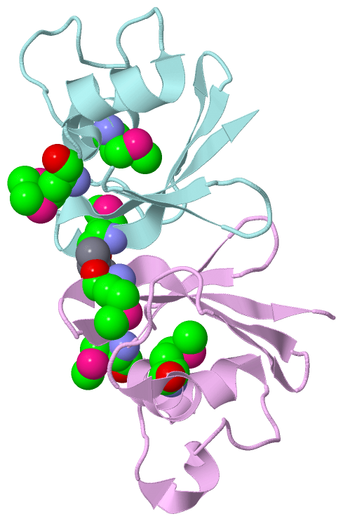
Chain E from PDB  Type:PROTEIN  Length:87
 aligned with Q7P1I2_CHRVO | Q7P1I2 from UniProtKB/TrEMBL  Length:436

    Alignment length:87
                                   350       360       370       380       390       400       410       420       
         Q7P1I2_CHRVO   341 DEIVQREDGSWLVDGMVSLDRFREFFELEAPLPGEAGGNIHTLAGVMLYQLGRVPSVTDRFEWNGFSFEVVDMDRTRVDKILVQRHH 427
               SCOP domains d3dede_ E: Probable hemolysin CV0231                                                    SCOP domains
               CATH domains 3dedE00 E:341-427  [code=3.30.465.10, no name defined]                                  CATH domains
               Pfam domains --------------------------------------------------------------------------------------- Pfam domains
         Sec.struct. author ..eee.....eeee...hhhhhhhhh.......hhhhh...hhhhhhhhhhh.......eeee..eeeeeeeee..eeeeeeeee.. Sec.struct. author
                 SAPs(SNPs) --------------------------------------------------------------------------------------- SAPs(SNPs)
                    PROSITE --------------------------------------------------------------------------------------- PROSITE
                 Transcript --------------------------------------------------------------------------------------- Transcript
                 3ded E 341 DEIVQREDGSWLVDGmVSLDRFREFFELEAPLPGEAGGNIHTLAGVmLYQLGRVPSVTDRFEWNGFSFEVVDmDRTRVDKILVQRHH 427
                                   350     | 360       370       380      |390       400       410  |    420       
                                         356-MSE                        387-MSE                   413-MSE          

Chain F from PDB  Type:PROTEIN  Length:86
 aligned with Q7P1I2_CHRVO | Q7P1I2 from UniProtKB/TrEMBL  Length:436

    Alignment length:86
                                   350       360       370       380       390       400       410       420      
         Q7P1I2_CHRVO   341 DEIVQREDGSWLVDGMVSLDRFREFFELEAPLPGEAGGNIHTLAGVMLYQLGRVPSVTDRFEWNGFSFEVVDMDRTRVDKILVQRH 426
               SCOP domains d3dedf_ F: Probable hemolysin CV0231                                                   SCOP domains
               CATH domains 3dedF00 F:341-426  [code=3.30.465.10, no name defined]                                 CATH domains
               Pfam domains -------------------------------------------------------------------------------------- Pfam domains
         Sec.struct. author ..eee.....eeee...hhhhhhhhhh..............hhhhhhhhhhh.......eeee..eeeeeeeee..eeeeeeeee. Sec.struct. author
                 SAPs(SNPs) -------------------------------------------------------------------------------------- SAPs(SNPs)
                    PROSITE -------------------------------------------------------------------------------------- PROSITE
                 Transcript -------------------------------------------------------------------------------------- Transcript
                 3ded F 341 DEIVQREDGSWLVDGmVSLDRFREFFELEAPLPGEAGGNIHTLAGVmLYQLGRVPSVTDRFEWNGFSFEVVDmDRTRVDKILVQRH 426
                                   350     | 360       370       380      |390       400       410  |    420      
                                         356-MSE                        387-MSE                   413-MSE         

   Legend:   → Mismatch (orange background)
  - → Gap (green background, '-', border residues have a numbering label)
    → Modified Residue (blue background, lower-case, 'x' indicates undefined single-letter code, labelled with number + name)
  x → Chemical Group (purple background, 'x', labelled with number + name, e.g. ACE or NH2)
  extra numbering lines below/above indicate numbering irregularities and modified residue names etc., number ends below/above '|'

 Classification and Annotation

(-) SCOP Domains  (1, 6)

Asymmetric Unit

(-) CATH Domains  (1, 6)

Asymmetric Unit
(-)
Class: Alpha Beta (26913)

(-) Pfam Domains  (0, 0)

(no "Pfam Domain" information available for 3DED)

(-) Gene Ontology  (7, 7)

Asymmetric Unit(hide GO term definitions)
Chain A,B,C,D,E,F   (Q7P1I2_CHRVO | Q7P1I2)
molecular function
    GO:0003824    catalytic activity    Catalysis of a biochemical reaction at physiological temperatures. In biologically catalyzed reactions, the reactants are known as substrates, and the catalysts are naturally occurring macromolecular substances known as enzymes. Enzymes possess specific binding sites for substrates, and are usually composed wholly or largely of protein, but RNA that has catalytic activity (ribozyme) is often also regarded as enzymatic.
    GO:0050660    flavin adenine dinucleotide binding    Interacting selectively and non-covalently with FAD, flavin-adenine dinucleotide, the coenzyme or the prosthetic group of various flavoprotein oxidoreductase enzymes, in either the oxidized form, FAD, or the reduced form, FADH2.
    GO:0046872    metal ion binding    Interacting selectively and non-covalently with any metal ion.
    GO:0016614    oxidoreductase activity, acting on CH-OH group of donors    Catalysis of an oxidation-reduction (redox) reaction in which a CH-OH group act as a hydrogen or electron donor and reduces a hydrogen or electron acceptor.
biological process
    GO:0055114    oxidation-reduction process    A metabolic process that results in the removal or addition of one or more electrons to or from a substance, with or without the concomitant removal or addition of a proton or protons.
cellular component
    GO:0016021    integral component of membrane    The component of a membrane consisting of the gene products and protein complexes having at least some part of their peptide sequence embedded in the hydrophobic region of the membrane.
    GO:0016020    membrane    A lipid bilayer along with all the proteins and protein complexes embedded in it an attached to it.

 Visualization

(-) Interactive Views

Asymmetric Unit
  Complete Structure
    Jena3D(integrated viewing of ligand, site, SAP, PROSITE, SCOP information)
    WebMol | AstexViewer[tm]@PDBe
(Java Applets, require no local installation except for Java; loading may be slow)
    STRAP
(Java WebStart application, automatic local installation, requires Java; full application with system access!)
    RasMol
(require local installation)
    Molscript (VRML)
(requires installation of a VRML viewer; select preferred view via VRML and generate a mono or stereo PDF format file)
 
  Ligands, Modified Residues, Ions
    CA  [ RasMol | Jena3D ]  +environment [ RasMol | Jena3D ]
    MSE  [ RasMol | Jena3D ]  +environment [ RasMol | Jena3D ]
 
  Sites
    AC1  [ RasMol ]  +environment [ RasMol ]
    AC2  [ RasMol ]  +environment [ RasMol ]
    AC3  [ RasMol ]  +environment [ RasMol ]
    AC4  [ RasMol ]  +environment [ RasMol ]
    AC5  [ RasMol ]  +environment [ RasMol ]
    AC6  [ RasMol ]  +environment [ RasMol ]
    AC7  [ RasMol ]  +environment [ RasMol ]
    AC8  [ RasMol ]  +environment [ RasMol ]
    AC9  [ RasMol ]  +environment [ RasMol ]
 
  Cis Peptide Bonds
    Gly C:338 - Glu C:339   [ RasMol ]  
 
Biological Units
  Complete Structure
    Biological Unit 1  [ Jena3D ]
    Biological Unit 2  [ Jena3D ]
    Biological Unit 3  [ Jena3D ]

(-) Still Images

Jmol
  protein: cartoon or spacefill or dots and stick; nucleic acid: cartoon and stick; ligands: spacefill; active site: stick
Molscript
  protein, nucleic acid: cartoon; ligands: spacefill; active site: ball and stick

 Databases and Analysis Tools

(-) Databases

Access by PDB/NDB ID
  3ded
    Family and Domain InformationProDom | SYSTERS
    General Structural InformationGlycoscienceDB | MMDB | NDB | OCA | PDB | PDBe | PDBj | PDBsum | PDBWiki | PQS | PROTEOPEDIA
    Orientation in MembranesOPM
    Protein SurfaceSURFACE
    Secondary StructureDSSP (structure derived) | HSSP (homology derived)
    Structural GenomicsGeneCensus
    Structural NeighboursCE | VAST
    Structure ClassificationCATH | Dali | SCOP
    Validation and Original DataBMRB Data View | BMRB Restraints Grid | EDS | PROCHECK | RECOORD | WHAT_CHECK
 
Access by UniProt ID/Accession number
  Q7P1I2_CHRVO | Q7P1I2
    Comparative Protein Structure ModelsModBase
    Genomic InformationEnsembl
    Protein-protein InteractionDIP
    Sequence, Family and Domain InformationInterPro | Pfam | SMART | UniProtKB/TrEMBL
 
Access by Enzyme Classificator   (EC Number)
  (no 'Enzyme Classificator' available)
    General Enzyme InformationBRENDA | EC-PDB | Enzyme | IntEnz
    PathwayKEGG | MetaCyc
 
Access by Disease Identifier   (MIM ID)
  (no 'MIM ID' available)
    Disease InformationOMIM
 
Access by GenAge ID
  (no 'GenAge ID' available)
    Age Related InformationGenAge

(-) Analysis Tools

Access by PDB/NDB ID
    Domain InformationXDom
    Interatomic Contacts of Structural UnitsCSU
    Ligand-protein ContactsLPC
    Protein CavitiescastP
    Sequence and Secondary StructurePDBCartoon
    Structure AlignmentSTRAP(Java WebStart application, automatic local installation, requires Java; full application with system access!)
    Structure and Sequence BrowserSTING
 
Access by UniProt ID/Accession number
  Q7P1I2_CHRVO | Q7P1I2
    Protein Disorder PredictionDisEMBL | FoldIndex | GLOBPLOT (for more information see DisProt)

 Related Entries

(-) Entries Sharing at Least One Protein Chain (UniProt ID)

(no "Entries Sharing at Least One Protein Chain" available for 3DED)

(-) Related Entries Specified in the PDB File

(no "Related Entries Specified in the PDB File" available for 3DED)