Show PDB file:   
         Plain Text   HTML   (compressed file size)
QuickSearch:   
by PDB,NDB,UniProt,PROSITE Code or Search Term(s)  
(-)Biol.Unit 1 - manually
(-)Asymmetric Unit
(-)Asym. Unit - sites
(-)Biological Unit 1
(-)Biol. Unit 1 - sites
collapse expand < >
Image Biol.Unit 1 - manually
Biol.Unit 1 - manually  (Jmol Viewer)
Image Asymmetric Unit
Asymmetric Unit  (Jmol Viewer)
Image Asym. Unit - sites
Asym. Unit - sites  (Jmol Viewer)
Image Biological Unit 1
Biological Unit 1  (Jmol Viewer)
Image Biol. Unit 1 - sites
Biol. Unit 1 - sites  (Jmol Viewer)

(-) Description

Title :  CRYSTAL STRUCTURE OF A MANGANESE SUPEROXIDE DISMUTASES FROM CAENORHABDITIS ELEGANS
 
Authors :  C. H. Trinh, T. Hunter, E. E. Stewart, S. E. V. Phillips, G. J. Hunter
Date :  03 Jun 08  (Deposition) - 09 Jun 09  (Release) - 13 Jul 11  (Revision)
Method :  X-RAY DIFFRACTION
Resolution :  1.80
Chains :  Asym. Unit :  A,C
Biol. Unit 1:  A,C  (2x)
Keywords :  Alpha Hairpin N Domain, Alpha/Beta C Domain, Oxidoreductase, Manganese, Metal-Binding, Mitochondrion, Transit Peptide (Keyword Search: [Gene Ontology, PubMed, Web (Google))
 
Reference :  C. H. Trinh, T. Hunter, E. E. Stewart, S. E. Phillips, G. J. Hunter
Purification, Crystallization And X-Ray Structures Of The Two Manganese Superoxide Dismutases From Caenorhabditis Elegans
Acta Crystallogr. , Sect. F V. 64 1110 2008
PubMed-ID: 19052361  |  Reference-DOI: 10.1107/S1744309108037056

(-) Compounds

Molecule 1 - SUPEROXIDE DISMUTASE [MN] 1
    ChainsA, C
    EC Number1.15.1.1
    EngineeredYES
    Expression SystemESCHERICHIA COLI
    Expression System PlasmidPTRC-99A
    Expression System StrainOX326A
    Expression System Taxid562
    Expression System Vector TypePLASMID
    GeneSOD-2, SDM-1, F10D11.1
    Organism CommonNEMATODE
    Organism ScientificCAENORHABDITIS ELEGANS
    Organism Taxid6239

 Structural Features

(-) Chains, Units

  12
Asymmetric Unit AC
Biological Unit 1 (2x)AC

Summary Information (see also Sequences/Alignments below)
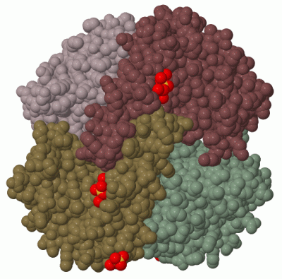
(-) Ligands, Modified Residues, Ions  (2, 5)

Asymmetric Unit (2, 5)
No.NameCountTypeFull Name
1MN2Ligand/IonMANGANESE (II) ION
2SO43Ligand/IonSULFATE ION
Biological Unit 1 (1, 6)
No.NameCountTypeFull Name
1MN-1Ligand/IonMANGANESE (II) ION
2SO46Ligand/IonSULFATE ION

(-) Sites  (5, 5)

Asymmetric Unit (5, 5)
No.NameEvidenceResiduesDescription
1AC1SOFTWAREHIS A:26 , HIS A:74 , ASP A:158 , HIS A:162 , HOH A:202BINDING SITE FOR RESIDUE MN A 198
2AC2SOFTWARETYR A:11 , GLN A:24 , GLN A:28 , HOH A:323 , LYS C:1 , SER C:3BINDING SITE FOR RESIDUE SO4 A 199
3AC3SOFTWAREHIS A:2 , SER A:3 , HIS A:71 , HOH A:288 , HOH A:419BINDING SITE FOR RESIDUE SO4 A 200
4AC4SOFTWAREHIS C:26 , HIS C:74 , ASP C:158 , HIS C:162 , HOH C:220BINDING SITE FOR RESIDUE MN C 198
5AC5SOFTWAREHIS C:2 , SER C:3 , HIS C:71 , HOH C:262 , HOH C:318 , HOH C:321 , HOH C:334BINDING SITE FOR RESIDUE SO4 C 199

(-) SS Bonds  (0, 0)

(no "SS Bond" information available for 3DC6)

(-) Cis Peptide Bonds  (2, 2)

Asymmetric Unit
No.Residues
1Glu A:15 -Pro A:16
2Glu C:15 -Pro C:16

 Sequence-Structure Mapping

(-) SAPs(SNPs)/Variants  (0, 0)

(no "SAP(SNP)/Variant" information available for 3DC6)

(-) PROSITE Motifs  (1, 2)

Asymmetric Unit (1, 2)
 PROSITEUniProtKBPDB
No.IDACDescriptionIDLocationCountLocation
1SOD_MNPS00088 Manganese and iron superoxide dismutases signature.SODM1_CAEEL182-189
 
  2A:158-165
C:158-165
Biological Unit 1 (1, 4)
 PROSITEUniProtKBPDB
No.IDACDescriptionIDLocationCountLocation
1SOD_MNPS00088 Manganese and iron superoxide dismutases signature.SODM1_CAEEL182-189
 
  4A:158-165
C:158-165

(-) Exons   (0, 0)

(no "Exon" information available for 3DC6)

(-) Sequences/Alignments

Asymmetric Unit
   Reformat: Number of residues per line =  ('0' or empty: single-line sequence representation)
  Number of residues per labelling interval =   
  UniProt sequence: complete  aligned part    
   Show mapping: SCOP domains CATH domains Pfam domains Secondary structure (by author)
SAPs(SNPs) PROSITE motifs Exons
(details for a mapped element are shown in a popup box when the mouse pointer rests over it)
Chain A from PDB  Type:PROTEIN  Length:197
 aligned with SODM1_CAEEL | P31161 from UniProtKB/Swiss-Prot  Length:221

    Alignment length:197
                                    34        44        54        64        74        84        94       104       114       124       134       144       154       164       174       184       194       204       214       
          SODM1_CAEEL    25 KHSLPDLPYDYADLEPVISHEIMQLHHQKHHATYVNNLNQIEEKLHEAVSKGNVKEAIALQPALKFNGGGHINHSIFWTNLAKDGGEPSAELLTAIKSDFGSLDNLQKQLSASTVAVQGSGWGWLGYCPKGKILKVATCANQDPLEATTGLVPLFGIDVWEHAYYLQYKNVRPDYVNAIWKIANWKNVSERFAKAQQ 221
               SCOP domains d3dc6a1 A:1-85 automated matches                                                     d3dc6a2 A:86-197 automated matches                                                                               SCOP domains
               CATH domains ----------------------------------------------------------------------------------------------------------------------------------------------------------------------------------------------------- CATH domains
               Pfam domains ----------------------------------------------------------------------------------------------------------------------------------------------------------------------------------------------------- Pfam domains
         Sec.struct. author ..................hhhhhhhhhhhhhhhhhhhhhhhhhhhhhhhhh.hhhhhhhhhhhhhhhhhhhhhhhhhhhh........hhhhhhhhhhhhhhhhhhhhhhhhhhhh....eeeeeeee....eeeeeeee...hhhhhhh.eeeeeee.hhhhhhhhhh.hhhhhhhhhhhhhhhhhhhhhhhhhh. Sec.struct. author
                 SAPs(SNPs) ----------------------------------------------------------------------------------------------------------------------------------------------------------------------------------------------------- SAPs(SNPs)
                    PROSITE -------------------------------------------------------------------------------------------------------------------------------------------------------------SOD_MN  -------------------------------- PROSITE
                 Transcript ----------------------------------------------------------------------------------------------------------------------------------------------------------------------------------------------------- Transcript
                 3dc6 A   1 KHSLPDLPYDYADLEPVISHEIMQLHHQKHHATYVNNLNQIEEKLHEAVSKGNVKEAIALQPALKFNGGGHINHSIFWTNLAKDGGEPSAELLTAIKSDFGSLDNLQKQLSASTVAVQGSGWGWLGYCPKGKILKVATCANQDPLEATTGLVPLFGIDVWEHAYYLQYKNVRPDYVNAIWKIANWKNVSERFAKAQQ 197
                                    10        20        30        40        50        60        70        80        90       100       110       120       130       140       150       160       170       180       190       

Chain C from PDB  Type:PROTEIN  Length:198
 aligned with SODM1_CAEEL | P31161 from UniProtKB/Swiss-Prot  Length:221

    Alignment length:198
                                    33        43        53        63        73        83        93       103       113       123       133       143       153       163       173       183       193       203       213        
          SODM1_CAEEL    24 SKHSLPDLPYDYADLEPVISHEIMQLHHQKHHATYVNNLNQIEEKLHEAVSKGNVKEAIALQPALKFNGGGHINHSIFWTNLAKDGGEPSAELLTAIKSDFGSLDNLQKQLSASTVAVQGSGWGWLGYCPKGKILKVATCANQDPLEATTGLVPLFGIDVWEHAYYLQYKNVRPDYVNAIWKIANWKNVSERFAKAQQ 221
               SCOP domains d3dc6c1 C:0-85 automated matches                                                      d3dc6c2 C:86-197 automated matches                                                                               SCOP domains
               CATH domains ------------------------------------------------------------------------------------------------------------------------------------------------------------------------------------------------------ CATH domains
               Pfam domains ------------------------------------------------------------------------------------------------------------------------------------------------------------------------------------------------------ Pfam domains
         Sec.struct. author ...................hhhhhhhhhhhhhhhhhhhhhhhhhhhhhhhhhhhhhhhhhhhhhhhhhhhhhhhhhhhhhh........hhhhhhhhhhhhhhhhhhhhhhhhhhhh....eeeeeeee....eeeeeeee...hhhhhhh.eeeeeee.hhhhhhhhhh.hhhhhhhhhhhhhhhhhhhhhhhhhh. Sec.struct. author
                 SAPs(SNPs) ------------------------------------------------------------------------------------------------------------------------------------------------------------------------------------------------------ SAPs(SNPs)
                    PROSITE --------------------------------------------------------------------------------------------------------------------------------------------------------------SOD_MN  -------------------------------- PROSITE
                 Transcript ------------------------------------------------------------------------------------------------------------------------------------------------------------------------------------------------------ Transcript
                 3dc6 C   0 MKHSLPDLPYDYADLEPVISHEIMQLHHQKHHATYVNNLNQIEEKLHEAVSKGNVKEAIALQPALKFNGGGHINHSIFWTNLAKDGGEPSAELLTAIKSDFGSLDNLQKQLSASTVAVQGSGWGWLGYCPKGKILKVATCANQDPLEATTGLVPLFGIDVWEHAYYLQYKNVRPDYVNAIWKIANWKNVSERFAKAQQ 197
                                     9        19        29        39        49        59        69        79        89        99       109       119       129       139       149       159       169       179       189        

   Legend:   → Mismatch (orange background)
  - → Gap (green background, '-', border residues have a numbering label)
    → Modified Residue (blue background, lower-case, 'x' indicates undefined single-letter code, labelled with number + name)
  x → Chemical Group (purple background, 'x', labelled with number + name, e.g. ACE or NH2)
  extra numbering lines below/above indicate numbering irregularities and modified residue names etc., number ends below/above '|'

 Classification and Annotation

(-) SCOP Domains  (2, 4)

Asymmetric Unit

(-) CATH Domains  (0, 0)

(no "CATH Domain" information available for 3DC6)

(-) Pfam Domains  (0, 0)

(no "Pfam Domain" information available for 3DC6)

(-) Gene Ontology  (10, 10)

Asymmetric Unit(hide GO term definitions)
Chain A,C   (SODM1_CAEEL | P31161)
molecular function
    GO:0046872    metal ion binding    Interacting selectively and non-covalently with any metal ion.
    GO:0016491    oxidoreductase activity    Catalysis of an oxidation-reduction (redox) reaction, a reversible chemical reaction in which the oxidation state of an atom or atoms within a molecule is altered. One substrate acts as a hydrogen or electron donor and becomes oxidized, while the other acts as hydrogen or electron acceptor and becomes reduced.
    GO:0042803    protein homodimerization activity    Interacting selectively and non-covalently with an identical protein to form a homodimer.
    GO:0004784    superoxide dismutase activity    Catalysis of the reaction: 2 superoxide + 2 H+ = O2 + hydrogen peroxide.
biological process
    GO:0055114    oxidation-reduction process    A metabolic process that results in the removal or addition of one or more electrons to or from a substance, with or without the concomitant removal or addition of a proton or protons.
    GO:0019430    removal of superoxide radicals    Any process, acting at the cellular level, involved in removing superoxide radicals (O2-) from a cell or organism, e.g. by conversion to dioxygen (O2) and hydrogen peroxide (H2O2).
    GO:0006801    superoxide metabolic process    The chemical reactions and pathways involving superoxide, the superoxide anion O2- (superoxide free radical), or any compound containing this species.
cellular component
    GO:0005759    mitochondrial matrix    The gel-like material, with considerable fine structure, that lies in the matrix space, or lumen, of a mitochondrion. It contains the enzymes of the tricarboxylic acid cycle and, in some organisms, the enzymes concerned with fatty acid oxidation.
    GO:0097249    mitochondrial respiratory chain supercomplex    A set of respiratory enzyme complexes of the mitochondrial inner membrane (including, for example, Complex II, Complex III, Complex IV, or F1-F0 ATPase) arranged to form a large supercomplex.
    GO:0005739    mitochondrion    A semiautonomous, self replicating organelle that occurs in varying numbers, shapes, and sizes in the cytoplasm of virtually all eukaryotic cells. It is notably the site of tissue respiration.

 Visualization

(-) Interactive Views

Asymmetric Unit
  Complete Structure
    Jena3D(integrated viewing of ligand, site, SAP, PROSITE, SCOP information)
    WebMol | AstexViewer[tm]@PDBe
(Java Applets, require no local installation except for Java; loading may be slow)
    STRAP
(Java WebStart application, automatic local installation, requires Java; full application with system access!)
    RasMol
(require local installation)
    Molscript (VRML)
(requires installation of a VRML viewer; select preferred view via VRML and generate a mono or stereo PDF format file)
 
  Ligands, Modified Residues, Ions
    MN  [ RasMol | Jena3D ]  +environment [ RasMol | Jena3D ]
    SO4  [ RasMol | Jena3D ]  +environment [ RasMol | Jena3D ]
 
  Sites
    AC1  [ RasMol ]  +environment [ RasMol ]
    AC2  [ RasMol ]  +environment [ RasMol ]
    AC3  [ RasMol ]  +environment [ RasMol ]
    AC4  [ RasMol ]  +environment [ RasMol ]
    AC5  [ RasMol ]  +environment [ RasMol ]
 
  Cis Peptide Bonds
    Glu A:15 - Pro A:16   [ RasMol ]  
    Glu C:15 - Pro C:16   [ RasMol ]  
 
Biological Unit
  Complete Structure
    Biological Unit 1  [ Jena3D ]

(-) Still Images

Jmol
  protein: cartoon or spacefill or dots and stick; nucleic acid: cartoon and stick; ligands: spacefill; active site: stick
Molscript
  protein, nucleic acid: cartoon; ligands: spacefill; active site: ball and stick

 Databases and Analysis Tools

(-) Databases

Access by PDB/NDB ID
  3dc6
    Family and Domain InformationProDom | SYSTERS
    General Structural InformationGlycoscienceDB | MMDB | NDB | OCA | PDB | PDBe | PDBj | PDBsum | PDBWiki | PQS | PROTEOPEDIA
    Orientation in MembranesOPM
    Protein SurfaceSURFACE
    Secondary StructureDSSP (structure derived) | HSSP (homology derived)
    Structural GenomicsGeneCensus
    Structural NeighboursCE | VAST
    Structure ClassificationCATH | Dali | SCOP
    Validation and Original DataBMRB Data View | BMRB Restraints Grid | EDS | PROCHECK | RECOORD | WHAT_CHECK
 
Access by UniProt ID/Accession number
  SODM1_CAEEL | P31161
    Comparative Protein Structure ModelsModBase
    Genomic InformationEnsembl
    Protein-protein InteractionDIP
    Sequence, Family and Domain InformationInterPro | Pfam | SMART | UniProtKB/SwissProt
 
Access by Enzyme Classificator   (EC Number)
  1.15.1.1
    General Enzyme InformationBRENDA | EC-PDB | Enzyme | IntEnz
    PathwayKEGG | MetaCyc
 
Access by Disease Identifier   (MIM ID)
  (no 'MIM ID' available)
    Disease InformationOMIM
 
Access by GenAge ID
  (no 'GenAge ID' available)
    Age Related InformationGenAge

(-) Analysis Tools

Access by PDB/NDB ID
    Domain InformationXDom
    Interatomic Contacts of Structural UnitsCSU
    Ligand-protein ContactsLPC
    Protein CavitiescastP
    Sequence and Secondary StructurePDBCartoon
    Structure AlignmentSTRAP(Java WebStart application, automatic local installation, requires Java; full application with system access!)
    Structure and Sequence BrowserSTING
 
Access by UniProt ID/Accession number
  SODM1_CAEEL | P31161
    Protein Disorder PredictionDisEMBL | FoldIndex | GLOBPLOT (for more information see DisProt)

 Related Entries

(-) Entries Sharing at Least One Protein Chain (UniProt ID)

(no "Entries Sharing at Least One Protein Chain" available for 3DC6)

(-) Related Entries Specified in the PDB File

3dc5 HOMOLOGOUS PROTEIN