Show PDB file:   
         Plain Text   HTML   (compressed file size)
QuickSearch:   
by PDB,NDB,UniProt,PROSITE Code or Search Term(s)  
(-)Biol.Unit 1 - manually
(-)Asymmetric Unit
(-)Asym. Unit - sites
(-)Biological Unit 1
(-)Biol. Unit 1 - sites
(-)Biological Unit 2
(-)Biological Unit 3
collapse expand < >
Image Biol.Unit 1 - manually
Biol.Unit 1 - manually  (Jmol Viewer)
Image Asymmetric Unit
Asymmetric Unit  (Jmol Viewer)
Image Asym. Unit - sites
Asym. Unit - sites  (Jmol Viewer)
Image Biological Unit 1
Biological Unit 1  (Jmol Viewer)
Image Biol. Unit 1 - sites
Biol. Unit 1 - sites  (Jmol Viewer)
Image Biological Unit 2
Biological Unit 2  (Jmol Viewer)
Image Biological Unit 3
Biological Unit 3  (Jmol Viewer)

(-) Description

Title :  CRYSTAL STRUCTURE OF THE CGMP-BOUND GAF A DOMAIN FROM THE PHOTORECEPTOR PHOSPHODIESTERASE 6C
 
Authors :  S. E. Martinez, C. C. Heikaus, R. E. Klevit, J. A. Beavo
Date :  30 May 08  (Deposition) - 01 Jul 08  (Release) - 13 Jul 11  (Revision)
Method :  X-RAY DIFFRACTION
Resolution :  2.57
Chains :  Asym. Unit :  A,B
Biol. Unit 1:  A,B  (2x)
Biol. Unit 2:  A  (1x)
Biol. Unit 3:  B  (1x)
Keywords :  3', 5' Cgmp, Gaf Domain, Cyclic Nucleotide Phosphodiesterase, Cgmp, Hydrolase, Lipoprotein, Membrane, Prenylation, Sensory Transduction, Vision (Keyword Search: [Gene Ontology, PubMed, Web (Google))
 
Reference :  S. E. Martinez, C. C. Heikaus, R. E. Klevit, J. A. Beavo
The Structure Of The Gaf A Domain From Phosphodiesterase 6C Reveals Determinants Of Cgmp Binding, A Conserved Binding Surface, And A Large Cgmp-Dependent Conformational Change.
J. Biol. Chem. V. 283 25913 2008
PubMed-ID: 18614542  |  Reference-DOI: 10.1074/JBC.M802891200

(-) Compounds

Molecule 1 - CONE CGMP-SPECIFIC 3',5'-CYCLIC PHOSPHODIESTERASE SUBUNIT ALPHA'
    ChainsA, B
    EC Number3.1.4.35
    EngineeredYES
    Expression SystemESCHERICHIA COLI
    Expression System PlasmidPRUNH
    Expression System StrainC41 CELLS
    Expression System Vector TypePLASMID
    FragmentGAF A DOMAIN (UNP RESIDUES 55-225)
    GenePDE6C
    Organism CommonBANTAM,CHICKENS
    Organism ScientificGALLUS GALLUS
    Organism Taxid9031
    StrainRHODE ISLAND RED
    SynonymCGMP PHOSPHODIESTERASE 6C

 Structural Features

(-) Chains, Units

  12
Asymmetric Unit AB
Biological Unit 1 (2x)AB
Biological Unit 2 (1x)A 
Biological Unit 3 (1x) B

Summary Information (see also Sequences/Alignments below)
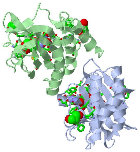
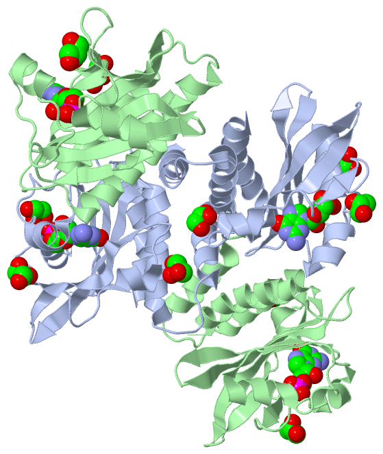
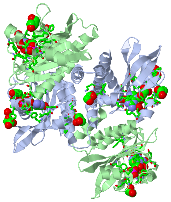
(-) Ligands, Modified Residues, Ions  (2, 7)

Asymmetric Unit (2, 7)
No.NameCountTypeFull Name
135G2Ligand/IonGUANOSINE-3',5'-MONOPHOSPHATE
2GOL5Ligand/IonGLYCEROL
Biological Unit 1 (2, 14)
No.NameCountTypeFull Name
135G4Ligand/IonGUANOSINE-3',5'-MONOPHOSPHATE
2GOL10Ligand/IonGLYCEROL
Biological Unit 2 (2, 4)
No.NameCountTypeFull Name
135G1Ligand/IonGUANOSINE-3',5'-MONOPHOSPHATE
2GOL3Ligand/IonGLYCEROL
Biological Unit 3 (2, 3)
No.NameCountTypeFull Name
135G1Ligand/IonGUANOSINE-3',5'-MONOPHOSPHATE
2GOL2Ligand/IonGLYCEROL

(-) Sites  (6, 6)

Asymmetric Unit (6, 6)
No.NameEvidenceResiduesDescription
1AC1SOFTWARESER A:97 , PHE A:99 , LEU A:115 , ASN A:116 , GLY A:141 , ILE A:142 , ALA A:143 , PHE A:164 , SER A:165 , LEU A:168 , ASP A:169 , THR A:172 , TYR A:174 , THR A:176 , LEU A:197 , HOH A:254BINDING SITE FOR RESIDUE 35G A 1
2AC2SOFTWARESER B:97 , PHE B:99 , LEU B:115 , ASN B:116 , PHE B:136 , GLY B:141 , ILE B:142 , ALA B:143 , PHE B:164 , SER B:165 , LEU B:168 , TYR B:174 , THR B:176 , MET B:179 , LEU B:197 , HOH B:246 , HOH B:248 , HOH B:276BINDING SITE FOR RESIDUE 35G B 2
3AC3SOFTWARETYR A:167 , HOH A:275BINDING SITE FOR RESIDUE GOL A 3
4AC4SOFTWARETRP A:145 , LYS A:151 , PHE A:153 , HIS A:163BINDING SITE FOR RESIDUE GOL A 4
5AC5SOFTWAREHIS A:81 , LYS A:82 , GLN A:85 , PHE A:123BINDING SITE FOR RESIDUE GOL A 5
6AC6SOFTWAREHIS B:81 , GLN B:85 , PHE B:123BINDING SITE FOR RESIDUE GOL B 7

(-) SS Bonds  (0, 0)

(no "SS Bond" information available for 3DBA)

(-) Cis Peptide Bonds  (2, 2)

Asymmetric Unit
No.Residues
1Ala A:73 -Gly A:74
2Arg B:104 -Asn B:105

 Sequence-Structure Mapping

(-) SAPs(SNPs)/Variants  (0, 0)

(no "SAP(SNP)/Variant" information available for 3DBA)

(-) PROSITE Motifs  (0, 0)

(no "PROSITE Motif" information available for 3DBA)

(-) Exons   (0, 0)

(no "Exon" information available for 3DBA)

(-) Sequences/Alignments

Asymmetric Unit
   Reformat: Number of residues per line =  ('0' or empty: single-line sequence representation)
  Number of residues per labelling interval =   
  UniProt sequence: complete  aligned part    
   Show mapping: SCOP domains CATH domains Pfam domains Secondary structure (by author)
SAPs(SNPs) PROSITE motifs Exons
(details for a mapped element are shown in a popup box when the mouse pointer rests over it)
Chain A from PDB  Type:PROTEIN  Length:171
 aligned with PDE6C_CHICK | P52731 from UniProtKB/Swiss-Prot  Length:862

    Alignment length:171
                                    64        74        84        94       104       114       124       134       144       154       164       174       184       194       204       214       224 
          PDE6C_CHICK    55 RLEECNILFELLTEIQDEAGSMEKIVHKTLQRLSQLLARDRCSMFICRSRNGIPEVATRLLNVTPTSKFEDNLVNPDKETVFPLDIGIAGWVAHTKKFFNIPDVKKNNHFSDYLDKKTGYTTVNMMAIPITQGKEVLAVVMALNKLNASEFSKEDEEVFKKYLNFISLVLR 225
               SCOP domains --------------------------------------------------------------------------------------------------------------------------------------------------------------------------- SCOP domains
               CATH domains --------------------------------------------------------------------------------------------------------------------------------------------------------------------------- CATH domains
               Pfam domains --------------------------------------------------------------------------------------------------------------------------------------------------------------------------- Pfam domains
     Sec.struct. author (1) hhhhhhhhhhhhh......hhhhhhhhhhhhhhhhhhh------------..eeeeeeeeeee....hhhhhh......eee....hhhhhhhhhh..eee..........hhhhhhhh....eeeeeeeee..eeeeeeeeeee......hhhhhhhhhhhhhhhhhhh. Sec.struct. author (1)
     Sec.struct. author (2) -------------------------------------eeeeeeeeeeeee------------------------------------------------------------------------------------------------------------------------- Sec.struct. author (2)
                 SAPs(SNPs) --------------------------------------------------------------------------------------------------------------------------------------------------------------------------- SAPs(SNPs)
                    PROSITE --------------------------------------------------------------------------------------------------------------------------------------------------------------------------- PROSITE
                 Transcript --------------------------------------------------------------------------------------------------------------------------------------------------------------------------- Transcript
                 3dba A  55 RLEECNILFELLTEIQDEAGSMEKIVHKTLQRLSQLLAADRCSMFICRSRNGIPEVATRLLNVTPTSKFEDNLVNPDKETVFPLDIGIAGWVAHTKKFFNIPDVKKNNHFSDYLDKKTGYTTVNMMAIPITQGKEVLAVVMALNKLNASEFSKEDEEVFKKYLNFISLVLR 225
                                    64        74        84        94       104       114       124       134       144       154       164       174       184       194       204       214       224 

Chain B from PDB  Type:PROTEIN  Length:171
 aligned with PDE6C_CHICK | P52731 from UniProtKB/Swiss-Prot  Length:862

    Alignment length:171
                                    64        74        84        94       104       114       124       134       144       154       164       174       184       194       204       214       224 
          PDE6C_CHICK    55 RLEECNILFELLTEIQDEAGSMEKIVHKTLQRLSQLLARDRCSMFICRSRNGIPEVATRLLNVTPTSKFEDNLVNPDKETVFPLDIGIAGWVAHTKKFFNIPDVKKNNHFSDYLDKKTGYTTVNMMAIPITQGKEVLAVVMALNKLNASEFSKEDEEVFKKYLNFISLVLR 225
               SCOP domains --------------------------------------------------------------------------------------------------------------------------------------------------------------------------- SCOP domains
               CATH domains --------------------------------------------------------------------------------------------------------------------------------------------------------------------------- CATH domains
               Pfam domains --------------------------------------------------------------------------------------------------------------------------------------------------------------------------- Pfam domains
     Sec.struct. author (1) ..hhhhhhhhhhhh.......hhhhhhhhhhhhhhhhh------------...eeeeeeeeee....hhhhhh......eee....hhhhhhhhhh..eee..........hhhhhhhh....eeeeeeeee..eeeeeeeeeee......hhhhhhhhhhhhhhhhhhh. Sec.struct. author (1)
     Sec.struct. author (2) -------------------------------------eeeeeeeeeeeee------------------------------------------------------------------------------------------------------------------------- Sec.struct. author (2)
                 SAPs(SNPs) --------------------------------------------------------------------------------------------------------------------------------------------------------------------------- SAPs(SNPs)
                    PROSITE --------------------------------------------------------------------------------------------------------------------------------------------------------------------------- PROSITE
                 Transcript --------------------------------------------------------------------------------------------------------------------------------------------------------------------------- Transcript
                 3dba B  55 RLEECNILFELLTEIQDEAGSMEKIVHKTLQRLSQLLAADRCSMFICRSRNGIPEVATRLLNVTPTSKFEDNLVNPDKETVFPLDIGIAGWVAHTKKFFNIPDVKKNNHFSDYLDKKTGYTTVNMMAIPITQGKEVLAVVMALNKLNASEFSKEDEEVFKKYLNFISLVLR 225
                                    64        74        84        94       104       114       124       134       144       154       164       174       184       194       204       214       224 

   Legend:   → Mismatch (orange background)
  - → Gap (green background, '-', border residues have a numbering label)
    → Modified Residue (blue background, lower-case, 'x' indicates undefined single-letter code, labelled with number + name)
  x → Chemical Group (purple background, 'x', labelled with number + name, e.g. ACE or NH2)
  extra numbering lines below/above indicate numbering irregularities and modified residue names etc., number ends below/above '|'

 Classification and Annotation

(-) SCOP Domains  (0, 0)

(no "SCOP Domain" information available for 3DBA)

(-) CATH Domains  (0, 0)

(no "CATH Domain" information available for 3DBA)

(-) Pfam Domains  (0, 0)

(no "Pfam Domain" information available for 3DBA)

(-) Gene Ontology  (12, 12)

Asymmetric Unit(hide GO term definitions)
Chain A,B   (PDE6C_CHICK | P52731)
molecular function
    GO:0047555    3',5'-cyclic-GMP phosphodiesterase activity    Catalysis of the reaction: guanosine 3',5'-cyclic phosphate + H2O = guanosine 5'-phosphate.
    GO:0004114    3',5'-cyclic-nucleotide phosphodiesterase activity    Catalysis of the reaction: nucleoside 3',5'-cyclic phosphate + H2O = nucleoside 5'-phosphate.
    GO:0030553    cGMP binding    Interacting selectively and non-covalently with cGMP, the nucleotide cyclic GMP (guanosine 3',5'-cyclophosphate).
    GO:0016787    hydrolase activity    Catalysis of the hydrolysis of various bonds, e.g. C-O, C-N, C-C, phosphoric anhydride bonds, etc. Hydrolase is the systematic name for any enzyme of EC class 3.
    GO:0046872    metal ion binding    Interacting selectively and non-covalently with any metal ion.
    GO:0000166    nucleotide binding    Interacting selectively and non-covalently with a nucleotide, any compound consisting of a nucleoside that is esterified with (ortho)phosphate or an oligophosphate at any hydroxyl group on the ribose or deoxyribose.
    GO:0008081    phosphoric diester hydrolase activity    Catalysis of the hydrolysis of a phosphodiester to give a phosphomonoester and a free hydroxyl group.
biological process
    GO:0050896    response to stimulus    Any process that results in a change in state or activity of a cell or an organism (in terms of movement, secretion, enzyme production, gene expression, etc.) as a result of a stimulus. The process begins with detection of the stimulus and ends with a change in state or activity or the cell or organism.
    GO:0007165    signal transduction    The cellular process in which a signal is conveyed to trigger a change in the activity or state of a cell. Signal transduction begins with reception of a signal (e.g. a ligand binding to a receptor or receptor activation by a stimulus such as light), or for signal transduction in the absence of ligand, signal-withdrawal or the activity of a constitutively active receptor. Signal transduction ends with regulation of a downstream cellular process, e.g. regulation of transcription or regulation of a metabolic process. Signal transduction covers signaling from receptors located on the surface of the cell and signaling via molecules located within the cell. For signaling between cells, signal transduction is restricted to events at and within the receiving cell.
    GO:0007601    visual perception    The series of events required for an organism to receive a visual stimulus, convert it to a molecular signal, and recognize and characterize the signal. Visual stimuli are detected in the form of photons and are processed to form an image.
cellular component
    GO:0016020    membrane    A lipid bilayer along with all the proteins and protein complexes embedded in it an attached to it.
    GO:0005886    plasma membrane    The membrane surrounding a cell that separates the cell from its external environment. It consists of a phospholipid bilayer and associated proteins.

 Visualization

(-) Interactive Views

Asymmetric Unit
  Complete Structure
    Jena3D(integrated viewing of ligand, site, SAP, PROSITE, SCOP information)
    WebMol | AstexViewer[tm]@PDBe
(Java Applets, require no local installation except for Java; loading may be slow)
    STRAP
(Java WebStart application, automatic local installation, requires Java; full application with system access!)
    RasMol
(require local installation)
    Molscript (VRML)
(requires installation of a VRML viewer; select preferred view via VRML and generate a mono or stereo PDF format file)
 
  Ligands, Modified Residues, Ions
    35G  [ RasMol | Jena3D ]  +environment [ RasMol | Jena3D ]
    GOL  [ RasMol | Jena3D ]  +environment [ RasMol | Jena3D ]
 
  Sites
    AC1  [ RasMol ]  +environment [ RasMol ]
    AC2  [ RasMol ]  +environment [ RasMol ]
    AC3  [ RasMol ]  +environment [ RasMol ]
    AC4  [ RasMol ]  +environment [ RasMol ]
    AC5  [ RasMol ]  +environment [ RasMol ]
    AC6  [ RasMol ]  +environment [ RasMol ]
 
  Cis Peptide Bonds
    Ala A:73 - Gly A:74   [ RasMol ]  
    Arg B:104 - Asn B:105   [ RasMol ]  
 
Biological Units
  Complete Structure
    Biological Unit 1  [ Jena3D ]
    Biological Unit 2  [ Jena3D ]
    Biological Unit 3  [ Jena3D ]

(-) Still Images

Jmol
  protein: cartoon or spacefill or dots and stick; nucleic acid: cartoon and stick; ligands: spacefill; active site: stick
Molscript
  protein, nucleic acid: cartoon; ligands: spacefill; active site: ball and stick

 Databases and Analysis Tools

(-) Databases

Access by PDB/NDB ID
  3dba
    Family and Domain InformationProDom | SYSTERS
    General Structural InformationGlycoscienceDB | MMDB | NDB | OCA | PDB | PDBe | PDBj | PDBsum | PDBWiki | PQS | PROTEOPEDIA
    Orientation in MembranesOPM
    Protein SurfaceSURFACE
    Secondary StructureDSSP (structure derived) | HSSP (homology derived)
    Structural GenomicsGeneCensus
    Structural NeighboursCE | VAST
    Structure ClassificationCATH | Dali | SCOP
    Validation and Original DataBMRB Data View | BMRB Restraints Grid | EDS | PROCHECK | RECOORD | WHAT_CHECK
 
Access by UniProt ID/Accession number
  PDE6C_CHICK | P52731
    Comparative Protein Structure ModelsModBase
    Genomic InformationEnsembl
    Protein-protein InteractionDIP
    Sequence, Family and Domain InformationInterPro | Pfam | SMART | UniProtKB/SwissProt
 
Access by Enzyme Classificator   (EC Number)
  3.1.4.35
    General Enzyme InformationBRENDA | EC-PDB | Enzyme | IntEnz
    PathwayKEGG | MetaCyc
 
Access by Disease Identifier   (MIM ID)
  (no 'MIM ID' available)
    Disease InformationOMIM
 
Access by GenAge ID
  (no 'GenAge ID' available)
    Age Related InformationGenAge

(-) Analysis Tools

Access by PDB/NDB ID
    Domain InformationXDom
    Interatomic Contacts of Structural UnitsCSU
    Ligand-protein ContactsLPC
    Protein CavitiescastP
    Sequence and Secondary StructurePDBCartoon
    Structure AlignmentSTRAP(Java WebStart application, automatic local installation, requires Java; full application with system access!)
    Structure and Sequence BrowserSTING
 
Access by UniProt ID/Accession number
  PDE6C_CHICK | P52731
    Protein Disorder PredictionDisEMBL | FoldIndex | GLOBPLOT (for more information see DisProt)

 Related Entries

(-) Entries Sharing at Least One Protein Chain (UniProt ID)

UniProtKB/Swiss-Prot
        PDE6C_CHICK | P527313jbq

(-) Related Entries Specified in the PDB File

(no "Related Entries Specified in the PDB File" available for 3DBA)