|
|
|
|
 Description
|
|
Compounds
|
||||||||||||||||||||||||||||||||||||||||||||||||||||||||
Chains, Units
Summary Information (see also Sequences/Alignments below) |
Ligands, Modified Residues, Ions (0, 0)| (no "Ligand,Modified Residues,Ions" information available for 3D9H) |
Sites (0, 0)| (no "Site" information available for 3D9H) |
SS Bonds (0, 0)| (no "SS Bond" information available for 3D9H) |
Cis Peptide Bonds (0, 0)| (no "Cis Peptide Bond" information available for 3D9H) |
SAPs(SNPs)/Variants (0, 0)| (no "SAP(SNP)/Variant" information available for 3D9H) |
PROSITE Motifs (3, 7)
Asymmetric/Biological Unit (3, 7)
|
||||||||||||||||||||||||||||||||||||||||
Exons (0, 0)| (no "Exon" information available for 3D9H) |
Sequences/Alignments
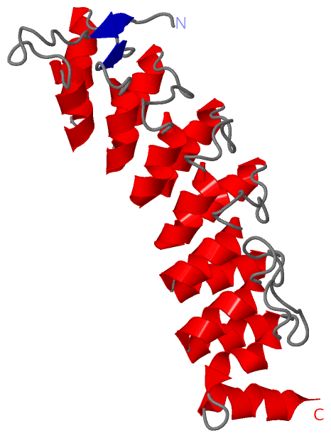
Asymmetric/Biological UnitChain A from PDB Type:PROTEIN Length:235 aligned with ASB9_HUMAN | Q96DX5 from UniProtKB/Swiss-Prot Length:294 Alignment length:235 28 38 48 58 68 78 88 98 108 118 128 138 148 158 168 178 188 198 208 218 228 238 248 ASB9_HUMAN 19 FPGIRLLSNPLMGDAVSDWSPMHEAAIHGHQLSLRNLISQGWAVNIITADHVSPLHEACLGGHLSCVKILLKHGAQVNGVTADWHTPLFNACVSGSWDCVNLLLQHGASVQPESDLASPIHEAARRGHVECVNSLIAYGGNIDHKISHLGTPLYLACENQQRACVKKLLESGADVNQGKGQDSPLHAVARTASEELACLLMDFGADTQAKNAEGKRPVELVPPESPLAQLFLERE 253 SCOP domains ------------------------------------------------------------------------------------------------------------------------------------------------------------------------------------------------------------------------------------------- SCOP domains CATH domains ------------------------------------------------------------------------------------------------------------------------------------------------------------------------------------------------------------------------------------------- CATH domains Pfam domains ------------------------------------------------------------------------------------------------------------------------------------------------------------------------------------------------------------------------------------------- Pfam domains SAPs(SNPs) ------------------------------------------------------------------------------------------------------------------------------------------------------------------------------------------------------------------------------------------- SAPs(SNPs) PROSITE (1) ----------------ANK_REP_REGION PDB: A:35-238 UniProt: 35-238 -----SOCS PROSITE (1) PROSITE (2) -------------------------------------------------ANK_REPEAT PDB: A:68-100 ANK_REPEAT PDB: A:101-133 --------------------------------ANK_REPEAT PDB: A:166-198 ------------------------------------------------------- PROSITE (2) PROSITE (3) ------------------------------------------------------------------------------------------------------------------ANK_REPEAT PDB: A:133-165 --------------------------------ANK_REPEAT PDB: A:198-230 ----------------------- PROSITE (3) Transcript ------------------------------------------------------------------------------------------------------------------------------------------------------------------------------------------------------------------------------------------- Transcript 3d9h A 19 FPGIRLLSNPLMGDAVSDWSPMHEAAIHGHQLSLRNLISQGWAVNIITADHVSPLHEACLGGHLSCVKILLKHGAQVNGVTADWHTPLFNACVSGSWDCVNLLLQHGASVQPESDLASPIHEAARRGHVECVNSLIAYGGNIDHKISHLGTPLYLACENQQRACVKKLLESGADVNQGKGQDSPLHAVVRTASEELACLLMDFGADTQAKNAEGKRPVELVPPESPLAQLFLERE 253 28 38 48 58 68 78 88 98 108 118 128 138 148 158 168 178 188 198 208 218 228 238 248
|
||||||||||||||||||||
SCOP Domains (0, 0)| (no "SCOP Domain" information available for 3D9H) |
CATH Domains (0, 0)| (no "CATH Domain" information available for 3D9H) |
Pfam Domains (0, 0)| (no "Pfam Domain" information available for 3D9H) |
Gene Ontology (5, 5)|
Asymmetric/Biological Unit(hide GO term definitions) Chain A (ASB9_HUMAN | Q96DX5)
|
||||||||||||||||||||||||||||||||||||||||||||||||
Interactive Views
|
||||||||||||||||||||||||||||||||||||||||||||||||||||||||||||||||||||||||||||||||||||||||||||||||||||||||||||||||||||
Still Images
|
||||||||||||||||
Databases
|
||||||||||||||||||||||||||||||||||||||||||||||||||||||||||||||||||||||||||||||||||||||||||||||||||||||||||||||||||||||||||||||||||||||||||||||||||||||||||||||||
Analysis Tools
|
|||||||||||||||||||||||||||||||||||||||||||||||||||||||||||||
Entries Sharing at Least One Protein Chain (UniProt ID)
Related Entries Specified in the PDB File
|
|