| molecular function |
|---|
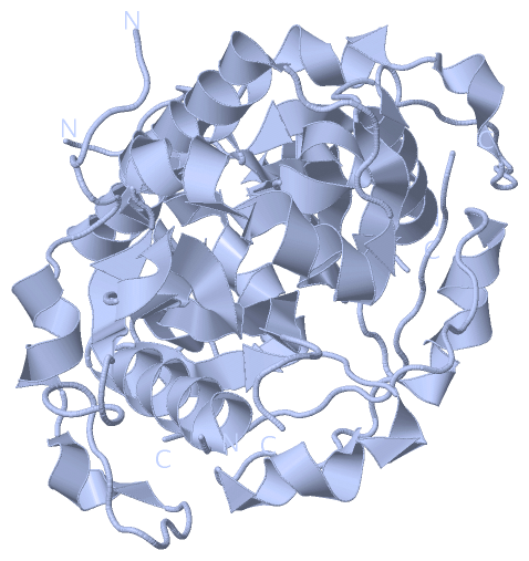
| | GO:0008080 | | N-acetyltransferase activity | | Catalysis of the transfer of an acetyl group to a nitrogen atom on the acceptor molecule. |
| | GO:0004343 | | glucosamine 6-phosphate N-acetyltransferase activity | | Catalysis of the reaction: D-glucosamine 6-phosphate + acetyl-CoA = N-acetyl-D-glucosamine 6-phosphate + CoA + H(+). |
| | GO:0042802 | | identical protein binding | | Interacting selectively and non-covalently with an identical protein or proteins. |
| | GO:0048029 | | monosaccharide binding | | Interacting selectively and non-covalently with any monosaccharide. Monosaccharides are the simplest carbohydrates; they are polyhydroxy aldehydes H[CH(OH)]nC(=O)H or polyhydroxy ketones H[CHOH]nC(=O)[CHOH]mH with three or more carbon atoms. They form the constitutional repeating units of oligo- and polysaccharides. |
| | GO:0016740 | | transferase activity | | Catalysis of the transfer of a group, e.g. a methyl group, glycosyl group, acyl group, phosphorus-containing, or other groups, from one compound (generally regarded as the donor) to another compound (generally regarded as the acceptor). Transferase is the systematic name for any enzyme of EC class 2. |
| | GO:0016746 | | transferase activity, transferring acyl groups | | Catalysis of the transfer of an acyl group from one compound (donor) to another (acceptor). |
| biological process |
|---|
| | GO:0006044 | | N-acetylglucosamine metabolic process | | The chemical reactions and pathways involving N-acetylglucosamine. The D isomer is a common structural unit of glycoproteins in plants, bacteria and animals; it is often the terminal sugar of an oligosaccharide group of a glycoprotein. |
| | GO:0006048 | | UDP-N-acetylglucosamine biosynthetic process | | The chemical reactions and pathways resulting in the formation of UDP-N-acetylglucosamine, a substance composed of N-acetylglucosamine, a common structural unit of oligosaccharides, in glycosidic linkage with uridine diphosphate. |
| | GO:0006041 | | glucosamine metabolic process | | The chemical reactions and pathways involving glucosamine (2-amino-2-deoxyglucopyranose), an aminodeoxysugar that occurs in combined form in chitin. |
| | GO:0001889 | | liver development | | The process whose specific outcome is the progression of the liver over time, from its formation to the mature structure. The liver is an exocrine gland which secretes bile and functions in metabolism of protein and carbohydrate and fat, synthesizes substances involved in the clotting of the blood, synthesizes vitamin A, detoxifies poisonous substances, stores glycogen, and breaks down worn-out erythrocytes. |
| cellular component |
|---|
| | GO:0005794 | | Golgi apparatus | | A compound membranous cytoplasmic organelle of eukaryotic cells, consisting of flattened, ribosome-free vesicles arranged in a more or less regular stack. The Golgi apparatus differs from the endoplasmic reticulum in often having slightly thicker membranes, appearing in sections as a characteristic shallow semicircle so that the convex side (cis or entry face) abuts the endoplasmic reticulum, secretory vesicles emerging from the concave side (trans or exit face). In vertebrate cells there is usually one such organelle, while in invertebrates and plants, where they are known usually as dictyosomes, there may be several scattered in the cytoplasm. The Golgi apparatus processes proteins produced on the ribosomes of the rough endoplasmic reticulum; such processing includes modification of the core oligosaccharides of glycoproteins, and the sorting and packaging of proteins for transport to a variety of cellular locations. Three different regions of the Golgi are now recognized both in terms of structure and function: cis, in the vicinity of the cis face, trans, in the vicinity of the trans face, and medial, lying between the cis and trans regions. |
| | GO:0000139 | | Golgi membrane | | The lipid bilayer surrounding any of the compartments of the Golgi apparatus. |
| | GO:0005829 | | cytosol | | The part of the cytoplasm that does not contain organelles but which does contain other particulate matter, such as protein complexes. |
| | GO:0005793 | | endoplasmic reticulum-Golgi intermediate compartment | | A complex system of membrane-bounded compartments located between endoplasmic reticulum (ER) and the Golgi complex, with a distinctive membrane protein composition; involved in ER-to-Golgi and Golgi-to-ER transport. |
| | GO:0005768 | | endosome | | A vacuole to which materials ingested by endocytosis are delivered. |
| | GO:0010008 | | endosome membrane | | The lipid bilayer surrounding an endosome. |
| | GO:0005770 | | late endosome | | A prelysosomal endocytic organelle differentiated from early endosomes by lower lumenal pH and different protein composition. Late endosomes are more spherical than early endosomes and are mostly juxtanuclear, being concentrated near the microtubule organizing center. |
| | GO:0016020 | | membrane | | A lipid bilayer along with all the proteins and protein complexes embedded in it an attached to it. |
 Description