Show PDB file:   
         Plain Text   HTML   (compressed file size)
QuickSearch:   
by PDB,NDB,UniProt,PROSITE Code or Search Term(s)  
(-)Biol.Unit 1 - manually
(-)Asymmetric Unit
(-)Asym. Unit - sites
(-)Biological Unit 1
(-)Biol. Unit 1 - sites
(-)Biological Unit 2
collapse expand < >
Image Biol.Unit 1 - manually
Biol.Unit 1 - manually  (Jmol Viewer)
Image Asymmetric Unit
Asymmetric Unit  (Jmol Viewer)
Image Asym. Unit - sites
Asym. Unit - sites  (Jmol Viewer)
Image Biological Unit 1
Biological Unit 1  (Jmol Viewer)
Image Biol. Unit 1 - sites
Biol. Unit 1 - sites  (Jmol Viewer)
Image Biological Unit 2
Biological Unit 2  (Jmol Viewer)

(-) Description

Title :  CRYSTAL STRUCTURE OF THE SPINACH AQUAPORIN SOPIP2;1 S274E MUTANT
 
Authors :  M. Nyblom, A. Alfredsson, K. Hallgren, K. Hedfalk, R. Neutze, S. Tornroth-Horsefield
Date :  25 Mar 08  (Deposition) - 24 Feb 09  (Release) - 26 May 09  (Revision)
Method :  X-RAY DIFFRACTION
Resolution :  2.95
Chains :  Asym. Unit :  A,B
Biol. Unit 1:  A  (4x)
Biol. Unit 2:  B  (4x)
Keywords :  Membrane Protein, Aquaporin, Transmembrane, Transport, Transport Protein (Keyword Search: [Gene Ontology, PubMed, Web (Google))
 
Reference :  M. Nyblom, A. Frick, Y. Wang, M. Ekvall, K. Hallgren, K. Hedfalk, R. Neutze, E. Tajkhorshid, S. Tornroth-Horsefield
Structural And Functional Analysis Of Sopip2; 1 Mutants Adds Insight Into Plant Aquaporin Gating.
J. Mol. Biol. V. 387 653 2009
PubMed-ID: 19302796  |  Reference-DOI: 10.1016/J.JMB.2009.01.065
(for further references see the PDB file header)

(-) Compounds

Molecule 1 - AQUAPORIN
    ChainsA, B
    EngineeredYES
    Expression SystemPICHIA PASTORIS
    Expression System PlasmidPPICZB
    Expression System StrainX33
    Expression System Taxid4922
    MutationYES
    Organism CommonSPINACH
    Organism ScientificSPINACIA OLERACEA
    Organism Taxid3562

 Structural Features

(-) Chains, Units

  12
Asymmetric Unit AB
Biological Unit 1 (4x)A 
Biological Unit 2 (4x) B

Summary Information (see also Sequences/Alignments below)

(-) Ligands, Modified Residues, Ions  (1, 2)

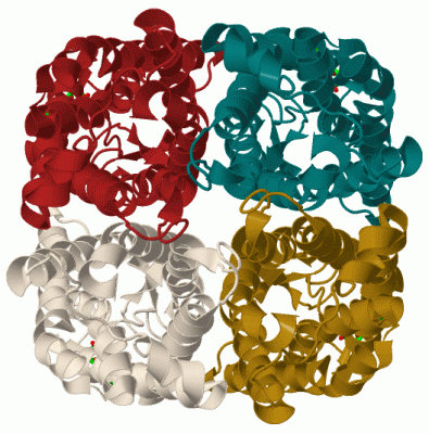
Asymmetric Unit (1, 2)
No.NameCountTypeFull Name
1CD2Ligand/IonCADMIUM ION
Biological Unit 1 (0, 0)
No.NameCountTypeFull Name
1CD-1Ligand/IonCADMIUM ION
Biological Unit 2 (0, 0)
No.NameCountTypeFull Name
1CD-1Ligand/IonCADMIUM ION

(-) Sites  (2, 2)

Asymmetric Unit (2, 2)
No.NameEvidenceResiduesDescription
1AC1SOFTWAREASP B:28 , GLU B:31BINDING SITE FOR RESIDUE CD B 282
2AC2SOFTWAREASP A:28 , GLU A:31BINDING SITE FOR RESIDUE CD A 282

(-) SS Bonds  (0, 0)

(no "SS Bond" information available for 3CN6)

(-) Cis Peptide Bonds  (0, 0)

(no "Cis Peptide Bond" information available for 3CN6)

 Sequence-Structure Mapping

(-) SAPs(SNPs)/Variants  (0, 0)

(no "SAP(SNP)/Variant" information available for 3CN6)

(-) PROSITE Motifs  (0, 0)

(no "PROSITE Motif" information available for 3CN6)

(-) Exons   (0, 0)

(no "Exon" information available for 3CN6)

(-) Sequences/Alignments

Asymmetric Unit
   Reformat: Number of residues per line =  ('0' or empty: single-line sequence representation)
  Number of residues per labelling interval =   
  UniProt sequence: complete  aligned part    
   Show mapping: SCOP domains CATH domains Pfam domains Secondary structure (by author)
SAPs(SNPs) PROSITE motifs Exons
(details for a mapped element are shown in a popup box when the mouse pointer rests over it)
Chain A from PDB  Type:PROTEIN  Length:243
 aligned with Q41372_SPIOL | Q41372 from UniProtKB/TrEMBL  Length:281

    Alignment length:243
                                    33        43        53        63        73        83        93       103       113       123       133       143       153       163       173       183       193       203       213       223       233       243       253       263   
         Q41372_SPIOL    24 APFFDLGELKLWSFWRAAIAEFIATLLFLYITVATVIGHSKETVVCGSVGLLGIAWAFGGMIFVLVYCTAGISGGHINPAVTFGLFLARKVSLLRALVYMIAQCLGAICGVGLVKAFMKGPYNQFGGGANSVALGYNKGTALGAEIIGTFVLVYTVFSATDPKRSARDSHVPILAPLPIGFAVFMVHLATIPITGTGINPARSFGAAVIFNSNKVWDDQWIFWVGPFIGAAVAAAYHQYVLRA 266
               SCOP domains d3cn6a_ A: automated matches                                                                                                                                                                                                                        SCOP domains
               CATH domains 3cn6A00 A:24-266 Glycerol uptake facilitator protein.                                                                                                                                                                                               CATH domains
               Pfam domains --------------------------------------------------------------------------------------------------------------------------------------------------------------------------------------------------------------------------------------------------- Pfam domains
         Sec.struct. author ....hhhhhhhhhhhhhhhhhhhhhhhhhhhhhhhhhhhhh.......hhhhhhhhhhhhhhhhhhhhhh.......hhhhhhhhhhh...hhhhhhhhhhhhhhhhhhhhhhhhhhhhhhhhhh...........hhhhhhhhhhhhhhhhhhhhhhheeeeee......eeehhhhhhhhhhhhhhhhhhhhh...hhhhhhhhhhhhhhhhhhhhhhhhhhhhhhhhhhhhhhhhhh... Sec.struct. author
                 SAPs(SNPs) --------------------------------------------------------------------------------------------------------------------------------------------------------------------------------------------------------------------------------------------------- SAPs(SNPs)
                    PROSITE --------------------------------------------------------------------------------------------------------------------------------------------------------------------------------------------------------------------------------------------------- PROSITE
                 Transcript --------------------------------------------------------------------------------------------------------------------------------------------------------------------------------------------------------------------------------------------------- Transcript
                 3cn6 A  24 APFFDLGELKLWSFWRAAIAEFIATLLFLYITVATVIGHSKETVVCGSVGLLGIAWAFGGMIFVLVYCTAGISGGHINPAVTFGLFLARKVSLLRALVYMIAQCLGAICGVGLVKAFMKGPYNQFGGGANSVALGYNKGTALGAEIIGTFVLVYTVFSATDPKRSARDSHVPILAPLPIGFAVFMVHLATIPITGTGINPARSFGAAVIFNSNKVWDDQWIFWVGPFIGAAVAAAYHQYVLRA 266
                                    33        43        53        63        73        83        93       103       113       123       133       143       153       163       173       183       193       203       213       223       233       243       253       263   

Chain B from PDB  Type:PROTEIN  Length:243
 aligned with Q41372_SPIOL | Q41372 from UniProtKB/TrEMBL  Length:281

    Alignment length:243
                                    33        43        53        63        73        83        93       103       113       123       133       143       153       163       173       183       193       203       213       223       233       243       253       263   
         Q41372_SPIOL    24 APFFDLGELKLWSFWRAAIAEFIATLLFLYITVATVIGHSKETVVCGSVGLLGIAWAFGGMIFVLVYCTAGISGGHINPAVTFGLFLARKVSLLRALVYMIAQCLGAICGVGLVKAFMKGPYNQFGGGANSVALGYNKGTALGAEIIGTFVLVYTVFSATDPKRSARDSHVPILAPLPIGFAVFMVHLATIPITGTGINPARSFGAAVIFNSNKVWDDQWIFWVGPFIGAAVAAAYHQYVLRA 266
               SCOP domains d3cn6b_ B: automated matches                                                                                                                                                                                                                        SCOP domains
               CATH domains 3cn6B00 B:24-266 Glycerol uptake facilitator protein.                                                                                                                                                                                               CATH domains
               Pfam domains --------------------------------------------------------------------------------------------------------------------------------------------------------------------------------------------------------------------------------------------------- Pfam domains
         Sec.struct. author ....hhhhhhhhhhhhhhhhhhhhhhhhhhhhhhhhhhhhh.......hhhhhhhhhhhhhhhhhhhhhh.......hhhhhhhhhhh...hhhhhhhhhhhhhhhhhhhhhhhhhhhhhhhhhh...........hhhhhhhhhhhhhhhhhhhhhhheeeeee......eeehhhhhhhhhhhhhhhhhhhhh...hhhhhhhhhhhhhhhhhhhhhhhhhhhhhhhhhhhhhhhhhh... Sec.struct. author
                 SAPs(SNPs) --------------------------------------------------------------------------------------------------------------------------------------------------------------------------------------------------------------------------------------------------- SAPs(SNPs)
                    PROSITE --------------------------------------------------------------------------------------------------------------------------------------------------------------------------------------------------------------------------------------------------- PROSITE
                 Transcript --------------------------------------------------------------------------------------------------------------------------------------------------------------------------------------------------------------------------------------------------- Transcript
                 3cn6 B  24 APFFDLGELKLWSFWRAAIAEFIATLLFLYITVATVIGHSKETVVCGSVGLLGIAWAFGGMIFVLVYCTAGISGGHINPAVTFGLFLARKVSLLRALVYMIAQCLGAICGVGLVKAFMKGPYNQFGGGANSVALGYNKGTALGAEIIGTFVLVYTVFSATDPKRSARDSHVPILAPLPIGFAVFMVHLATIPITGTGINPARSFGAAVIFNSNKVWDDQWIFWVGPFIGAAVAAAYHQYVLRA 266
                                    33        43        53        63        73        83        93       103       113       123       133       143       153       163       173       183       193       203       213       223       233       243       253       263   

   Legend:   → Mismatch (orange background)
  - → Gap (green background, '-', border residues have a numbering label)
    → Modified Residue (blue background, lower-case, 'x' indicates undefined single-letter code, labelled with number + name)
  x → Chemical Group (purple background, 'x', labelled with number + name, e.g. ACE or NH2)
  extra numbering lines below/above indicate numbering irregularities and modified residue names etc., number ends below/above '|'

 Classification and Annotation

(-) SCOP Domains  (1, 2)

Asymmetric Unit

(-) CATH Domains  (1, 2)

Asymmetric Unit

(-) Pfam Domains  (0, 0)

(no "Pfam Domain" information available for 3CN6)

(-) Gene Ontology  (4, 4)

Asymmetric Unit(hide GO term definitions)
Chain A,B   (Q41372_SPIOL | Q41372)
molecular function
    GO:0005215    transporter activity    Enables the directed movement of substances (such as macromolecules, small molecules, ions) into, out of or within a cell, or between cells.
biological process
    GO:0006810    transport    The directed movement of substances (such as macromolecules, small molecules, ions) or cellular components (such as complexes and organelles) into, out of or within a cell, or between cells, or within a multicellular organism by means of some agent such as a transporter, pore or motor protein.
cellular component
    GO:0016021    integral component of membrane    The component of a membrane consisting of the gene products and protein complexes having at least some part of their peptide sequence embedded in the hydrophobic region of the membrane.
    GO:0016020    membrane    A lipid bilayer along with all the proteins and protein complexes embedded in it an attached to it.

 Visualization

(-) Interactive Views

Asymmetric Unit
  Complete Structure
    Jena3D(integrated viewing of ligand, site, SAP, PROSITE, SCOP information)
    WebMol | AstexViewer[tm]@PDBe
(Java Applets, require no local installation except for Java; loading may be slow)
    STRAP
(Java WebStart application, automatic local installation, requires Java; full application with system access!)
    RasMol
(require local installation)
    Molscript (VRML)
(requires installation of a VRML viewer; select preferred view via VRML and generate a mono or stereo PDF format file)
 
  Ligands, Modified Residues, Ions
    CD  [ RasMol | Jena3D ]  +environment [ RasMol | Jena3D ]
 
  Sites
    AC1  [ RasMol ]  +environment [ RasMol ]
    AC2  [ RasMol ]  +environment [ RasMol ]
 
  Cis Peptide Bonds
(no "Cis Peptide Bonds" information available for 3cn6)
 
Biological Units
  Complete Structure
    Biological Unit 1  [ Jena3D ]
    Biological Unit 2  [ Jena3D ]

(-) Still Images

Jmol
  protein: cartoon or spacefill or dots and stick; nucleic acid: cartoon and stick; ligands: spacefill; active site: stick
Molscript
  protein, nucleic acid: cartoon; ligands: spacefill; active site: ball and stick

 Databases and Analysis Tools

(-) Databases

Access by PDB/NDB ID
  3cn6
    Family and Domain InformationProDom | SYSTERS
    General Structural InformationGlycoscienceDB | MMDB | NDB | OCA | PDB | PDBe | PDBj | PDBsum | PDBWiki | PQS | PROTEOPEDIA
    Orientation in MembranesOPM
    Protein SurfaceSURFACE
    Secondary StructureDSSP (structure derived) | HSSP (homology derived)
    Structural GenomicsGeneCensus
    Structural NeighboursCE | VAST
    Structure ClassificationCATH | Dali | SCOP
    Validation and Original DataBMRB Data View | BMRB Restraints Grid | EDS | PROCHECK | RECOORD | WHAT_CHECK
 
Access by UniProt ID/Accession number
  Q41372_SPIOL | Q41372
    Comparative Protein Structure ModelsModBase
    Genomic InformationEnsembl
    Protein-protein InteractionDIP
    Sequence, Family and Domain InformationInterPro | Pfam | SMART | UniProtKB/TrEMBL
 
Access by Enzyme Classificator   (EC Number)
  (no 'Enzyme Classificator' available)
    General Enzyme InformationBRENDA | EC-PDB | Enzyme | IntEnz
    PathwayKEGG | MetaCyc
 
Access by Disease Identifier   (MIM ID)
  (no 'MIM ID' available)
    Disease InformationOMIM
 
Access by GenAge ID
  (no 'GenAge ID' available)
    Age Related InformationGenAge

(-) Analysis Tools

Access by PDB/NDB ID
    Domain InformationXDom
    Interatomic Contacts of Structural UnitsCSU
    Ligand-protein ContactsLPC
    Protein CavitiescastP
    Sequence and Secondary StructurePDBCartoon
    Structure AlignmentSTRAP(Java WebStart application, automatic local installation, requires Java; full application with system access!)
    Structure and Sequence BrowserSTING
 
Access by UniProt ID/Accession number
  Q41372_SPIOL | Q41372
    Protein Disorder PredictionDisEMBL | FoldIndex | GLOBPLOT (for more information see DisProt)

 Related Entries

(-) Entries Sharing at Least One Protein Chain (UniProt ID)

UniProtKB/TrEMBL
        Q41372_SPIOL | Q413721z98 2b5f 3cll 3cn5 4ia4 4jc6

(-) Related Entries Specified in the PDB File

1z98 CLOSED STRUCTURE OF WILD-TYPE SOPIP2;1
2b5f OPEN STRUCTURE OF WILD-TYPE SOPIP2;1
3cll STRUCTURE OF OF THE SOPIP2;1 S115E MUTANT
3cn5 STRUCTURE OF OF THE SOPIP2;1 S274E:S115E MUTANT