|
|
|
|
 Description
|
|
Compounds
|
||||||||||||||||||||||||||||||||||||||||||||||||
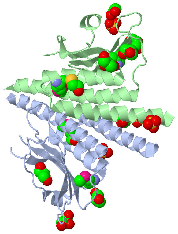
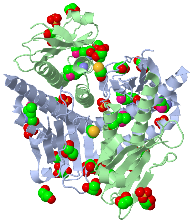
Chains, Units
Summary Information (see also Sequences/Alignments below) |
Ligands, Modified Residues, Ions (5, 17)| Asymmetric Unit (5, 17) Biological Unit 1 (5, 34) |
Sites (13, 13)
Asymmetric Unit (13, 13)
|
SS Bonds (0, 0)| (no "SS Bond" information available for 3CIT) |
Cis Peptide Bonds (0, 0)| (no "Cis Peptide Bond" information available for 3CIT) |
SAPs(SNPs)/Variants (0, 0)| (no "SAP(SNP)/Variant" information available for 3CIT) |
PROSITE Motifs (0, 0)| (no "PROSITE Motif" information available for 3CIT) |
Exons (0, 0)| (no "Exon" information available for 3CIT) |
Sequences/Alignments
Asymmetric UnitChain A from PDB Type:PROTEIN Length:155 aligned with Q884G2_PSESM | Q884G2 from UniProtKB/TrEMBL Length:394 Alignment length:157 25 35 45 55 65 75 85 95 105 115 125 135 145 155 165 Q884G2_PSESM 16 NHYRQSQSRAARLRLLVDTGQELIQLPPEAMRKCVLQRACAFVAMDHGLLLEWGADNGVQTTARHGSKERLSTLETTADPLAIGPQWLERPGTHLPCVLLLPLRGADEGSFGTLVLANSVAISAPDGEDIESLQLLATLLAAHLENNRLLEALVARD 172 SCOP domains ------------------------------------------------------------------------------------------------------------------------------------------------------------- SCOP domains CATH domains 3citA00 A:16-172 [code=3.30.450.40, no name defined] CATH domains Pfam domains ------------------------------------------------------------------------------------------------------------------------------------------------------------- Pfam domains SAPs(SNPs) ------------------------------------------------------------------------------------------------------------------------------------------------------------- SAPs(SNPs) PROSITE ------------------------------------------------------------------------------------------------------------------------------------------------------------- PROSITE Transcript ------------------------------------------------------------------------------------------------------------------------------------------------------------- Transcript 3cit A 16 NAYRQSQSRAARLRLLVDTGQELIQLPPEAmRKCVLQRACAFVAmDHGLLLEWG--NGVQTTARHGSKERLSTLETTADPLAIGPQWLERPGTHLPCVLLLPLRGADEGSFGTLVLANSVAISAPDGEDIESLQLLATLLAAHLENNRLLEALVARD 172 25 35 45| 55 | 65 | | 75 85 95 105 115 125 135 145 155 165 46-MSE 60-MSE 69 72 Chain B from PDB Type:PROTEIN Length:150 aligned with Q884G2_PSESM | Q884G2 from UniProtKB/TrEMBL Length:394 Alignment length:154 25 35 45 55 65 75 85 95 105 115 125 135 145 155 165 Q884G2_PSESM 16 NHYRQSQSRAARLRLLVDTGQELIQLPPEAMRKCVLQRACAFVAMDHGLLLEWGADNGVQTTARHGSKERLSTLETTADPLAIGPQWLERPGTHLPCVLLLPLRGADEGSFGTLVLANSVAISAPDGEDIESLQLLATLLAAHLENNRLLEALV 169 SCOP domains ---------------------------------------------------------------------------------------------------------------------------------------------------------- SCOP domains CATH domains 3citB00 B:16-169 [code=3.30.450.40, no name defined] CATH domains Pfam domains ---------------------------------------------------------------------------------------------------------------------------------------------------------- Pfam domains SAPs(SNPs) ---------------------------------------------------------------------------------------------------------------------------------------------------------- SAPs(SNPs) PROSITE ---------------------------------------------------------------------------------------------------------------------------------------------------------- PROSITE Transcript ---------------------------------------------------------------------------------------------------------------------------------------------------------- Transcript 3cit B 16 NAYRQSQSRAARLRLLVDTGQELIQLPPEAmRKCVLQRACAFVAmDHGLLLEWGA-NGVQTTARHGSKERLSTL---ADPLAIGPQWLERPGTHLPCVLLLPLRGADEGSFGTLVLANSVAISAPDGEDIESLQLLATLLAAHLENNRLLEALV 169 25 35 45| 55 | 65 | | 75 85 | |95 105 115 125 135 145 155 165 46-MSE 60-MSE 70 | 89 93 72
|
||||||||||||||||||||
SCOP Domains (0, 0)| (no "SCOP Domain" information available for 3CIT) |
CATH Domains (1, 2)
Asymmetric Unit
 |
Pfam Domains (0, 0)| (no "Pfam Domain" information available for 3CIT) |
Gene Ontology (9, 9)|
Asymmetric Unit(hide GO term definitions) Chain A,B (Q884G2_PSESM | Q884G2)
|
||||||||||||||||||||||||||||||||||||||||||||||||||||||||||||||||||||||||
Interactive Views
|
||||||||||||||||||||||||||||||||||||||||||||||||||||||||||||||||||||||||||||||||||||||||||||||||||||||||||||||||||||||||||||||||||||||||||||||||||||||||||||||||||||||||||||||||||||||||||||||||||||||||||||||||||||||||||||||||||||||||||||||||||||||||
Still Images
|
||||||||||||||||
Databases
|
||||||||||||||||||||||||||||||||||||||||||||||||||||||||||||||||||||||||||||||||||||||||||||||||||||||||||||||||||||||||||||||||||||||||||||||||||||||||||||||||
Analysis Tools
|
|||||||||||||||||||||||||||||||||||||||||||||||||||||||||||||
Entries Sharing at Least One Protein Chain (UniProt ID)
Related Entries Specified in the PDB File
|
|