|
|
|
|
 Description
|
|
Compounds
|
||||||||||||||||||||||||||||||||||||||||||||||||||||
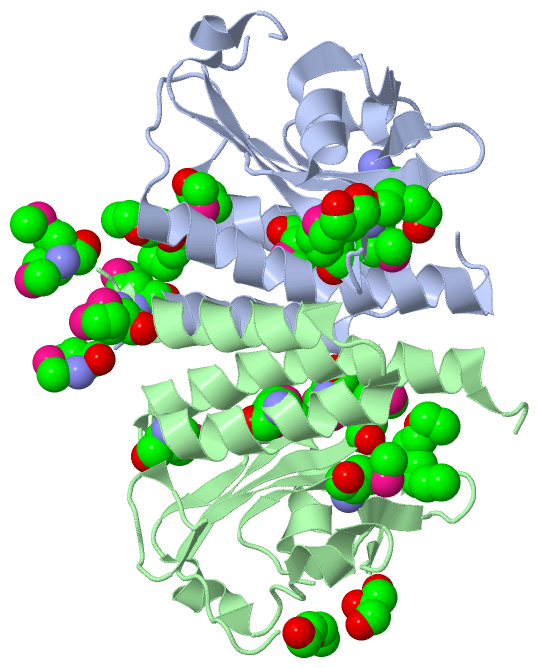
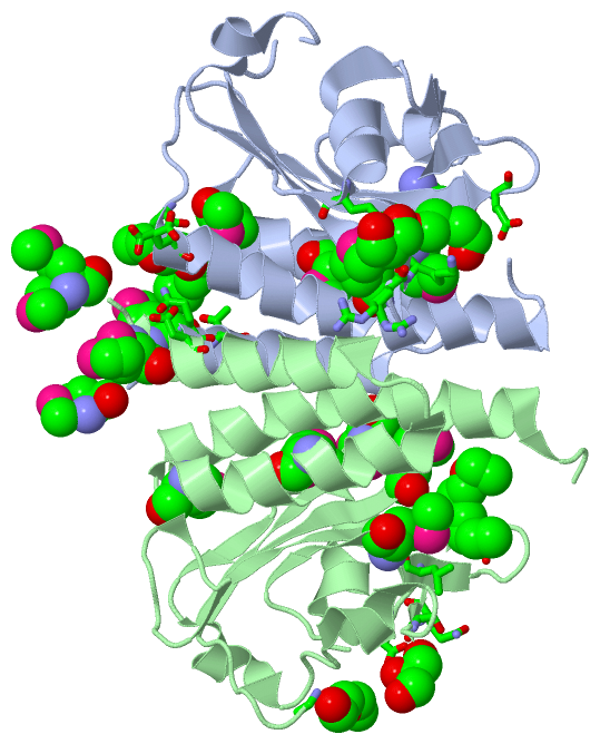
Chains, Units
Summary Information (see also Sequences/Alignments below) |
Ligands, Modified Residues, Ions (4, 20)| Asymmetric/Biological Unit (4, 20) |
Sites (6, 6)
Asymmetric Unit (6, 6)
|
SS Bonds (0, 0)| (no "SS Bond" information available for 3CI6) |
Cis Peptide Bonds (0, 0)| (no "Cis Peptide Bond" information available for 3CI6) |
SAPs(SNPs)/Variants (0, 0)| (no "SAP(SNP)/Variant" information available for 3CI6) |
PROSITE Motifs (0, 0)| (no "PROSITE Motif" information available for 3CI6) |
Exons (0, 0)| (no "Exon" information available for 3CI6) |
Sequences/Alignments
Asymmetric/Biological UnitChain A from PDB Type:PROTEIN Length:158 aligned with Q6FEW8_ACIAD | Q6FEW8 from UniProtKB/TrEMBL Length:764 Alignment length:167 10 20 30 40 50 60 70 80 90 100 110 120 130 140 150 160 Q6FEW8_ACIAD 1 MSNMQLDTLRRIVQEINSSVSLHDSLDIMVNQVADAMKVDVCSIYLLDERNQRYLLMASKGLNPESVGHVSLQLSEGLVGLVGQREEIVNLENASKHERFAYLPETGEEIYNSFLGVPVMYRRKVMGVLVVQNKQPQDFSEAAESFLVTLCAQLSGVIAHAHAVGNI 167 SCOP domains ----------------------------------------------------------------------------------------------------------------------------------------------------------------------- SCOP domains CATH domains -3ci6A00 A:2-167 [code=3.30.450.40, no name defined] CATH domains Pfam domains ----------------------------------------------------------------------------------------------------------------------------------------------------------------------- Pfam domains SAPs(SNPs) ----------------------------------------------------------------------------------------------------------------------------------------------------------------------- SAPs(SNPs) PROSITE ----------------------------------------------------------------------------------------------------------------------------------------------------------------------- PROSITE Transcript ----------------------------------------------------------------------------------------------------------------------------------------------------------------------- Transcript 3ci6 A 1 mSNmQLDTLRRIVQEINSSVSLHDSLDImVNQVADAmKVDVCSIYLLDERNQRYLLmASKGLNPESVGHVSLQLSEGLVGLVGQREEIVNLENASKHERF---------IYNSFLGVPVmYRRKVmGVLVVQNKQPQDFSEAAESFLVTLCAQLSGVIAHAHAVGNI 167 | | 10 20 30 | 40 50 | 60 70 80 90 100 110 120 | 130 140 150 160 | | 29-MSE 37-MSE 57-MSE 100 110 120-MSE | 1-MSE 126-MSE 4-MSE Chain B from PDB Type:PROTEIN Length:166 aligned with Q6FEW8_ACIAD | Q6FEW8 from UniProtKB/TrEMBL Length:764 Alignment length:168 10 20 30 40 50 60 70 80 90 100 110 120 130 140 150 160 Q6FEW8_ACIAD 1 MSNMQLDTLRRIVQEINSSVSLHDSLDIMVNQVADAMKVDVCSIYLLDERNQRYLLMASKGLNPESVGHVSLQLSEGLVGLVGQREEIVNLENASKHERFAYLPETGEEIYNSFLGVPVMYRRKVMGVLVVQNKQPQDFSEAAESFLVTLCAQLSGVIAHAHAVGNID 168 SCOP domains ------------------------------------------------------------------------------------------------------------------------------------------------------------------------ SCOP domains CATH domains -3ci6B00 B:2-168 [code=3.30.450.40, no name defined] CATH domains Pfam domains ------------------------------------------------------------------------------------------------------------------------------------------------------------------------ Pfam domains SAPs(SNPs) ------------------------------------------------------------------------------------------------------------------------------------------------------------------------ SAPs(SNPs) PROSITE ------------------------------------------------------------------------------------------------------------------------------------------------------------------------ PROSITE Transcript ------------------------------------------------------------------------------------------------------------------------------------------------------------------------ Transcript 3ci6 B 1 mSNmQLDTLRRIVQEINSSVSLHDSLDImVNQVADAmKVDVCSIYLLDERNQRYLLmASKGLNPESVGHVSLQLSEGLVGLVGQREEIVNLENASKHERFAYLP--GEEIYNSFLGVPVmYRRKVmGVLVVQNKQPQDFSEAAESFLVTLCAQLSGVIAHAHAVGNID 168 | | 10 20 30 | 40 50 | 60 70 80 90 100 | |110 120 | 130 140 150 160 1-MSE 29-MSE 37-MSE 57-MSE 104 | 120-MSE | 4-MSE 107 126-MSE
|
||||||||||||||||||||
SCOP Domains (0, 0)| (no "SCOP Domain" information available for 3CI6) |
CATH Domains (1, 2)
Asymmetric/Biological Unit
 |
Pfam Domains (0, 0)| (no "Pfam Domain" information available for 3CI6) |
Gene Ontology (6, 6)|
Asymmetric/Biological Unit(hide GO term definitions) Chain A,B (Q6FEW8_ACIAD | Q6FEW8)
|
||||||||||||||||||||||||||||||||||||||||||||||||
Interactive Views
|
||||||||||||||||||||||||||||||||||||||||||||||||||||||||||||||||||||||||||||||||||||||||||||||||||||||||||||||||||||||||||||||||||||||||||||||||||||||||||||||||||||||||||||||
Still Images
|
||||||||||||||||
Databases
|
||||||||||||||||||||||||||||||||||||||||||||||||||||||||||||||||||||||||||||||||||||||||||||||||||||||||||||||||||||||||||||||||||||||||||||||||||||||||||||||||
Analysis Tools
|
|||||||||||||||||||||||||||||||||||||||||||||||||||||||||||||
Entries Sharing at Least One Protein Chain (UniProt ID)
Related Entries Specified in the PDB File
|
|