|
|
|
|
 Description
|
|
Compounds
|
||||||||||||||||||||||||||||||||||||||||||||||||
Chains, Units
Summary Information (see also Sequences/Alignments below) |
Ligands, Modified Residues, Ions (1, 3)
Asymmetric Unit (1, 3)
|
Sites (0, 0)| (no "Site" information available for 3CAN) |
SS Bonds (0, 0)| (no "SS Bond" information available for 3CAN) |
Cis Peptide Bonds (0, 0)| (no "Cis Peptide Bond" information available for 3CAN) |
SAPs(SNPs)/Variants (0, 0)| (no "SAP(SNP)/Variant" information available for 3CAN) |
PROSITE Motifs (0, 0)| (no "PROSITE Motif" information available for 3CAN) |
Exons (0, 0)| (no "Exon" information available for 3CAN) |
Sequences/Alignments
Asymmetric UnitChain A from PDB Type:PROTEIN Length:161 aligned with A6L094_BACV8 | A6L094 from UniProtKB/TrEMBL Length:302 Alignment length:179 133 143 153 163 173 183 193 203 213 223 233 243 253 263 273 283 293 A6L094_BACV8 124 GGGVTFCGGEPLLHPEFLIDILKRCGQQGIHRAVDTTLLARKETVDEVMRNCELLLIDLKSMDSTVHQTFCDVPNELILKNIRRVAEADFPYYIRIPLIEGVNADEKNIKLSAEFLASLPRHPEIINLLPYHDIGKGKHAKLGSIYNPKGYKMQTPSEEVQQQCIQILTDYGLKATIGG 302 SCOP domains ----------------------------------------------------------------------------------------------------------------------------------------------------------------------------------- SCOP domains CATH domains 3canA00 A:7-185 pyruvate-formate lyase- activating enzyme CATH domains Pfam domains ----------------------------------------------------------------------------------------------------------------------------------------------------------------------------------- Pfam domains SAPs(SNPs) ----------------------------------------------------------------------------------------------------------------------------------------------------------------------------------- SAPs(SNPs) PROSITE ----------------------------------------------------------------------------------------------------------------------------------------------------------------------------------- PROSITE Transcript ----------------------------------------------------------------------------------------------------------------------------------------------------------------------------------- Transcript 3can A 7 GGGVTFCGGEPLLHPEFLIDILKRCGQQGIHRAVDTTLLARKETVDEVmRNCELLLIDLKSmDSTVHQTFCDVPNELILKNIRRVAEADFPYYIRIPLIEGVNADEKNIKLSAEFLASLPRHPEIINLLPYHD------------------KmQTPSEEVQQQCIQILTDYGLKATIGG 185 16 26 36 46 56 66 | 76 86 96 106 116 126 136 | - - || 166 176 55-MSE 68-MSE 139 158| 159-MSE
|
||||||||||||||||||||
SCOP Domains (0, 0)| (no "SCOP Domain" information available for 3CAN) |
CATH Domains (1, 1)
Asymmetric Unit
 |
Pfam Domains (0, 0)| (no "Pfam Domain" information available for 3CAN) |
Gene Ontology (6, 6)|
Asymmetric Unit(hide GO term definitions) Chain A (A6L094_BACV8 | A6L094)
|
||||||||||||||||||||||||||||||||||||||||||||||||
Interactive Views
|
|||||||||||||||||||||||||||||||||||||||||||||||||||||||||||||||||||||||||||||||||||||||||||||||||||||||||||||||||||||||||||||||||||||||
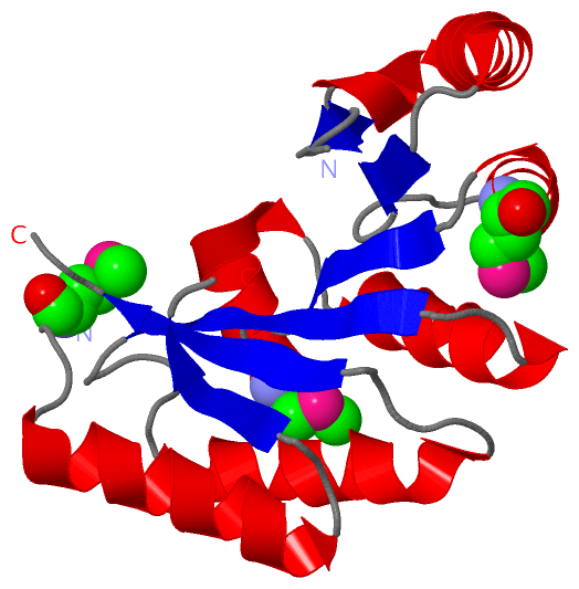
Still Images
|
||||||||||||||||
Databases
|
||||||||||||||||||||||||||||||||||||||||||||||||||||||||||||||||||||||||||||||||||||||||||||||||||||||||||||||||||||||||||||||||||||||||||||||||||||||||||||||||
Analysis Tools
|
|||||||||||||||||||||||||||||||||||||||||||||||||||||||||||||
Entries Sharing at Least One Protein Chain (UniProt ID)
Related Entries Specified in the PDB File
|
|