|
|
|
|
 Description
|
|
Compounds
|
||||||||||||||||||||||||||||||||||||||||||||
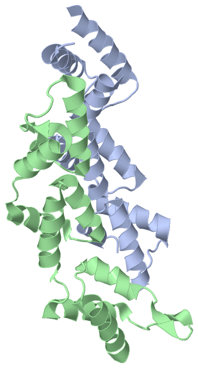
Chains, Units
Summary Information (see also Sequences/Alignments below) |
Ligands, Modified Residues, Ions (0, 0)| (no "Ligand,Modified Residues,Ions" information available for 3C1D) |
Sites (0, 0)| (no "Site" information available for 3C1D) |
SS Bonds (0, 0)| (no "SS Bond" information available for 3C1D) |
Cis Peptide Bonds (2, 2)
Asymmetric Unit
|
||||||||||||
SAPs(SNPs)/Variants (0, 0)| (no "SAP(SNP)/Variant" information available for 3C1D) |
PROSITE Motifs (0, 0)| (no "PROSITE Motif" information available for 3C1D) |
Exons (0, 0)| (no "Exon" information available for 3C1D) |
Sequences/Alignments
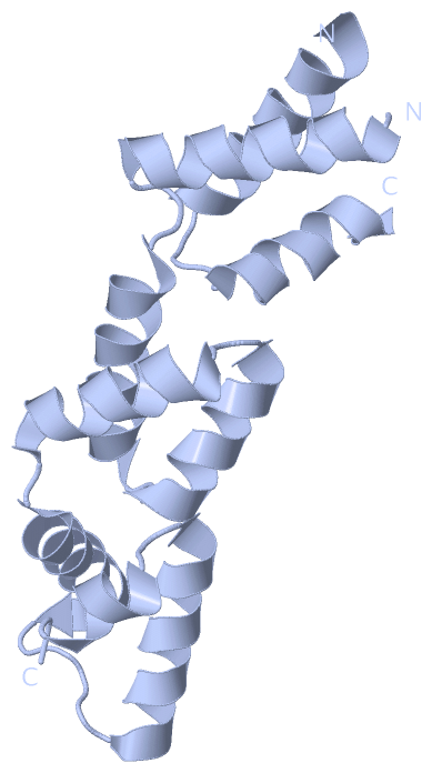
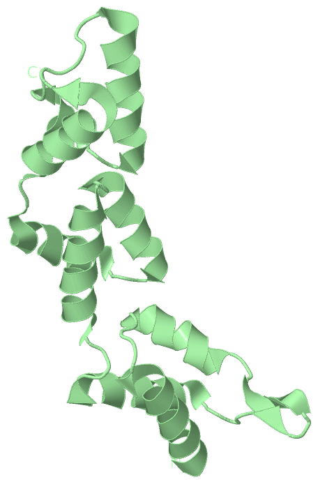
Asymmetric UnitChain A from PDB Type:PROTEIN Length:143 aligned with RECX_ECO57 | P66000 from UniProtKB/Swiss-Prot Length:166 Alignment length:154 17 27 37 47 57 67 77 87 97 107 117 127 137 147 157 RECX_ECO57 8 RPAYARLLDRAVRILAVRDHSEQELRRKLAAPIMGKNGPEEIDATAEDYERVIAWCHEHGYLDDSRFVARFIASRSRKGYGPARIRQELNQKGISREATEKAMRECDIDWCALARDQATRKYGEPLPTVFSEKVKIQRFLLYRGYLMEDIQDIW 161 SCOP domains ---------------------------------------------------------------------------------------------------------------------------------------------------------- SCOP domains CATH domains ---------------------------------------------------------------------------------------------------------------------------------------------------------- CATH domains Pfam domains ---------------------------------------------------------------------------------------------------------------------------------------------------------- Pfam domains SAPs(SNPs) ---------------------------------------------------------------------------------------------------------------------------------------------------------- SAPs(SNPs) PROSITE ---------------------------------------------------------------------------------------------------------------------------------------------------------- PROSITE Transcript ---------------------------------------------------------------------------------------------------------------------------------------------------------- Transcript 3c1d A 8 GPAYARLLDRAVRILAVRDHSEQELRRKLAAP-----------ATAEDYERVIAWCHEHGYLDDSRFVARFIASRSRKGYGPARIRQELNQKGISREATEKAMREADIDWAALARDQATRKYGEPLPTVFSEKVKIQRFLLYRGYLMEDIQDIW 161 17 27 37 | - | 57 67 77 87 97 107 117 127 137 147 157 39 51 Chain B from PDB Type:PROTEIN Length:154 aligned with RECX_ECO57 | P66000 from UniProtKB/Swiss-Prot Length:166 Alignment length:154 17 27 37 47 57 67 77 87 97 107 117 127 137 147 157 RECX_ECO57 8 RPAYARLLDRAVRILAVRDHSEQELRRKLAAPIMGKNGPEEIDATAEDYERVIAWCHEHGYLDDSRFVARFIASRSRKGYGPARIRQELNQKGISREATEKAMRECDIDWCALARDQATRKYGEPLPTVFSEKVKIQRFLLYRGYLMEDIQDIW 161 SCOP domains ---------------------------------------------------------------------------------------------------------------------------------------------------------- SCOP domains CATH domains ---------------------------------------------------------------------------------------------------------------------------------------------------------- CATH domains Pfam domains ---------------------------------------------------------------------------------------------------------------------------------------------------------- Pfam domains SAPs(SNPs) ---------------------------------------------------------------------------------------------------------------------------------------------------------- SAPs(SNPs) PROSITE ---------------------------------------------------------------------------------------------------------------------------------------------------------- PROSITE Transcript ---------------------------------------------------------------------------------------------------------------------------------------------------------- Transcript 3c1d B 8 GPAYARLLDRAVRILAVRDHSEQELRRKLAAPIMGKNGPEEIDATAEDYERVIAWCHEHGYLDDSRFVARFIASRSRKGYGPARIRQELNQKGISREATEKAMREADIDWAALARDQATRKYGEPLPTVFSEKVKIQRFLLYRGYLMEDIQDIW 161 17 27 37 47 57 67 77 87 97 107 117 127 137 147 157
|
||||||||||||||||||||
SCOP Domains (0, 0)| (no "SCOP Domain" information available for 3C1D) |
CATH Domains (0, 0)| (no "CATH Domain" information available for 3C1D) |
Pfam Domains (0, 0)| (no "Pfam Domain" information available for 3C1D) |
Gene Ontology (5, 5)|
Asymmetric Unit(hide GO term definitions) Chain A,B (RECX_ECO57 | P66000)
|
||||||||||||||||||||||||||||||||||||||||||
Interactive Views
|
||||||||||||||||||||||||||||||||||||||||||||||||||||||||||||||||||||||||||||||||||||||||||||||||||||||||||||||||||||||||||||||||||||||||||||||||||||||||
Still Images
|
||||||||||||||||
Databases
|
||||||||||||||||||||||||||||||||||||||||||||||||||||||||||||||||||||||||||||||||||||||||||||||||||||||||||||||||||||||||||||||||||||||||||||||||||||||||||||||||
Analysis Tools
|
|||||||||||||||||||||||||||||||||||||||||||||||||||||||||||||
Entries Sharing at Least One Protein Chain (UniProt ID)
Related Entries Specified in the PDB File
|
|