Show PDB file:   
         Plain Text   HTML   (compressed file size)
QuickSearch:   
by PDB,NDB,UniProt,PROSITE Code or Search Term(s)  
(-)Asymmetric Unit
(-)Asym. Unit - sites
(-)Biological Unit 1
(-)Biol. Unit 1 - sites
(-)Biological Unit 2
collapse expand < >
Image Asymmetric Unit
Asymmetric Unit  (Jmol Viewer)
Image Asym. Unit - sites
Asym. Unit - sites  (Jmol Viewer)
Image Biological Unit 1
Biological Unit 1  (Jmol Viewer)
Image Biol. Unit 1 - sites
Biol. Unit 1 - sites  (Jmol Viewer)
Image Biological Unit 2
Biological Unit 2  (Jmol Viewer)

(-) Description

Title :  CRYSTAL STRUCTURE OF ACOSTATIN FROM AGKISTRODON CONTORTRIX CONTORTRIX
 
Authors :  N. Moiseeva, R. Bau, M. Allaire
Date :  18 Jan 08  (Deposition) - 29 Apr 08  (Release) - 24 Feb 09  (Revision)
Method :  X-RAY DIFFRACTION
Resolution :  1.70
Chains :  Asym. Unit :  A,B,C,D
Biol. Unit 1:  A,B  (1x)
Biol. Unit 2:  C,D  (1x)
Keywords :  Beta-Sheets, Disulfide Bridges, Blood Coagulation, Cell Adhesion, Secreted, Hydrolase, Metal-Binding, Metalloprotease, Protease, Zinc, Zymogen, Blood Clotting/Antitumor Protein Complex (Keyword Search: [Gene Ontology, PubMed, Web (Google))
 
Reference :  N. Moiseeva, R. Bau, S. D. Swenson, F. S. Markland, J. -Y. Choe, Z. -J. Liu, M. Allaire
Crystal Structure Of Acostatin, A Dimeric Disintegrin From Southern Copperhead (Agkistrodon Contortrix Contortrix) At 1. 7 A Resolution
Acta Crystallogr. , Sect. D V. 64 466 2008
PubMed-ID: 18391413  |  Reference-DOI: 10.1107/S0907444908002370
(for further references see the PDB file header)

(-) Compounds

Molecule 1 - DISINTEGRIN ACOSTATIN ALPHA
    ChainsA, C
    FragmentUNP RESIDUES 48-109
    Organism CommonSOUTHERN COPPERHEAD
    Organism ScientificAGKISTRODON CONTORTRIX CONTORTRIX
 
Molecule 2 - DISINTEGRIN ACOSTATIN-BETA
    ChainsB, D
    FragmentUNP RESIDUES 419-482
    Organism CommonSOUTHERN COPPERHEAD
    Organism ScientificAGKISTRODON CONTORTRIX CONTORTRIX

 Structural Features

(-) Chains, Units

  1234
Asymmetric Unit ABCD
Biological Unit 1 (1x)AB  
Biological Unit 2 (1x)  CD

Summary Information (see also Sequences/Alignments below)

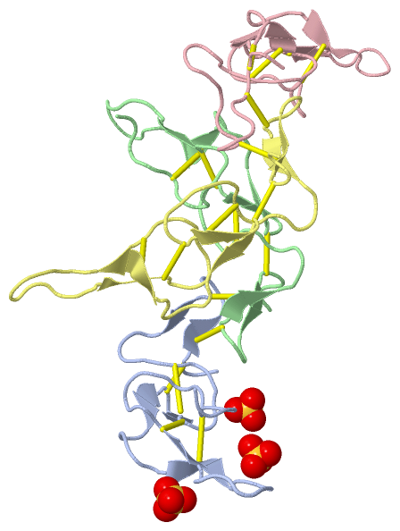
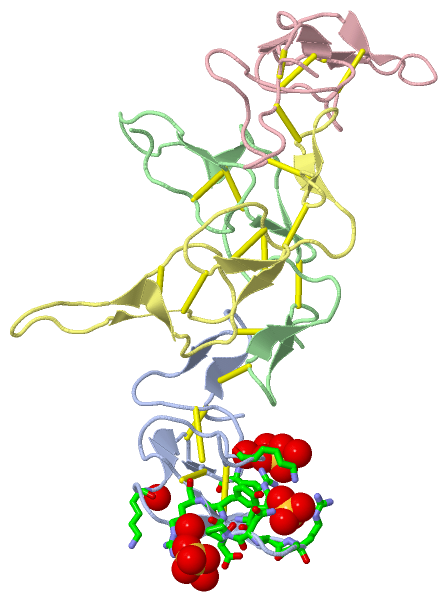
(-) Ligands, Modified Residues, Ions  (1, 3)

Asymmetric Unit (1, 3)
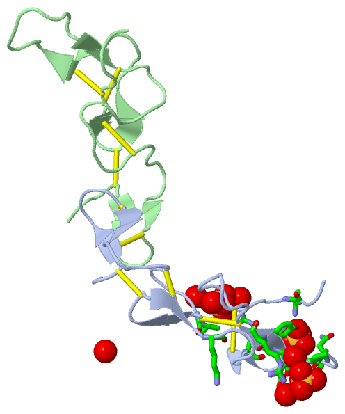
No.NameCountTypeFull Name
1SO43Ligand/IonSULFATE ION
Biological Unit 1 (1, 3)
No.NameCountTypeFull Name
1SO43Ligand/IonSULFATE ION
Biological Unit 2 (0, 0)
No.NameCountTypeFull Name
1SO4-1Ligand/IonSULFATE ION

(-) Sites  (3, 3)

Asymmetric Unit (3, 3)
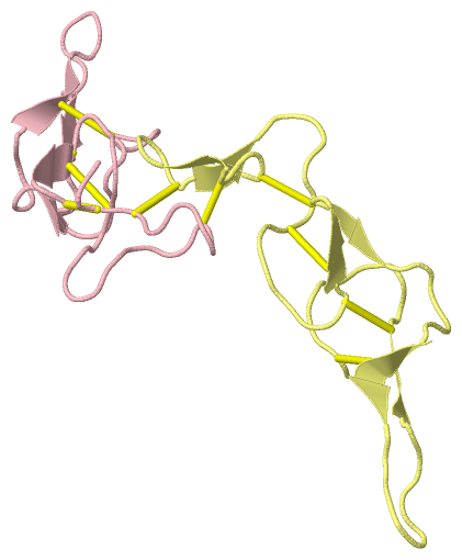
No.NameEvidenceResiduesDescription
1AC1SOFTWARELEU A:25 , LYS A:34 , ARG A:40 , HOH A:79 , HOH A:104 , HOH A:115BINDING SITE FOR RESIDUE SO4 A 64
2AC2SOFTWAREALA A:42 , ARG A:43 , GLY A:44 , ASN A:46 , TYR A:49 , LYS A:61 , HOH A:96 , HOH A:111BINDING SITE FOR RESIDUE SO4 A 65
3AC3SOFTWAREPRO A:47 , ASP A:48 , ARG A:50 , HOH A:89 , HOH A:100BINDING SITE FOR RESIDUE SO4 A 66

(-) SS Bonds  (20, 20)

Asymmetric Unit
No.Residues
1A:7 -A:30
2A:8 -B:13
3A:13 -B:8
4A:21 -A:27
5A:26 -A:51
6A:39 -A:58
7B:7 -B:30
8B:21 -B:27
9B:26 -B:51
10B:39 -B:58
11C:7 -C:30
12C:8 -D:13
13C:13 -D:8
14C:21 -C:27
15C:26 -C:51
16C:39 -C:58
17D:7 -D:30
18D:21 -D:27
19D:26 -D:51
20D:39 -D:58

(-) Cis Peptide Bonds  (0, 0)

(no "Cis Peptide Bond" information available for 3C05)

 Sequence-Structure Mapping

(-) SAPs(SNPs)/Variants  (0, 0)

(no "SAP(SNP)/Variant" information available for 3C05)

(-) PROSITE Motifs  (2, 6)

Asymmetric Unit (2, 6)
 PROSITEUniProtKBPDB
No.IDACDescriptionIDLocationCountLocation
1DISINTEGRIN_2PS50214 Disintegrin domain profile.DIDA_AGKCO51-111
 
  2A:5-63
C:5-62
2DISINTEGRIN_1PS00427 Disintegrins signature.DIDA_AGKCO67-86
 
  2A:21-40
C:21-40
VM2AB_AGKCO439-458
 
  2B:21-40
D:21-40
Biological Unit 1 (2, 3)
 PROSITEUniProtKBPDB
No.IDACDescriptionIDLocationCountLocation
1DISINTEGRIN_2PS50214 Disintegrin domain profile.DIDA_AGKCO51-111
 
  1A:5-63
-
2DISINTEGRIN_1PS00427 Disintegrins signature.DIDA_AGKCO67-86
 
  1A:21-40
-
VM2AB_AGKCO439-458
 
  1B:21-40
-
Biological Unit 2 (2, 3)
 PROSITEUniProtKBPDB
No.IDACDescriptionIDLocationCountLocation
1DISINTEGRIN_2PS50214 Disintegrin domain profile.DIDA_AGKCO51-111
 
  1-
C:5-62
2DISINTEGRIN_1PS00427 Disintegrins signature.DIDA_AGKCO67-86
 
  1-
C:21-40
VM2AB_AGKCO439-458
 
  1-
D:21-40

(-) Exons   (0, 0)

(no "Exon" information available for 3C05)

(-) Sequences/Alignments

Asymmetric Unit
   Reformat: Number of residues per line =  ('0' or empty: single-line sequence representation)
  Number of residues per labelling interval =   
  UniProt sequence: complete  aligned part    
   Show mapping: SCOP domains CATH domains Pfam domains Secondary structure (by author)
SAPs(SNPs) PROSITE motifs Exons
(details for a mapped element are shown in a popup box when the mouse pointer rests over it)
Chain A from PDB  Type:PROTEIN  Length:59
 aligned with DIDA_AGKCO | Q805F7 from UniProtKB/Swiss-Prot  Length:111

    Alignment length:59
                                    60        70        80        90       100         
           DIDA_AGKCO    51 NPCCDAATCKLTPGSQCAEGLCCDQCKFIKAGKICRRARGDNPDYRCTGQSGDCPRKHF 109
               SCOP domains d3c05a_ A: automated matches                                SCOP domains
               CATH domains 3c05A00 A:5-63 Echistatin                                   CATH domains
               Pfam domains ----------------------------------------------------------- Pfam domains
         Sec.struct. author ...ee....ee...........ee..ee.....eee........ee............. Sec.struct. author
                 SAPs(SNPs) ----------------------------------------------------------- SAPs(SNPs)
                PROSITE (1) DISINTEGRIN_2  PDB: A:5-63 UniProt: 51-111                  PROSITE (1)
                PROSITE (2) ----------------DISINTEGRIN_1       ----------------------- PROSITE (2)
                 Transcript ----------------------------------------------------------- Transcript
                 3c05 A   5 NPCCDAATCKLTPGSQCAEGLCCDQCKFIKAGKICRRARGDNPDYRCTGQSGDCPRKHF  63
                                    14        24        34        44        54         

Chain B from PDB  Type:PROTEIN  Length:59
 aligned with VM2AB_AGKCO | Q805F6 from UniProtKB/Swiss-Prot  Length:483

    Alignment length:59
                                   431       441       451       461       471         
          VM2AB_AGKCO   422 ANPCCDAATCKLTTGSQCADGLCCDQCKFMKEGTVCRRARGDDLDDYCNGISAGCPRNP 480
               SCOP domains d3c05b_ B: automated matches                                SCOP domains
               CATH domains 3c05B00 B:4-62 Echistatin                                   CATH domains
               Pfam domains ----------------------------------------------------------- Pfam domains
         Sec.struct. author ....ee....ee...........ee..ee.....eee........ee............ Sec.struct. author
                 SAPs(SNPs) ----------------------------------------------------------- SAPs(SNPs)
                    PROSITE -----------------DISINTEGRIN_1       ---------------------- PROSITE
                 Transcript ----------------------------------------------------------- Transcript
                 3c05 B   4 ANPCCDAATCKLTTGSQCADGLCCDQCKFMKEGTVCRRARGDDLDDYCNGISAGCPRNP  62
                                    13        23        33        43        53         

Chain C from PDB  Type:PROTEIN  Length:58
 aligned with DIDA_AGKCO | Q805F7 from UniProtKB/Swiss-Prot  Length:111

    Alignment length:58
                                    60        70        80        90       100        
           DIDA_AGKCO    51 NPCCDAATCKLTPGSQCAEGLCCDQCKFIKAGKICRRARGDNPDYRCTGQSGDCPRKH 108
               SCOP domains d3c05c_ C: automated matches                               SCOP domains
               CATH domains 3c05C00 C:5-62 Echistatin                                  CATH domains
               Pfam domains ---------------------------------------------------------- Pfam domains
         Sec.struct. author ......................ee..ee.....eee........ee............ Sec.struct. author
                 SAPs(SNPs) ---------------------------------------------------------- SAPs(SNPs)
                PROSITE (1) DISINTEGRIN_2  PDB: C:5-62 UniProt: 51-111                 PROSITE (1)
                PROSITE (2) ----------------DISINTEGRIN_1       ---------------------- PROSITE (2)
                 Transcript ---------------------------------------------------------- Transcript
                 3c05 C   5 NPCCDAATCKLTPGSQCAEGLCCDQCKFIKAGKICRRARGDNPDYRCTGQSGDCPRKH  62
                                    14        24        34        44        54        

Chain D from PDB  Type:PROTEIN  Length:56
 aligned with VM2AB_AGKCO | Q805F6 from UniProtKB/Swiss-Prot  Length:483

    Alignment length:56
                                   431       441       451       461       471      
          VM2AB_AGKCO   422 ANPCCDAATCKLTTGSQCADGLCCDQCKFMKEGTVCRRARGDDLDDYCNGISAGCP 477
               SCOP domains d3c05d_ D: automated matches                             SCOP domains
               CATH domains 3c05D00 D:4-59 Echistatin                                CATH domains
               Pfam domains -------------------------------------------------------- Pfam domains
         Sec.struct. author ....ee....ee...........ee..ee.....eee........ee......... Sec.struct. author
                 SAPs(SNPs) -------------------------------------------------------- SAPs(SNPs)
                    PROSITE -----------------DISINTEGRIN_1       ------------------- PROSITE
                 Transcript -------------------------------------------------------- Transcript
                 3c05 D   4 ANPCCDAATCKLTTGSQCADGLCCDQCKFMKEGTVCRRARGDDLDDYCNGISAGCP  59
                                    13        23        33        43        53      

   Legend:   → Mismatch (orange background)
  - → Gap (green background, '-', border residues have a numbering label)
    → Modified Residue (blue background, lower-case, 'x' indicates undefined single-letter code, labelled with number + name)
  x → Chemical Group (purple background, 'x', labelled with number + name, e.g. ACE or NH2)
  extra numbering lines below/above indicate numbering irregularities and modified residue names etc., number ends below/above '|'

 Classification and Annotation

(-) SCOP Domains  (1, 4)

Asymmetric Unit

(-) CATH Domains  (1, 4)

Asymmetric Unit

(-) Pfam Domains  (0, 0)

(no "Pfam Domain" information available for 3C05)

(-) Gene Ontology  (11, 12)

Asymmetric Unit(hide GO term definitions)
Chain A,C   (DIDA_AGKCO | Q805F7)
biological process
    GO:0007155    cell adhesion    The attachment of a cell, either to another cell or to an underlying substrate such as the extracellular matrix, via cell adhesion molecules.
    GO:0030195    negative regulation of blood coagulation    Any process that stops, prevents, or reduces the frequency, rate or extent of blood coagulation.
cellular component
    GO:0005575    cellular_component    The part of a cell, extracellular environment or virus in which a gene product is located. A gene product may be located in one or more parts of a cell and its location may be as specific as a particular macromolecular complex, that is, a stable, persistent association of macromolecules that function together.
    GO:0005576    extracellular region    The space external to the outermost structure of a cell. For cells without external protective or external encapsulating structures this refers to space outside of the plasma membrane. This term covers the host cell environment outside an intracellular parasite.

Chain B,D   (VM2AB_AGKCO | Q805F6)
molecular function
    GO:0016787    hydrolase activity    Catalysis of the hydrolysis of various bonds, e.g. C-O, C-N, C-C, phosphoric anhydride bonds, etc. Hydrolase is the systematic name for any enzyme of EC class 3.
    GO:0046872    metal ion binding    Interacting selectively and non-covalently with any metal ion.
    GO:0004222    metalloendopeptidase activity    Catalysis of the hydrolysis of internal, alpha-peptide bonds in a polypeptide chain by a mechanism in which water acts as a nucleophile, one or two metal ions hold the water molecule in place, and charged amino acid side chains are ligands for the metal ions.
    GO:0008237    metallopeptidase activity    Catalysis of the hydrolysis of peptide bonds by a mechanism in which water acts as a nucleophile, one or two metal ions hold the water molecule in place, and charged amino acid side chains are ligands for the metal ions.
    GO:0008233    peptidase activity    Catalysis of the hydrolysis of a peptide bond. A peptide bond is a covalent bond formed when the carbon atom from the carboxyl group of one amino acid shares electrons with the nitrogen atom from the amino group of a second amino acid.
    GO:0008270    zinc ion binding    Interacting selectively and non-covalently with zinc (Zn) ions.
biological process
    GO:0006508    proteolysis    The hydrolysis of proteins into smaller polypeptides and/or amino acids by cleavage of their peptide bonds.
cellular component
    GO:0005576    extracellular region    The space external to the outermost structure of a cell. For cells without external protective or external encapsulating structures this refers to space outside of the plasma membrane. This term covers the host cell environment outside an intracellular parasite.

 Visualization

(-) Interactive Views

Asymmetric Unit
  Complete Structure
    Jena3D(integrated viewing of ligand, site, SAP, PROSITE, SCOP information)
    WebMol | AstexViewer[tm]@PDBe
(Java Applets, require no local installation except for Java; loading may be slow)
    STRAP
(Java WebStart application, automatic local installation, requires Java; full application with system access!)
    RasMol
(require local installation)
    Molscript (VRML)
(requires installation of a VRML viewer; select preferred view via VRML and generate a mono or stereo PDF format file)
 
  Ligands, Modified Residues, Ions
    SO4  [ RasMol | Jena3D ]  +environment [ RasMol | Jena3D ]
 
  Sites
    AC1  [ RasMol ]  +environment [ RasMol ]
    AC2  [ RasMol ]  +environment [ RasMol ]
    AC3  [ RasMol ]  +environment [ RasMol ]
 
  Cis Peptide Bonds
(no "Cis Peptide Bonds" information available for 3c05)
 
Biological Units
  Complete Structure
    Biological Unit 1  [ Jena3D ]
    Biological Unit 2  [ Jena3D ]

(-) Still Images

Jmol
  protein: cartoon or spacefill or dots and stick; nucleic acid: cartoon and stick; ligands: spacefill; active site: stick
Molscript
  protein, nucleic acid: cartoon; ligands: spacefill; active site: ball and stick

 Databases and Analysis Tools

(-) Databases

Access by PDB/NDB ID
  3c05
    Family and Domain InformationProDom | SYSTERS
    General Structural InformationGlycoscienceDB | MMDB | NDB | OCA | PDB | PDBe | PDBj | PDBsum | PDBWiki | PQS | PROTEOPEDIA
    Orientation in MembranesOPM
    Protein SurfaceSURFACE
    Secondary StructureDSSP (structure derived) | HSSP (homology derived)
    Structural GenomicsGeneCensus
    Structural NeighboursCE | VAST
    Structure ClassificationCATH | Dali | SCOP
    Validation and Original DataBMRB Data View | BMRB Restraints Grid | EDS | PROCHECK | RECOORD | WHAT_CHECK
 
Access by UniProt ID/Accession number
  DIDA_AGKCO | Q805F7
    Comparative Protein Structure ModelsModBase
    Genomic InformationEnsembl
    Protein-protein InteractionDIP
    Sequence, Family and Domain InformationInterPro | Pfam | SMART | UniProtKB/SwissProt
  VM2AB_AGKCO | Q805F6
    Comparative Protein Structure ModelsModBase
    Genomic InformationEnsembl
    Protein-protein InteractionDIP
    Sequence, Family and Domain InformationInterPro | Pfam | SMART | UniProtKB/SwissProt
 
Access by Enzyme Classificator   (EC Number)
  (no 'Enzyme Classificator' available)
    General Enzyme InformationBRENDA | EC-PDB | Enzyme | IntEnz
    PathwayKEGG | MetaCyc
 
Access by Disease Identifier   (MIM ID)
  (no 'MIM ID' available)
    Disease InformationOMIM
 
Access by GenAge ID
  (no 'GenAge ID' available)
    Age Related InformationGenAge

(-) Analysis Tools

Access by PDB/NDB ID
    Domain InformationXDom
    Interatomic Contacts of Structural UnitsCSU
    Ligand-protein ContactsLPC
    Protein CavitiescastP
    Sequence and Secondary StructurePDBCartoon
    Structure AlignmentSTRAP(Java WebStart application, automatic local installation, requires Java; full application with system access!)
    Structure and Sequence BrowserSTING
 
Access by UniProt ID/Accession number
  DIDA_AGKCO | Q805F7
    Protein Disorder PredictionDisEMBL | FoldIndex | GLOBPLOT (for more information see DisProt)
  VM2AB_AGKCO | Q805F6
    Protein Disorder PredictionDisEMBL | FoldIndex | GLOBPLOT (for more information see DisProt)

 Related Entries

(-) Entries Sharing at Least One Protein Chain (UniProt ID)

(no "Entries Sharing at Least One Protein Chain" available for 3C05)

(-) Related Entries Specified in the PDB File

(no "Related Entries Specified in the PDB File" available for 3C05)