|
|
|
|
 Description
|
|
Compounds
|
||||||||||||||||||||||||||||||||||||||||||||||||||||||||||||
Chains, Units
Summary Information (see also Sequences/Alignments below) |
Ligands, Modified Residues, Ions (4, 5)| Asymmetric/Biological Unit (4, 5) |
Sites (5, 5)
Asymmetric Unit (5, 5)
|
SS Bonds (0, 0)| (no "SS Bond" information available for 3BXD) |
Cis Peptide Bonds (0, 0)| (no "Cis Peptide Bond" information available for 3BXD) |
SAPs(SNPs)/Variants (0, 0)| (no "SAP(SNP)/Variant" information available for 3BXD) |
PROSITE Motifs (0, 0)| (no "PROSITE Motif" information available for 3BXD) |
Exons (0, 0)| (no "Exon" information available for 3BXD) |
Sequences/Alignments
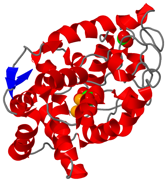
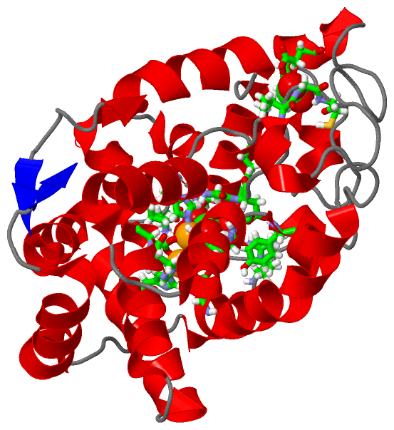
Asymmetric/Biological UnitChain A from PDB Type:PROTEIN Length:258 aligned with MIOX_MOUSE | Q9QXN5 from UniProtKB/Swiss-Prot Length:285 Alignment length:258 37 47 57 67 77 87 97 107 117 127 137 147 157 167 177 187 197 207 217 227 237 247 257 267 277 MIOX_MOUSE 28 FRNYTSGPLLDRVFTTYKLMHTHQTVDFVSRKRIQYGSFSYKKMTIMEAVGMLDDLVDESDPDVDFPNSFHAFQTAEGIRKAHPDKDWFHLVGLLHDLGKIMALWGEPQWAVVGDTFPVGCRPQASVVFCDSTFQDNPDLQDPRYSTELGMYQPHCGLENVLMSWGHDEYLYQMMKFNKFSLPSEAFYMIRFHSFYPWHTGGDYRQLCSQQDLDMLPWVQEFNKFDLYTKCPDLPDVESLRPYYQGLIDKYCPGTLSW 285 SCOP domains d3bxda_ A: Myo-inositol oxygenase MioX SCOP domains CATH domains ------------------------------------------------------------------------------------------------------------------------------------------------------------------------------------------------------------------------------------------------------------------ CATH domains Pfam domains ------------------------------------------------------------------------------------------------------------------------------------------------------------------------------------------------------------------------------------------------------------------ Pfam domains SAPs(SNPs) ------------------------------------------------------------------------------------------------------------------------------------------------------------------------------------------------------------------------------------------------------------------ SAPs(SNPs) PROSITE ------------------------------------------------------------------------------------------------------------------------------------------------------------------------------------------------------------------------------------------------------------------ PROSITE Transcript ------------------------------------------------------------------------------------------------------------------------------------------------------------------------------------------------------------------------------------------------------------------ Transcript 3bxd A 28 FRNYTSGPLLDRVFTTYKLMHTHQTVDFVSRKRIQYGSFSYKKMTIMEAVGMLDDLVDESDPDVDFPNSFHAFQTAEGIRKAHPDKDWFHLVGLLHDLGKIMALWGEPQWAVVGDTFPVGCRPQASVVFCDSTFQDNPDLQDPRYSTELGMYQPHCGLENVLMSWGHDEYLYQMMKFNKFSLPSEAFYMIRFHSFYPWHTGGDYRQLCSQQDLDMLPWVQEFNKFDLYTKCPDLPDVESLRPYYQGLIDKYCPGTLSW 285 37 47 57 67 77 87 97 107 117 127 137 147 157 167 177 187 197 207 217 227 237 247 257 267 277
|
||||||||||||||||||||
SCOP Domains (1, 1)
Asymmetric/Biological Unit
 |
CATH Domains (0, 0)| (no "CATH Domain" information available for 3BXD) |
Pfam Domains (0, 0)| (no "Pfam Domain" information available for 3BXD) |
Gene Ontology (13, 13)|
Asymmetric/Biological Unit(hide GO term definitions) Chain A (MIOX_MOUSE | Q9QXN5)
|
||||||||||||||||||||||||||||||||||||||||||||||||||||||||||||||||||||||||||||||||||||||||||||||||
Interactive Views
|
|||||||||||||||||||||||||||||||||||||||||||||||||||||||||||||||||||||||||||||||||||||||||||||||||||||||||||||||||||||||||||||||||||||||||||||||||||||||||||||||||||||||
Still Images
|
||||||||||||||||
Databases
|
||||||||||||||||||||||||||||||||||||||||||||||||||||||||||||||||||||||||||||||||||||||||||||||||||||||||||||||||||||||||||||||||||||||||||||||||||||||||||||||||
Analysis Tools
|
|||||||||||||||||||||||||||||||||||||||||||||||||||||||||||||
Entries Sharing at Least One Protein Chain (UniProt ID)
Related Entries Specified in the PDB File
|
|