Show PDB file:   
         Plain Text   HTML   (compressed file size)
QuickSearch:   
by PDB,NDB,UniProt,PROSITE Code or Search Term(s)  
(-)Asymmetric Unit
(-)Biological Unit 1
(-)Biological Unit 2
(-)Biological Unit 3
collapse expand < >
Image Asymmetric Unit
Asymmetric Unit  (Jmol Viewer)
Image Biological Unit 1
Biological Unit 1  (Jmol Viewer)
Image Biological Unit 2
Biological Unit 2  (Jmol Viewer)
Image Biological Unit 3
Biological Unit 3  (Jmol Viewer)

(-) Description

Title :  STRUCTURE OF MOUSE BETA-NEUREXIN 2D4
 
Authors :  J. Koehnke, X. Jin, L. Shapiro
Date :  17 Dec 07  (Deposition) - 25 Mar 08  (Release) - 13 Jul 11  (Revision)
Method :  X-RAY DIFFRACTION
Resolution :  3.00
Chains :  Asym. Unit :  A,B,C
Biol. Unit 1:  A  (1x)
Biol. Unit 2:  B  (1x)
Biol. Unit 3:  C  (1x)
Keywords :  Beta-Neurexin 2D4, Lns6, Lectin, Cell Adhesion (Keyword Search: [Gene Ontology, PubMed, Web (Google))
 
Reference :  J. Koehnke, X. Jin, N. Trbovic, P. S. Katsamba, J. Brasch, G. Ahlsen, P. Scheiffele, B. Honig, A. G. Palmer, L. Shapiro
Crystal Structures Of Beta-Neurexin 1 And Beta-Neurexin 2 Ectodomains And Dynamics Of Splice Insertion Sequence 4.
Structure V. 16 410 2008
PubMed-ID: 18334216  |  Reference-DOI: 10.1016/J.STR.2007.12.024

(-) Compounds

Molecule 1 - BETA-NEUREXIN 2D4
    ChainsA, B, C
    EngineeredYES
    Expression SystemESCHERICHIA COLI BL21
    Expression System PlasmidPGEX-NRX2D4
    Expression System StrainBL21
    Expression System Taxid511693
    Expression System Vector TypePLASMID
    FragmentLNS DOMAIN
    GeneNRXN2
    Organism CommonHOUSE MOUSE
    Organism ScientificMUS MUSCULUS
    Organism Taxid10090

 Structural Features

(-) Chains, Units

  123
Asymmetric Unit ABC
Biological Unit 1 (1x)A  
Biological Unit 2 (1x) B 
Biological Unit 3 (1x)  C

Summary Information (see also Sequences/Alignments below)

(-) Ligands, Modified Residues, Ions  (0, 0)

(no "Ligand,Modified Residues,Ions" information available for 3BOP)

(-) Sites  (0, 0)

(no "Site" information available for 3BOP)

(-) SS Bonds  (0, 0)

(no "SS Bond" information available for 3BOP)

(-) Cis Peptide Bonds  (4, 4)

Asymmetric Unit
No.Residues
1Gly A:223 -Arg A:224
2Ser B:135 -Gly B:136
3Gly C:94 -Gly C:95
4Gly C:136 -Leu C:137

 Sequence-Structure Mapping

(-) SAPs(SNPs)/Variants  (0, 0)

(no "SAP(SNP)/Variant" information available for 3BOP)

(-) PROSITE Motifs  (0, 0)

(no "PROSITE Motif" information available for 3BOP)

(-) Exons   (0, 0)

(no "Exon" information available for 3BOP)

(-) Sequences/Alignments

Asymmetric Unit
   Reformat: Number of residues per line =  ('0' or empty: single-line sequence representation)
  Number of residues per labelling interval =   
  UniProt sequence: complete  aligned part    
   Show mapping: SCOP domains CATH domains Pfam domains Secondary structure (by author)
SAPs(SNPs) PROSITE motifs Exons
(details for a mapped element are shown in a popup box when the mouse pointer rests over it)
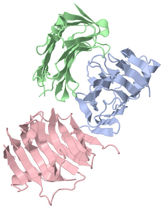
Chain A from PDB  Type:PROTEIN  Length:168
 aligned with Q3TQ54_MOUSE | Q3TQ54 from UniProtKB/TrEMBL  Length:353

    Alignment length:175
                                    95       105       115       125       135       145       155       165       175       185       195       205       215       225       235       245       255     
         Q3TQ54_MOUSE    86 GTTYIFGKGGALITYTWPPNDRPSTRMDRLAVGFSTHQRSAVLVRVDSASGLGDYLQLHIDQGTVGVIFNVGTDDITIDEPNAIVSDGKYHVVRFTRSGGNATLQVDSWPVNERYPAGRQLTIFNSQAAIKIGGRDQGRPFQGQVSGLYYNGLKVLALAAESDPNVRTEGHLRLV 260
               SCOP domains d3bopa_ A: automated matches                                                                                                                                                    SCOP domains
               CATH domains 3bopA00 A:86-260  [code=2.60.120.200, no name defined]                                                                                                                          CATH domains
               Pfam domains ------------------------------------------------------------------------------------------------------------------------------------------------------------------------------- Pfam domains
         Sec.struct. author ..eeee.....eeeee............eeeeeee.....eeeeeeee.....eeeeeeee..eeeeeeee..eeeeeee..........eeeeeeee..eeeeee..eeeee....-------..eeeeeee.........eeeeeeee...hhhhhhhh....eeee..eee. Sec.struct. author
                 SAPs(SNPs) ------------------------------------------------------------------------------------------------------------------------------------------------------------------------------- SAPs(SNPs)
                    PROSITE ------------------------------------------------------------------------------------------------------------------------------------------------------------------------------- PROSITE
                 Transcript ------------------------------------------------------------------------------------------------------------------------------------------------------------------------------- Transcript
                 3bop A  86 ATTYIFGKGGALITYTWPPNDRPSTRMDRLAVGFSTHQRSAVLVRVDSASGLGDYLQLHIDQGTVGVIFNVGTDDITIDEPNAIVSDGKYHVVRFTRSGGNATLQVDSWPVNERYPA-------NSQAAIKIGGRDQGRPFQGQVSGLYYNGLKVLALAAESDPNVRTEGHLRLV 260
                                    95       105       115       125       135       145       155       165       175       185       195      |  -    |  215       225       235       245       255     
                                                                                                                                              202     210                                                  

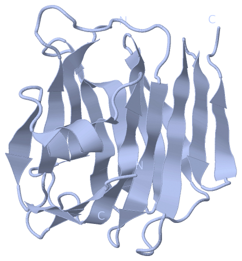
Chain B from PDB  Type:PROTEIN  Length:168
 aligned with Q3TQ54_MOUSE | Q3TQ54 from UniProtKB/TrEMBL  Length:353

    Alignment length:174
                                    96       106       116       126       136       146       156       166       176       186       196       206       216       226       236       246       256    
         Q3TQ54_MOUSE    87 TTYIFGKGGALITYTWPPNDRPSTRMDRLAVGFSTHQRSAVLVRVDSASGLGDYLQLHIDQGTVGVIFNVGTDDITIDEPNAIVSDGKYHVVRFTRSGGNATLQVDSWPVNERYPAGRQLTIFNSQAAIKIGGRDQGRPFQGQVSGLYYNGLKVLALAAESDPNVRTEGHLRLV 260
               SCOP domains ------------------------------------------------------------------------------------------------------------------------------------------------------------------------------ SCOP domains
               CATH domains 3bopB00 B:87-260  [code=2.60.120.200, no name defined]                                                                                                                         CATH domains
               Pfam domains ------------------------------------------------------------------------------------------------------------------------------------------------------------------------------ Pfam domains
         Sec.struct. author .eeeeeeeeeeeeee............eeeeeee.....eeeeeeee.....eeeeeeee..eeeeeeee..eeeeeeeeeee......eeeeeeee..eeeeee..eeeee...------....eeeeeee.........eeeeeeee...hhhhhhhh....eeeeeeeee. Sec.struct. author
                 SAPs(SNPs) ------------------------------------------------------------------------------------------------------------------------------------------------------------------------------ SAPs(SNPs)
                    PROSITE ------------------------------------------------------------------------------------------------------------------------------------------------------------------------------ PROSITE
                 Transcript ------------------------------------------------------------------------------------------------------------------------------------------------------------------------------ Transcript
                 3bop B  87 TTYIFGKGGALITYTWPPNDRPSTRMDRLAVGFSTHQRSAVLVRVDSASGLGDYLQLHIDQGTVGVIFNVGTDDITIDEPNAIVSDGKYHVVRFTRSGGNATLQVDSWPVNERYP------IFNSQAAIKIGGRDQGRPFQGQVSGLYYNGLKVLALAAESDPNVRTEGHLRLV 260
                                    96       106       116       126       136       146       156       166       176       186       196    |    - |     216       226       236       246       256    
                                                                                                                                            201    208                                                    

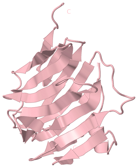
Chain C from PDB  Type:PROTEIN  Length:169
 aligned with Q3TQ54_MOUSE | Q3TQ54 from UniProtKB/TrEMBL  Length:353

    Alignment length:175
                                    95       105       115       125       135       145       155       165       175       185       195       205       215       225       235       245       255     
         Q3TQ54_MOUSE    86 GTTYIFGKGGALITYTWPPNDRPSTRMDRLAVGFSTHQRSAVLVRVDSASGLGDYLQLHIDQGTVGVIFNVGTDDITIDEPNAIVSDGKYHVVRFTRSGGNATLQVDSWPVNERYPAGRQLTIFNSQAAIKIGGRDQGRPFQGQVSGLYYNGLKVLALAAESDPNVRTEGHLRLV 260
               SCOP domains d3bopc_ C: automated matches                                                                                                                                                    SCOP domains
               CATH domains 3bopC00 C:86-260  [code=2.60.120.200, no name defined]                                                                                                                          CATH domains
               Pfam domains ------------------------------------------------------------------------------------------------------------------------------------------------------------------------------- Pfam domains
         Sec.struct. author ..eeee.....eeeee...........eeeeeeeee....eeeeeeee.......eeeeee..eeeeeeee..eeeeeee.........eeeeeeee....eeeee..eeeee.....------..eeeeeee.ee..ee..eee...ee...hhhhhhhh....eeee..eeee Sec.struct. author
                 SAPs(SNPs) ------------------------------------------------------------------------------------------------------------------------------------------------------------------------------- SAPs(SNPs)
                    PROSITE ------------------------------------------------------------------------------------------------------------------------------------------------------------------------------- PROSITE
                 Transcript ------------------------------------------------------------------------------------------------------------------------------------------------------------------------------- Transcript
                 3bop C  86 ATTYIFGKGGALITYTWPPNDRPSTRMDRLAVGFSTHQRSAVLVRVDSASGLGDYLQLHIDQGTVGVIFNVGTDDITIDEPNAIVSDGKYHVVRFTRSGGNATLQVDSWPVNERYPAG------NSQAAIKIGGRDQGRPFQGQVSGLYYNGLKVLALAAESDPNVRTEGHLRLV 260
                                    95       105       115       125       135       145       155       165       175       185       195       | -    |  215       225       235       245       255     
                                                                                                                                               203    210                                                  

   Legend:   → Mismatch (orange background)
  - → Gap (green background, '-', border residues have a numbering label)
    → Modified Residue (blue background, lower-case, 'x' indicates undefined single-letter code, labelled with number + name)
  x → Chemical Group (purple background, 'x', labelled with number + name, e.g. ACE or NH2)
  extra numbering lines below/above indicate numbering irregularities and modified residue names etc., number ends below/above '|'

 Classification and Annotation

(-) SCOP Domains  (1, 2)

Asymmetric Unit

(-) CATH Domains  (1, 3)

Asymmetric Unit
(-)
Class: Mainly Beta (13760)

(-) Pfam Domains  (0, 0)

(no "Pfam Domain" information available for 3BOP)

(-) Gene Ontology  (3, 3)

Asymmetric Unit(hide GO term definitions)
Chain A,B,C   (Q3TQ54_MOUSE | Q3TQ54)
molecular function
    GO:0046872    metal ion binding    Interacting selectively and non-covalently with any metal ion.
biological process
    GO:0007268    chemical synaptic transmission    The vesicular release of classical neurotransmitter molecules from a presynapse, across a chemical synapse, the subsequent activation of neurotransmitter receptors at the postsynapse of a target cell (neuron, muscle, or secretory cell) and the effects of this activation on the postsynaptic membrane potential and ionic composition of the postsynaptic cytosol. This process encompasses both spontaneous and evoked release of neurotransmitter and all parts of synaptic vesicle exocytosis. Evoked transmission starts with the arrival of an action potential at the presynapse.
cellular component
    GO:0016021    integral component of membrane    The component of a membrane consisting of the gene products and protein complexes having at least some part of their peptide sequence embedded in the hydrophobic region of the membrane.

 Visualization

(-) Interactive Views

Asymmetric Unit
  Complete Structure
    Jena3D(integrated viewing of ligand, site, SAP, PROSITE, SCOP information)
    WebMol | AstexViewer[tm]@PDBe
(Java Applets, require no local installation except for Java; loading may be slow)
    STRAP
(Java WebStart application, automatic local installation, requires Java; full application with system access!)
    RasMol
(require local installation)
    Molscript (VRML)
(requires installation of a VRML viewer; select preferred view via VRML and generate a mono or stereo PDF format file)
 
  Ligands, Modified Residues, Ions
(no "Ligands, Modified Residues, Ions" information available for 3bop)
 
  Sites
(no "Sites" information available for 3bop)
 
  Cis Peptide Bonds
    Gly A:223 - Arg A:224   [ RasMol ]  
    Gly C:136 - Leu C:137   [ RasMol ]  
    Gly C:94 - Gly C:95   [ RasMol ]  
    Ser B:135 - Gly B:136   [ RasMol ]  
 
Biological Units
  Complete Structure
    Biological Unit 1  [ Jena3D ]
    Biological Unit 2  [ Jena3D ]
    Biological Unit 3  [ Jena3D ]

(-) Still Images

Jmol
  protein: cartoon or spacefill or dots and stick; nucleic acid: cartoon and stick; ligands: spacefill; active site: stick
Molscript
  protein, nucleic acid: cartoon; ligands: spacefill; active site: ball and stick

 Databases and Analysis Tools

(-) Databases

Access by PDB/NDB ID
  3bop
    Family and Domain InformationProDom | SYSTERS
    General Structural InformationGlycoscienceDB | MMDB | NDB | OCA | PDB | PDBe | PDBj | PDBsum | PDBWiki | PQS | PROTEOPEDIA
    Orientation in MembranesOPM
    Protein SurfaceSURFACE
    Secondary StructureDSSP (structure derived) | HSSP (homology derived)
    Structural GenomicsGeneCensus
    Structural NeighboursCE | VAST
    Structure ClassificationCATH | Dali | SCOP
    Validation and Original DataBMRB Data View | BMRB Restraints Grid | EDS | PROCHECK | RECOORD | WHAT_CHECK
 
Access by UniProt ID/Accession number
  Q3TQ54_MOUSE | Q3TQ54
    Comparative Protein Structure ModelsModBase
    Genomic InformationEnsembl
    Protein-protein InteractionDIP
    Sequence, Family and Domain InformationInterPro | Pfam | SMART | UniProtKB/TrEMBL
 
Access by Enzyme Classificator   (EC Number)
  (no 'Enzyme Classificator' available)
    General Enzyme InformationBRENDA | EC-PDB | Enzyme | IntEnz
    PathwayKEGG | MetaCyc
 
Access by Disease Identifier   (MIM ID)
  (no 'MIM ID' available)
    Disease InformationOMIM
 
Access by GenAge ID
  (no 'GenAge ID' available)
    Age Related InformationGenAge

(-) Analysis Tools

Access by PDB/NDB ID
    Domain InformationXDom
    Interatomic Contacts of Structural UnitsCSU
    Ligand-protein ContactsLPC
    Protein CavitiescastP
    Sequence and Secondary StructurePDBCartoon
    Structure AlignmentSTRAP(Java WebStart application, automatic local installation, requires Java; full application with system access!)
    Structure and Sequence BrowserSTING
 
Access by UniProt ID/Accession number
  Q3TQ54_MOUSE | Q3TQ54
    Protein Disorder PredictionDisEMBL | FoldIndex | GLOBPLOT (for more information see DisProt)

 Related Entries

(-) Entries Sharing at Least One Protein Chain (UniProt ID)

(no "Entries Sharing at Least One Protein Chain" available for 3BOP)

(-) Related Entries Specified in the PDB File

3bod STRUCTURE OF NEUREXIN 1D4