Show PDB file:   
         Plain Text   HTML   (compressed file size)
QuickSearch:   
by PDB,NDB,UniProt,PROSITE Code or Search Term(s)  
(-)Asym./Biol. Unit
(-)Asym./Biol. Unit - sites
collapse expand < >
Image Asym./Biol. Unit
Asym./Biol. Unit  (Jmol Viewer)
Image Asym./Biol. Unit - sites
Asym./Biol. Unit - sites  (Jmol Viewer)

(-) Description

Title :  CARBONIC ANHYDRASE FROM MARINE DIATOM THALASSIOSIRA WEISSFLOGII-CADMIUM BOUND DOMAIN 2 WITH ACETATE (CDCA1-R2)
 
Authors :  Y. Xu, L. Feng, P. D. Jeffrey, Y. Shi, F. M. M. Morel
Date :  17 Dec 07  (Deposition) - 22 Jan 08  (Release) - 26 May 10  (Revision)
Method :  X-RAY DIFFRACTION
Resolution :  1.40
Chains :  Asym./Biol. Unit :  A
Keywords :  Carbonic Anhydrase, Marine Diatom, Cadmium-Bound, Acetate Ligand, Lyase (Keyword Search: [Gene Ontology, PubMed, Web (Google))
 
Reference :  Y. Xu, L. Feng, P. D. Jeffrey, Y. Shi, F. M. Morel
Structure And Metal Exchange In The Cadmium Carbonic Anhydrase Of Marine Diatoms.
Nature V. 452 56 2008
PubMed-ID: 18322527  |  Reference-DOI: 10.1038/NATURE06636

(-) Compounds

Molecule 1 - CADMIUM-SPECIFIC CARBONIC ANHYDRASE
    ChainsA
    EC Number4.2.1.1
    EngineeredYES
    Expression SystemESCHERICHIA COLI
    Expression System Taxid562
    FragmentDOMAIN 2, CDCA1-R2
    GeneCDCA1
    Organism ScientificTHALASSIOSIRA WEISSFLOGII
    Organism Taxid67004

 Structural Features

(-) Chains, Units

  1
Asymmetric/Biological Unit A

Summary Information (see also Sequences/Alignments below)
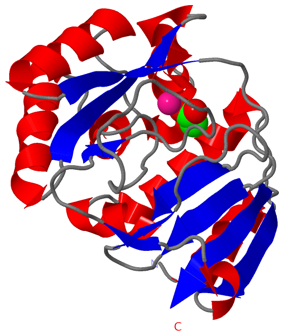
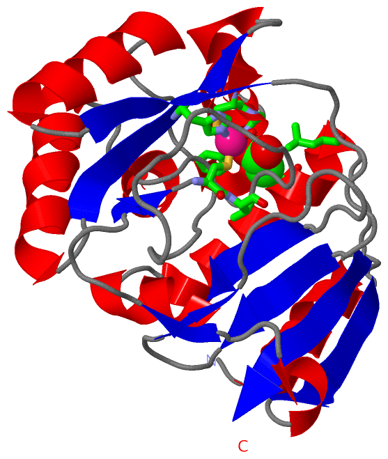
(-) Ligands, Modified Residues, Ions  (2, 2)

Asymmetric/Biological Unit (2, 2)
No.NameCountTypeFull Name
1ACT1Ligand/IonACETATE ION
2CD1Ligand/IonCADMIUM ION

(-) Sites  (2, 2)

Asymmetric Unit (2, 2)
No.NameEvidenceResiduesDescription
1AC1SOFTWARECYS A:263 , HIS A:315 , CYS A:325 , ACT A:1002 , HOH A:1003BINDING SITE FOR RESIDUE CD A 1001
2AC2SOFTWARECYS A:263 , VAL A:264 , GLY A:326 , PHE A:327 , THR A:417 , LEU A:421 , CD A:1001 , HOH A:1003BINDING SITE FOR RESIDUE ACT A 1002

(-) SS Bonds  (0, 0)

(no "SS Bond" information available for 3BOE)

(-) Cis Peptide Bonds  (0, 0)

(no "Cis Peptide Bond" information available for 3BOE)

 Sequence-Structure Mapping

(-) SAPs(SNPs)/Variants  (0, 0)

(no "SAP(SNP)/Variant" information available for 3BOE)

(-) PROSITE Motifs  (0, 0)

(no "PROSITE Motif" information available for 3BOE)

(-) Exons   (0, 0)

(no "Exon" information available for 3BOE)

(-) Sequences/Alignments

Asymmetric/Biological Unit
   Reformat: Number of residues per line =  ('0' or empty: single-line sequence representation)
  Number of residues per labelling interval =   
  UniProt sequence: complete  aligned part    
   Show mapping: SCOP domains CATH domains Pfam domains Secondary structure (by author)
SAPs(SNPs) PROSITE motifs Exons
(details for a mapped element are shown in a popup box when the mouse pointer rests over it)
Chain A from PDB  Type:PROTEIN  Length:209
 aligned with Q50EL4_THAWE | Q50EL4 from UniProtKB/TrEMBL  Length:616

    Alignment length:209
                                   207       217       227       237       247       257       267       277       287       297       307       317       327       337       347       357       367       377       387       397         
         Q50EL4_THAWE   198 ISPAQIAEALQGRGWDAEIVTDASMAGQLVDVRPEGILKCVDGRGSDNTRMGGPKMPGGIYAIAHNRGVTSIEGLKQITKEVASKGHLPSVHGDHSSDMLGCGFFKLWVTGRFDDMGYPRPQFDADQGANAVKDAGGIIEMHHGSHTEKVVYINLLANKTLEPNENDQRFIVDGWAADKFGLDVPKFLIAAAATVEMLGGPKNAKIVVP 406
               SCOP domains d3boea_ A: Cadmium-specific carbonic anhydrase CdCA1                                                                                                                                                              SCOP domains
               CATH domains ----------------------------------------------------------------------------------------------------------------------------------------------------------------------------------------------------------------- CATH domains
               Pfam domains ----------------------------------------------------------------------------------------------------------------------------------------------------------------------------------------------------------------- Pfam domains
         Sec.struct. author .hhhhhhhhhhh...eeeeee.hhhh...ee.....ee.....................hhhhhhhhh..hhhhhhhhhhhhhhh..eeeeeee..hhhhhhhhhhhhhh..hhhhh......hhhhhhhhhhhh..eeeee........eeee....ee.........eeeehhhhhhh..hhhhhhhhhhhhhhhh....eeeeee. Sec.struct. author
                 SAPs(SNPs) ----------------------------------------------------------------------------------------------------------------------------------------------------------------------------------------------------------------- SAPs(SNPs)
                    PROSITE ----------------------------------------------------------------------------------------------------------------------------------------------------------------------------------------------------------------- PROSITE
                 Transcript ----------------------------------------------------------------------------------------------------------------------------------------------------------------------------------------------------------------- Transcript
                 3boe A 224 ISPAQIAEALQGRGWDAEIVTDASMAGQLVDVRPEGILKCVDGRGSDNTRMGGPKMPGGIYAIAHNRGVTSIEGLKQITKEVASKGHLPSVHGDHSSDMLGCGFFKLWVTGRFDDMGYPRPQFDADQGANAVKDAGGIIEMHHGSHTEKVVYINLLANKTLEPNENDQRFIVDGWAADKFGLDVPKFLIAAAATVEMLGGPKNAKIVVP 432
                                   233       243       253       263       273       283       293       303       313       323       333       343       353       363       373       383       393       403       413       423         

   Legend:   → Mismatch (orange background)
  - → Gap (green background, '-', border residues have a numbering label)
    → Modified Residue (blue background, lower-case, 'x' indicates undefined single-letter code, labelled with number + name)
  x → Chemical Group (purple background, 'x', labelled with number + name, e.g. ACE or NH2)
  extra numbering lines below/above indicate numbering irregularities and modified residue names etc., number ends below/above '|'

 Classification and Annotation

(-) SCOP Domains  (1, 1)

Asymmetric/Biological Unit

(-) CATH Domains  (0, 0)

(no "CATH Domain" information available for 3BOE)

(-) Pfam Domains  (0, 0)

(no "Pfam Domain" information available for 3BOE)

(-) Gene Ontology  (1, 1)

Asymmetric/Biological Unit(hide GO term definitions)
Chain A   (Q50EL4_THAWE | Q50EL4)
molecular function
    GO:0046872    metal ion binding    Interacting selectively and non-covalently with any metal ion.

 Visualization

(-) Interactive Views

Asymmetric/Biological Unit
  Complete Structure
    Jena3D(integrated viewing of ligand, site, SAP, PROSITE, SCOP information)
    WebMol | AstexViewer[tm]@PDBe
(Java Applets, require no local installation except for Java; loading may be slow)
    STRAP
(Java WebStart application, automatic local installation, requires Java; full application with system access!)
    RasMol
(require local installation)
    Molscript (VRML)
(requires installation of a VRML viewer; select preferred view via VRML and generate a mono or stereo PDF format file)
 
  Ligands, Modified Residues, Ions
    ACT  [ RasMol | Jena3D ]  +environment [ RasMol | Jena3D ]
    CD  [ RasMol | Jena3D ]  +environment [ RasMol | Jena3D ]
 
  Sites
    AC1  [ RasMol ]  +environment [ RasMol ]
    AC2  [ RasMol ]  +environment [ RasMol ]
 
  Cis Peptide Bonds
(no "Cis Peptide Bonds" information available for 3boe)
 

(-) Still Images

Jmol
  protein: cartoon or spacefill or dots and stick; nucleic acid: cartoon and stick; ligands: spacefill; active site: stick
Molscript
  protein, nucleic acid: cartoon; ligands: spacefill; active site: ball and stick

 Databases and Analysis Tools

(-) Databases

Access by PDB/NDB ID
  3boe
    Family and Domain InformationProDom | SYSTERS
    General Structural InformationGlycoscienceDB | MMDB | NDB | OCA | PDB | PDBe | PDBj | PDBsum | PDBWiki | PQS | PROTEOPEDIA
    Orientation in MembranesOPM
    Protein SurfaceSURFACE
    Secondary StructureDSSP (structure derived) | HSSP (homology derived)
    Structural GenomicsGeneCensus
    Structural NeighboursCE | VAST
    Structure ClassificationCATH | Dali | SCOP
    Validation and Original DataBMRB Data View | BMRB Restraints Grid | EDS | PROCHECK | RECOORD | WHAT_CHECK
 
Access by UniProt ID/Accession number
  Q50EL4_THAWE | Q50EL4
    Comparative Protein Structure ModelsModBase
    Genomic InformationEnsembl
    Protein-protein InteractionDIP
    Sequence, Family and Domain InformationInterPro | Pfam | SMART | UniProtKB/TrEMBL
 
Access by Enzyme Classificator   (EC Number)
  4.2.1.1
    General Enzyme InformationBRENDA | EC-PDB | Enzyme | IntEnz
    PathwayKEGG | MetaCyc
 
Access by Disease Identifier   (MIM ID)
  (no 'MIM ID' available)
    Disease InformationOMIM
 
Access by GenAge ID
  (no 'GenAge ID' available)
    Age Related InformationGenAge

(-) Analysis Tools

Access by PDB/NDB ID
    Domain InformationXDom
    Interatomic Contacts of Structural UnitsCSU
    Ligand-protein ContactsLPC
    Protein CavitiescastP
    Sequence and Secondary StructurePDBCartoon
    Structure AlignmentSTRAP(Java WebStart application, automatic local installation, requires Java; full application with system access!)
    Structure and Sequence BrowserSTING
 
Access by UniProt ID/Accession number
  Q50EL4_THAWE | Q50EL4
    Protein Disorder PredictionDisEMBL | FoldIndex | GLOBPLOT (for more information see DisProt)

 Related Entries

(-) Entries Sharing at Least One Protein Chain (UniProt ID)

UniProtKB/TrEMBL
        Q50EL4_THAWE | Q50EL43bob 3boc 3boh 3boj 3uk8

(-) Related Entries Specified in the PDB File

3bob CDCA1-R2 WITH CADMIUM
3boc CDCA1-R2 WITH ZINC