Show PDB file:   
         Plain Text   HTML   (compressed file size)
QuickSearch:   
by PDB,NDB,UniProt,PROSITE Code or Search Term(s)  
(-)Asymmetric Unit
(-)Biological Unit 1
(-)Biological Unit 2
collapse expand < >
Image Asymmetric Unit
Asymmetric Unit  (Jmol Viewer)
Image Biological Unit 1
Biological Unit 1  (Jmol Viewer)
Image Biological Unit 2
Biological Unit 2  (Jmol Viewer)

(-) Description

Title :  CRYSTAL STRUCTURE OF THE PD-L1
 
Authors :  D. Y. Lin, Y. Tanaka, M. Iwasaki, A. G. Gittis, H. P. Su, B. Mikami, T. Okaz T. Honjo, N. Minato, D. N. Garboczi
Date :  30 Nov 07  (Deposition) - 26 Feb 08  (Release) - 13 Jul 11  (Revision)
Method :  X-RAY DIFFRACTION
Resolution :  2.64
Chains :  Asym. Unit :  A,B
Biol. Unit 1:  A  (1x)
Biol. Unit 2:  B  (1x)
Keywords :  Co-Stimulation, Immunoglobulin-Like Beta-Sandwich, T Cell, B Cell, Programmed Death, Immune System, Transmembrane, Inhibitory Receptor, Glycoprotein, Immunoglobulin Domain (Keyword Search: [Gene Ontology, PubMed, Web (Google))
 
Reference :  D. Y. Lin, Y. Tanaka, M. Iwasaki, A. G. Gittis, H. P. Su, B. Mikami, T. Okazaki, T. Honjo, N. Minato, D. N. Garboczi
The Pd-1/Pd-L1 Complex Resembles The Antigen-Binding Fv Domains Of Antibodies And T Cell Receptors.
Proc. Natl. Acad. Sci. Usa V. 105 3011 2008
PubMed-ID: 18287011  |  Reference-DOI: 10.1073/PNAS.0712278105

(-) Compounds

Molecule 1 - PROGRAMMED CELL DEATH 1 LIGAND 1
    ChainsA, B
    EngineeredYES
    Expression SystemESCHERICHIA COLI
    Expression System PlasmidPLM1
    Expression System StrainROSETTA 2 (DE3)
    Expression System Taxid562
    Expression System Vector TypePLASMID
    FragmentEXTRACELLULAR REGION
    GeneCD274, B7H1, PDCD1L1, PDCD1LG1, PDL1
    Organism CommonHUMAN
    Organism ScientificHOMO SAPIENS
    Organism Taxid9606
    SynonymPROGRAMMED DEATH LIGAND 1, PD-L1, PDCD1 LIGAND 1, B7 HOMOLOG 1, B7-H1, CD274 ANTIGEN

 Structural Features

(-) Chains, Units

  12
Asymmetric Unit AB
Biological Unit 1 (1x)A 
Biological Unit 2 (1x) B

Summary Information (see also Sequences/Alignments below)

(-) Ligands, Modified Residues, Ions  (0, 0)

(no "Ligand,Modified Residues,Ions" information available for 3BIS)

(-) Sites  (0, 0)

(no "Site" information available for 3BIS)

(-) SS Bonds  (4, 4)

Asymmetric Unit
No.Residues
1A:40 -A:114
2A:155 -A:209
3B:40 -B:114
4B:155 -B:209

(-) Cis Peptide Bonds  (3, 3)

Asymmetric Unit
No.Residues
1Tyr A:160 -Pro A:161
2Asp A:215 -Pro A:216
3Tyr B:160 -Pro B:161

 Sequence-Structure Mapping

(-) SAPs(SNPs)/Variants  (0, 0)

(no "SAP(SNP)/Variant" information available for 3BIS)

(-) PROSITE Motifs  (0, 0)

(no "PROSITE Motif" information available for 3BIS)

(-) Exons   (4, 7)

Asymmetric Unit (4, 7)
 ENSEMBLUniProtKBPDB
No.Transcript IDExonExon IDGenome LocationLengthIDLocationLengthCountLocationLength
1.1aENST000003815771aENSE00001892659chr9:5450525-545059672PD1L1_HUMAN-00--
1.2aENST000003815772aENSE00000813155chr9:5456100-545616566PD1L1_HUMAN1-18181-
B:18-18
-
1
1.3aENST000003815773aENSE00000813156chr9:5457079-5457420342PD1L1_HUMAN18-1321152A:19-132
B:18-132
114
115
1.4bENST000003815774bENSE00000813157chr9:5462834-5463121288PD1L1_HUMAN132-228972A:132-228
B:132-228
97
97
1.5bENST000003815775bENSE00000813158chr9:5465499-5465606108PD1L1_HUMAN228-264372A:228-230
B:228-229
3
2
1.6ENST000003815776ENSE00001410545chr9:5466770-546682960PD1L1_HUMAN264-284210--
1.7cENST000003815777cENSE00001889081chr9:5467840-54705472708PD1L1_HUMAN284-29070--

(-) Sequences/Alignments

Asymmetric Unit
   Reformat: Number of residues per line =  ('0' or empty: single-line sequence representation)
  Number of residues per labelling interval =   
  UniProt sequence: complete  aligned part    
   Show mapping: SCOP domains CATH domains Pfam domains Secondary structure (by author)
SAPs(SNPs) PROSITE motifs Exons
(details for a mapped element are shown in a popup box when the mouse pointer rests over it)
Chain A from PDB  Type:PROTEIN  Length:212
 aligned with PD1L1_HUMAN | Q9NZQ7 from UniProtKB/Swiss-Prot  Length:290

    Alignment length:212
                                    28        38        48        58        68        78        88        98       108       118       128       138       148       158       168       178       188       198       208       218       228  
          PD1L1_HUMAN    19 FTVTVPKDLYVVEYGSNMTIECKFPVEKQLDLAALIVYWEMEDKNIIQFVHGEEDLKVQHSSYRQRARLLKDQLSLGNAALQITDVKLQDAGVYRCMISYGGADYKRITVKVNAPYNKINQRILVVDPVTSEHELTCQAEGYPKAEVIWTSSDHQVLSGKTTTTNSKREEKLFNVTSTLRINTTTNEIFYCTFRRLDPEENHTAELVIPELP 230
               SCOP domains -------------------------------------------------------------------------------------------------------------------------------------------------------------------------------------------------------------------- SCOP domains
               CATH domains -------------------------------------------------------------------------------------------------------------------------------------------------------------------------------------------------------------------- CATH domains
               Pfam domains -------------------------------------------------------------------------------------------------------------------------------------------------------------------------------------------------------------------- Pfam domains
         Sec.struct. author ........eeeee....eeeeee............eeeeee..eeeeeee..ee.hhhh.......eee.hhhhhh.eeeeee...hhhhheeeeeeee...eeeeeeeeeee......eeeeeeee....eeeeeeeeee....eeeeee....ee..eeeeee.......eeeeeeeeee.....eeeeeeee....eeeeeeee..... Sec.struct. author
                 SAPs(SNPs) -------------------------------------------------------------------------------------------------------------------------------------------------------------------------------------------------------------------- SAPs(SNPs)
                    PROSITE -------------------------------------------------------------------------------------------------------------------------------------------------------------------------------------------------------------------- PROSITE
           Transcript 1 (1) -----------------------------------------------------------------------------------------------------------------Exon 1.4b  PDB: A:132-228 UniProt: 132-228                                                       -- Transcript 1 (1)
           Transcript 1 (2) Exon 1.3a  PDB: A:19-132 UniProt: 18-132 [INCOMPLETE]                                                             -----------------------------------------------------------------------------------------------1.5 Transcript 1 (2)
                 3bis A  19 FTVTVPKDLYVVEYGSNMTIECKFPVEKQLDLAALIVYWEMEDKNIIQFVHGEEDLKVQHSSYRQRARLLKDQLSLGNAALQITDVKLQDAGVYRCMISYGGADYKRITVKVNAPYNKINQRILVVDPVTSEHELTCQAEGYPKAEVIWTSSDHQVLSGKTTTTNSKREEKLFNVTSTLRINTTTNEIFYCTFRRLDPEENHTAELVIPELP 230
                                    28        38        48        58        68        78        88        98       108       118       128       138       148       158       168       178       188       198       208       218       228  

Chain B from PDB  Type:PROTEIN  Length:212
 aligned with PD1L1_HUMAN | Q9NZQ7 from UniProtKB/Swiss-Prot  Length:290

    Alignment length:212
                                    27        37        47        57        67        77        87        97       107       117       127       137       147       157       167       177       187       197       207       217       227  
          PD1L1_HUMAN    18 AFTVTVPKDLYVVEYGSNMTIECKFPVEKQLDLAALIVYWEMEDKNIIQFVHGEEDLKVQHSSYRQRARLLKDQLSLGNAALQITDVKLQDAGVYRCMISYGGADYKRITVKVNAPYNKINQRILVVDPVTSEHELTCQAEGYPKAEVIWTSSDHQVLSGKTTTTNSKREEKLFNVTSTLRINTTTNEIFYCTFRRLDPEENHTAELVIPEL 229
               SCOP domains -------------------------------------------------------------------------------------------------------------------------------------------------------------------------------------------------------------------- SCOP domains
               CATH domains -------------------------------------------------------------------------------------------------------------------------------------------------------------------------------------------------------------------- CATH domains
               Pfam domains -------------------------------------------------------------------------------------------------------------------------------------------------------------------------------------------------------------------- Pfam domains
         Sec.struct. author .........eeeee....eeeeee.......hhhhheeeee....eeeeee..ee.....hhhhh..eee..hhhhh.eeeeee........eeeeeeee...eeeeeeeeeee.....eeeeeeeee....eeeeeeeeee....eeeeee....ee..eeeeee.......eeeeeeeeee.....eeeeeeee....eeeeeeee.... Sec.struct. author
                 SAPs(SNPs) -------------------------------------------------------------------------------------------------------------------------------------------------------------------------------------------------------------------- SAPs(SNPs)
                    PROSITE -------------------------------------------------------------------------------------------------------------------------------------------------------------------------------------------------------------------- PROSITE
           Transcript 1 (1) 1-----------------------------------------------------------------------------------------------------------------Exon 1.4b  PDB: B:132-228 UniProt: 132-228                                                       - Transcript 1 (1)
           Transcript 1 (2) Exon 1.3a  PDB: B:18-132 UniProt: 18-132                                                                           -----------------------------------------------------------------------------------------------1. Transcript 1 (2)
                 3bis B  18 AFTVTVPKDLYVVEYGSNMTIECKFPVEKQLDLAALIVYWEMEDKNIIQFVHGEEDLKVQHSSYRQRARLLKDQLSLGNAALQITDVKLQDAGVYRCMISYGGADYKRITVKVNAPYNKINQRILVVDPVTSEHELTCQAEGYPKAEVIWTSSDHQVLSGKTTTTNSKREEKLFNVTSTLRINTTTNEIFYCTFRRLDPEENHTAELVIPEL 229
                                    27        37        47        57        67        77        87        97       107       117       127       137       147       157       167       177       187       197       207       217       227  

   Legend:   → Mismatch (orange background)
  - → Gap (green background, '-', border residues have a numbering label)
    → Modified Residue (blue background, lower-case, 'x' indicates undefined single-letter code, labelled with number + name)
  x → Chemical Group (purple background, 'x', labelled with number + name, e.g. ACE or NH2)
  extra numbering lines below/above indicate numbering irregularities and modified residue names etc., number ends below/above '|'

 Classification and Annotation

(-) SCOP Domains  (0, 0)

(no "SCOP Domain" information available for 3BIS)

(-) CATH Domains  (0, 0)

(no "CATH Domain" information available for 3BIS)

(-) Pfam Domains  (0, 0)

(no "Pfam Domain" information available for 3BIS)

(-) Gene Ontology  (24, 24)

Asymmetric Unit(hide GO term definitions)
Chain A,B   (PD1L1_HUMAN | Q9NZQ7)
molecular function
    GO:0005515    protein binding    Interacting selectively and non-covalently with any protein or protein complex (a complex of two or more proteins that may include other nonprotein molecules).
biological process
    GO:0031295    T cell costimulation    The process of providing, via surface-bound receptor-ligand pairs, a second, antigen-independent, signal in addition to that provided by the T cell receptor to augment T cell activation.
    GO:0007166    cell surface receptor signaling pathway    A series of molecular signals initiated by activation of a receptor on the surface of a cell. The pathway begins with binding of an extracellular ligand to a cell surface receptor, or for receptors that signal in the absence of a ligand, by ligand-withdrawal or the activity of a constitutively active receptor. The pathway ends with regulation of a downstream cellular process, e.g. transcription.
    GO:0006955    immune response    Any immune system process that functions in the calibrated response of an organism to a potential internal or invasive threat.
    GO:0042130    negative regulation of T cell proliferation    Any process that stops, prevents or reduces the rate or extent of T cell proliferation.
    GO:0046007    negative regulation of activated T cell proliferation    Any process that stops, prevents or reduces the rate or extent of activated T cell proliferation.
    GO:0032689    negative regulation of interferon-gamma production    Any process that stops, prevents, or reduces the frequency, rate, or extent of interferon-gamma production. Interferon-gamma is also known as type II interferon.
    GO:0032693    negative regulation of interleukin-10 production    Any process that stops, prevents, or reduces the frequency, rate, or extent of interleukin-10 production.
    GO:1903556    negative regulation of tumor necrosis factor superfamily cytokine production    Any process that stops, prevents or reduces the frequency, rate or extent of tumor necrosis factor superfamily cytokine production.
    GO:0042102    positive regulation of T cell proliferation    Any process that activates or increases the rate or extent of T cell proliferation.
    GO:0030335    positive regulation of cell migration    Any process that activates or increases the frequency, rate or extent of cell migration.
    GO:2001181    positive regulation of interleukin-10 secretion    Any process that activates or increases the frequency, rate or extent of interleukin-10 secretion.
    GO:0070232    regulation of T cell apoptotic process    Any process that modulates the occurrence or rate of T cell death by apoptotic process.
    GO:0046006    regulation of activated T cell proliferation    Any process that modulates the frequency, rate or extent of activated T cell proliferation.
    GO:0034097    response to cytokine    Any process that results in a change in state or activity of a cell or an organism (in terms of movement, secretion, enzyme production, gene expression, etc.) as a result of a cytokine stimulus.
    GO:0007165    signal transduction    The cellular process in which a signal is conveyed to trigger a change in the activity or state of a cell. Signal transduction begins with reception of a signal (e.g. a ligand binding to a receptor or receptor activation by a stimulus such as light), or for signal transduction in the absence of ligand, signal-withdrawal or the activity of a constitutively active receptor. Signal transduction ends with regulation of a downstream cellular process, e.g. regulation of transcription or regulation of a metabolic process. Signal transduction covers signaling from receptors located on the surface of the cell and signaling via molecules located within the cell. For signaling between cells, signal transduction is restricted to events at and within the receiving cell.
    GO:1901998    toxin transport    The directed movement of a toxin into, out of or within a cell, or between cells, by means of some agent such as a transporter or pore.
cellular component
    GO:0009986    cell surface    The external part of the cell wall and/or plasma membrane.
    GO:0012505    endomembrane system    A collection of membranous structures involved in transport within the cell. The main components of the endomembrane system are endoplasmic reticulum, Golgi bodies, vesicles, cell membrane and nuclear envelope. Members of the endomembrane system pass materials through each other or though the use of vesicles.
    GO:0009897    external side of plasma membrane    The leaflet of the plasma membrane that faces away from the cytoplasm and any proteins embedded or anchored in it or attached to its surface.
    GO:0070062    extracellular exosome    A vesicle that is released into the extracellular region by fusion of the limiting endosomal membrane of a multivesicular body with the plasma membrane. Extracellular exosomes, also simply called exosomes, have a diameter of about 40-100 nm.
    GO:0016021    integral component of membrane    The component of a membrane consisting of the gene products and protein complexes having at least some part of their peptide sequence embedded in the hydrophobic region of the membrane.
    GO:0016020    membrane    A lipid bilayer along with all the proteins and protein complexes embedded in it an attached to it.
    GO:0005886    plasma membrane    The membrane surrounding a cell that separates the cell from its external environment. It consists of a phospholipid bilayer and associated proteins.
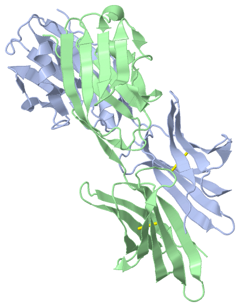
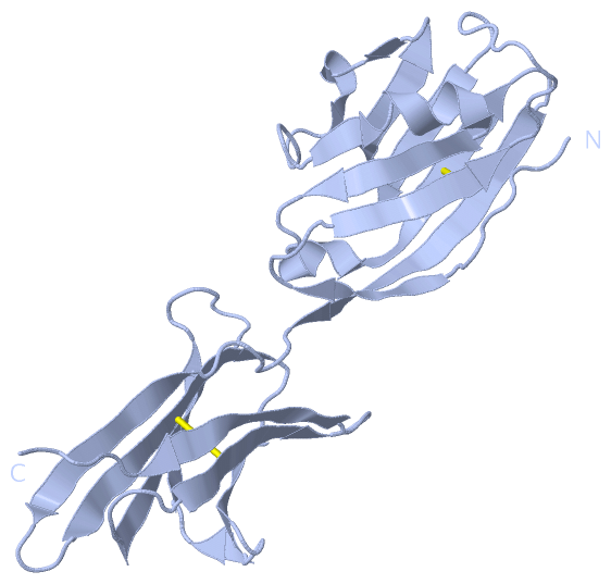
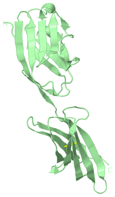
 Visualization

(-) Interactive Views

Asymmetric Unit
  Complete Structure
    Jena3D(integrated viewing of ligand, site, SAP, PROSITE, SCOP information)
    WebMol | AstexViewer[tm]@PDBe
(Java Applets, require no local installation except for Java; loading may be slow)
    STRAP
(Java WebStart application, automatic local installation, requires Java; full application with system access!)
    RasMol
(require local installation)
    Molscript (VRML)
(requires installation of a VRML viewer; select preferred view via VRML and generate a mono or stereo PDF format file)
 
  Ligands, Modified Residues, Ions
(no "Ligands, Modified Residues, Ions" information available for 3bis)
 
  Sites
(no "Sites" information available for 3bis)
 
  Cis Peptide Bonds
    Asp A:215 - Pro A:216   [ RasMol ]  
    Tyr A:160 - Pro A:161   [ RasMol ]  
    Tyr B:160 - Pro B:161   [ RasMol ]  
 
Biological Units
  Complete Structure
    Biological Unit 1  [ Jena3D ]
    Biological Unit 2  [ Jena3D ]

(-) Still Images

Jmol
  protein: cartoon or spacefill or dots and stick; nucleic acid: cartoon and stick; ligands: spacefill; active site: stick
Molscript
  protein, nucleic acid: cartoon; ligands: spacefill; active site: ball and stick

 Databases and Analysis Tools

(-) Databases

Access by PDB/NDB ID
  3bis
    Family and Domain InformationProDom | SYSTERS
    General Structural InformationGlycoscienceDB | MMDB | NDB | OCA | PDB | PDBe | PDBj | PDBsum | PDBWiki | PQS | PROTEOPEDIA
    Orientation in MembranesOPM
    Protein SurfaceSURFACE
    Secondary StructureDSSP (structure derived) | HSSP (homology derived)
    Structural GenomicsGeneCensus
    Structural NeighboursCE | VAST
    Structure ClassificationCATH | Dali | SCOP
    Validation and Original DataBMRB Data View | BMRB Restraints Grid | EDS | PROCHECK | RECOORD | WHAT_CHECK
 
Access by UniProt ID/Accession number
  PD1L1_HUMAN | Q9NZQ7
    Comparative Protein Structure ModelsModBase
    Genomic InformationEnsembl
    Protein-protein InteractionDIP
    Sequence, Family and Domain InformationInterPro | Pfam | SMART | UniProtKB/SwissProt
 
Access by Enzyme Classificator   (EC Number)
  (no 'Enzyme Classificator' available)
    General Enzyme InformationBRENDA | EC-PDB | Enzyme | IntEnz
    PathwayKEGG | MetaCyc
 
Access by Disease Identifier   (MIM ID)
  (no 'MIM ID' available)
    Disease InformationOMIM
 
Access by GenAge ID
  (no 'GenAge ID' available)
    Age Related InformationGenAge

(-) Analysis Tools

Access by PDB/NDB ID
    Domain InformationXDom
    Interatomic Contacts of Structural UnitsCSU
    Ligand-protein ContactsLPC
    Protein CavitiescastP
    Sequence and Secondary StructurePDBCartoon
    Structure AlignmentSTRAP(Java WebStart application, automatic local installation, requires Java; full application with system access!)
    Structure and Sequence BrowserSTING
 
Access by UniProt ID/Accession number
  PD1L1_HUMAN | Q9NZQ7
    Protein Disorder PredictionDisEMBL | FoldIndex | GLOBPLOT (for more information see DisProt)

 Related Entries

(-) Entries Sharing at Least One Protein Chain (UniProt ID)

UniProtKB/Swiss-Prot
        PD1L1_HUMAN | Q9NZQ73bik 3fn3 3sbw 4z18 4zqk 5c3t 5ggt 5grj 5ius 5j89 5j8o 5jdr 5jds 5n2d 5n2f

(-) Related Entries Specified in the PDB File

3bik THE SAME PROTEIN COMPLEXED WITH PD-1 (CD279)