Show PDB file:   
         Plain Text   HTML   (compressed file size)
QuickSearch:   
by PDB,NDB,UniProt,PROSITE Code or Search Term(s)  
(-)Asymmetric Unit
(-)Asym. Unit - sites
(-)Biological Unit 1
(-)Biol. Unit 1 - sites
(-)Biological Unit 2
collapse expand < >
Image Asymmetric Unit
Asymmetric Unit  (Jmol Viewer)
Image Asym. Unit - sites
Asym. Unit - sites  (Jmol Viewer)
Image Biological Unit 1
Biological Unit 1  (Jmol Viewer)
Image Biol. Unit 1 - sites
Biol. Unit 1 - sites  (Jmol Viewer)
Image Biological Unit 2
Biological Unit 2  (Jmol Viewer)

(-) Description

Title :  CRYSTAL STRUCTURE OF A PUTATIVE MULTIPLE ANTIBIOTIC-RESISTANCE REPRESSOR (SSU05_1136) FROM STREPTOCOCCUS SUIS 89/1591 AT 2.20 A RESOLUTION
 
Authors :  Joint Center For Structural Genomics (Jcsg)
Date :  14 Nov 07  (Deposition) - 04 Dec 07  (Release) - 13 Jul 11  (Revision)
Method :  X-RAY DIFFRACTION
Resolution :  2.20
Chains :  Asym. Unit :  A,B,C,D
Biol. Unit 1:  A,B  (1x)
Biol. Unit 2:  C,D  (1x)
Keywords :  Putative Multiple Antibiotic-Resistance Repressor, Structural Genomics, Joint Center For Structural Genomics, Jcsg, Protein Structure Initiative, Psi-2, Dna-Binding, Transcription Regulation, Transcription (Keyword Search: [Gene Ontology, PubMed, Web (Google))
 
Reference :  Joint Center For Structural Genomics (Jcsg)
Crystal Structure Of Putative Multiple Antibiotic-Resistanc Repressor (Marr) (Zp_00875883. 1) From Streptococcus Suis 89/1591 At 2. 20 A Resolution
To Be Published
PubMed: search

(-) Compounds

Molecule 1 - REGULATORY PROTEIN MARR
    ChainsA, B, C, D
    EngineeredYES
    Expression SystemESCHERICHIA COLI
    Expression System PlasmidSPEEDET
    Expression System StrainHK100
    Expression System Taxid562
    Expression System Vector TypePLASMID
    GeneZP_00875883.1, SSUIDRAFT_0346
    Organism ScientificSTREPTOCOCCUS SUIS
    Organism Taxid286604
    Strain89/1591

 Structural Features

(-) Chains, Units

  1234
Asymmetric Unit ABCD
Biological Unit 1 (1x)AB  
Biological Unit 2 (1x)  CD

Summary Information (see also Sequences/Alignments below)

(-) Ligands, Modified Residues, Ions  (3, 14)

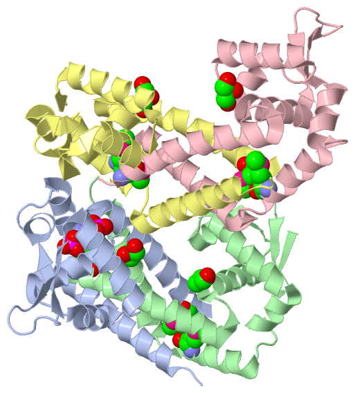
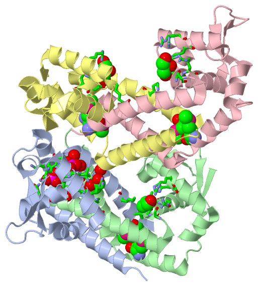
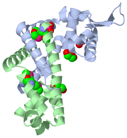
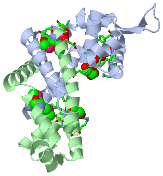
Asymmetric Unit (3, 14)
No.NameCountTypeFull Name
1GOL4Ligand/IonGLYCEROL
2MSE8Mod. Amino AcidSELENOMETHIONINE
3PO42Ligand/IonPHOSPHATE ION
Biological Unit 1 (3, 8)
No.NameCountTypeFull Name
1GOL2Ligand/IonGLYCEROL
2MSE4Mod. Amino AcidSELENOMETHIONINE
3PO42Ligand/IonPHOSPHATE ION
Biological Unit 2 (2, 6)
No.NameCountTypeFull Name
1GOL2Ligand/IonGLYCEROL
2MSE4Mod. Amino AcidSELENOMETHIONINE
3PO4-1Ligand/IonPHOSPHATE ION

(-) Sites  (6, 6)

Asymmetric Unit (6, 6)
No.NameEvidenceResiduesDescription
1AC1SOFTWAREILE A:28 , ARG A:32 , LEU A:67 , LEU A:68 , SER A:71 , TYR A:73 , HOH A:145 , HOH A:211BINDING SITE FOR RESIDUE PO4 A 142
2AC2SOFTWARELYS A:110 , MSE A:113 , ASN A:114 , GLN B:2 , GLU B:5BINDING SITE FOR RESIDUE PO4 A 143
3AC3SOFTWARESER C:29 , THR C:31 , ARG C:32 , ARG C:63 , HIS C:64 , LYS D:12BINDING SITE FOR RESIDUE GOL C 142
4AC4SOFTWARELYS A:12 , SER B:29 , LEU B:30 , THR B:31 , ARG B:32 , ARG B:63 , HIS B:64BINDING SITE FOR RESIDUE GOL B 142
5AC5SOFTWARESER A:29 , THR A:31 , ARG A:32 , ARG A:63 , HIS A:64 , HOH A:155 , LYS B:12BINDING SITE FOR RESIDUE GOL A 144
6AC6SOFTWARELYS C:12 , SER D:29 , THR D:31 , ARG D:32 , ARG D:63BINDING SITE FOR RESIDUE GOL D 142

(-) SS Bonds  (0, 0)

(no "SS Bond" information available for 3BDD)

(-) Cis Peptide Bonds  (4, 4)

Asymmetric Unit
No.Residues
1Ala A:43 -Pro A:44
2Ala B:43 -Pro B:44
3Ala C:43 -Pro C:44
4Ala D:43 -Pro D:44

 Sequence-Structure Mapping

(-) SAPs(SNPs)/Variants  (0, 0)

(no "SAP(SNP)/Variant" information available for 3BDD)

(-) PROSITE Motifs  (0, 0)

(no "PROSITE Motif" information available for 3BDD)

(-) Exons   (0, 0)

(no "Exon" information available for 3BDD)

(-) Sequences/Alignments

Asymmetric Unit
   Reformat: Number of residues per line =  ('0' or empty: single-line sequence representation)
  Number of residues per labelling interval =   
  UniProt sequence: complete  aligned part    
   Show mapping: SCOP domains CATH domains Pfam domains Secondary structure (by author)
SAPs(SNPs) PROSITE motifs Exons
(details for a mapped element are shown in a popup box when the mouse pointer rests over it)
Chain A from PDB  Type:PROTEIN  Length:140
                                                                                                                                                                            
               SCOP domains -------------------------------------------------------------------------------------------------------------------------------------------- SCOP domains
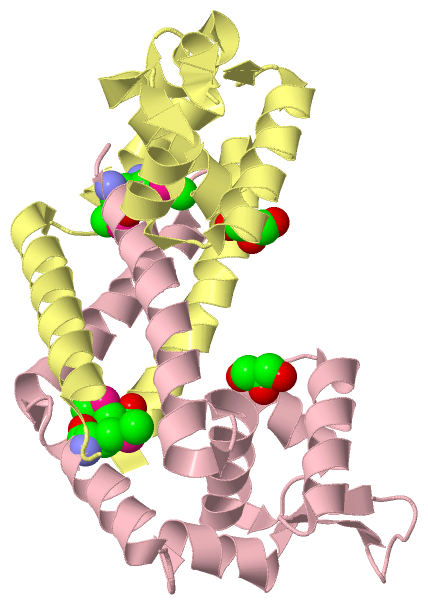
               CATH domains 3bddA00 A:2-141 'winged helix' repressor DNA binding domain                                                                                  CATH domains
               Pfam domains -------------------------------------------------------------------------------------------------------------------------------------------- Pfam domains
         Sec.struct. author hhhhhhhhhhhhhhhhhhhhhhhhhh.hhhhhhhhhhhhhhh..hhhhhhhhhh.hhhhhhhhhhhhhhh..eeeee.......eeeeehhhhhhhhh...hhhhhhhhhhhhh..hhhhhhhhhhhhhhhhhhhhh... Sec.struct. author
                 SAPs(SNPs) -------------------------------------------------------------------------------------------------------------------------------------------- SAPs(SNPs)
                    PROSITE -------------------------------------------------------------------------------------------------------------------------------------------- PROSITE
                 Transcript -------------------------------------------------------------------------------------------------------------------------------------------- Transcript
                 3bdd A   2 QEmEDLLYRLKVADETISNLFEKQLGISLTRYSILQTLLKDAPLHQLALQERLQIDRAAVTRHLKLLEESGYIIRKRNPDNQREVLVWPTEQAREALITNPSAHHQAIKTSmNQILTVEESEQFLATLDKLLIGLQNLPI 141
                              |     11        21        31        41        51        61        71        81        91       101       111 |     121       131       141
                              |                                                                                                          113-MSE                        
                              4-MSE                                                                                                                                     

Chain B from PDB  Type:PROTEIN  Length:133
                                                                                                                                                                     
               SCOP domains ------------------------------------------------------------------------------------------------------------------------------------- SCOP domains
               CATH domains 3bddB00 B:2-141 'winged helix' repressor DNA binding domain                                                                           CATH domains
               Pfam domains ------------------------------------------------------------------------------------------------------------------------------------- Pfam domains
         Sec.struct. author hhhhhhhhhhhhhhhhhhhhhhhhhh.hhhhhhhhhhhhhh..eehhhhhhhhh.hhhhhhhhhhhhhhh..eeee..eeeehhhhhhhhh...hhhhhhhhhhhhh..hhhhhhhhhhhhhhhhhhhhh... Sec.struct. author
                 SAPs(SNPs) ------------------------------------------------------------------------------------------------------------------------------------- SAPs(SNPs)
                    PROSITE ------------------------------------------------------------------------------------------------------------------------------------- PROSITE
                 Transcript ------------------------------------------------------------------------------------------------------------------------------------- Transcript
                 3bdd B   2 QEmEDLLYRLKVADETISNLFEKQLGISLTRYSILQTLLKDAPLHQLALQERLQIDRAAVTRHLKLLEESGYIIRKEVLVWPTEQAREALITNPSAHHQAIKTSmNQILTVEESEQFLATLDKLLIGLQNLPI 141
                              |     11        21        31        41        51        61        71     || 88        98       108    |  118       128       138   
                              4-MSE                                                                   77|                         113-MSE                        
                                                                                                       85                                                        

Chain C from PDB  Type:PROTEIN  Length:140
                                                                                                                                                                            
               SCOP domains -------------------------------------------------------------------------------------------------------------------------------------------- SCOP domains
               CATH domains 3bddC00 C:2-141 'winged helix' repressor DNA binding domain                                                                                  CATH domains
               Pfam domains -------------------------------------------------------------------------------------------------------------------------------------------- Pfam domains
         Sec.struct. author .hhhhhhhhhhhhhhhhhhhhhhhhh.hhhhhhhhhhhhhhh..hhhhhhhhhh.hhhhhhhhhhhhhhh..eeeee.......eeeeehhhhhhhhh...hhhhhhhhhhhh...hhhhhhhhhhhhhhhhhhhhh... Sec.struct. author
                 SAPs(SNPs) -------------------------------------------------------------------------------------------------------------------------------------------- SAPs(SNPs)
                    PROSITE -------------------------------------------------------------------------------------------------------------------------------------------- PROSITE
                 Transcript -------------------------------------------------------------------------------------------------------------------------------------------- Transcript
                 3bdd C   2 QEmEDLLYRLKVADETISNLFEKQLGISLTRYSILQTLLKDAPLHQLALQERLQIDRAAVTRHLKLLEESGYIIRKRNPDNQREVLVWPTEQAREALITNPSAHHQAIKTSmNQILTVEESEQFLATLDKLLIGLQNLPI 141
                              |     11        21        31        41        51        61        71        81        91       101       111 |     121       131       141
                              4-MSE                                                                                                      113-MSE                        

Chain D from PDB  Type:PROTEIN  Length:132
                                                                                                                                                                    
               SCOP domains ------------------------------------------------------------------------------------------------------------------------------------ SCOP domains
               CATH domains 3bddD00 D:2-141 'winged helix' repressor DNA binding domain                                                                          CATH domains
               Pfam domains ------------------------------------------------------------------------------------------------------------------------------------ Pfam domains
         Sec.struct. author hhhhhhhhhhhhhhhhhhhhhhhhhh.hhhhhhhhhhhhhhh.eehhhhhhhhh.hhhhhhhhhhhhhhh..eee..eeeehhhhhhhhh...hhhhhhhhhhhhh..hhhhhhhhhhhhhhhhhhhhh... Sec.struct. author
                 SAPs(SNPs) ------------------------------------------------------------------------------------------------------------------------------------ SAPs(SNPs)
                    PROSITE ------------------------------------------------------------------------------------------------------------------------------------ PROSITE
                 Transcript ------------------------------------------------------------------------------------------------------------------------------------ Transcript
                 3bdd D   2 QEmEDLLYRLKVADETISNLFEKQLGISLTRYSILQTLLKDAPLHQLALQERLQIDRAAVTRHLKLLEESGYIIRKVLVWPTEQAREALITNPSAHHQAIKTSmNQILTVEESEQFLATLDKLLIGLQNLPI 141
                              |     11        21        31        41        51        61        71     || 89        99       109   |   119       129       139  
                              4-MSE                                                                   77|                        113-MSE                        
                                                                                                       86                                                       

   Legend:   → Mismatch (orange background)
  - → Gap (green background, '-', border residues have a numbering label)
    → Modified Residue (blue background, lower-case, 'x' indicates undefined single-letter code, labelled with number + name)
  x → Chemical Group (purple background, 'x', labelled with number + name, e.g. ACE or NH2)
  extra numbering lines below/above indicate numbering irregularities and modified residue names etc., number ends below/above '|'

 Classification and Annotation

(-) SCOP Domains  (0, 0)

(no "SCOP Domain" information available for 3BDD)

(-) CATH Domains  (1, 4)

Asymmetric Unit

(-) Pfam Domains  (0, 0)

(no "Pfam Domain" information available for 3BDD)

(-) Gene Ontology  (0, 0)

Asymmetric Unit(hide GO term definitions)
    (no "Gene Ontology" information available for 3BDD)

 Visualization

(-) Interactive Views

Asymmetric Unit
  Complete Structure
    Jena3D(integrated viewing of ligand, site, SAP, PROSITE, SCOP information)
    WebMol | AstexViewer[tm]@PDBe
(Java Applets, require no local installation except for Java; loading may be slow)
    STRAP
(Java WebStart application, automatic local installation, requires Java; full application with system access!)
    RasMol
(require local installation)
    Molscript (VRML)
(requires installation of a VRML viewer; select preferred view via VRML and generate a mono or stereo PDF format file)
 
  Ligands, Modified Residues, Ions
    GOL  [ RasMol | Jena3D ]  +environment [ RasMol | Jena3D ]
    MSE  [ RasMol | Jena3D ]  +environment [ RasMol | Jena3D ]
    PO4  [ RasMol | Jena3D ]  +environment [ RasMol | Jena3D ]
 
  Sites
    AC1  [ RasMol ]  +environment [ RasMol ]
    AC2  [ RasMol ]  +environment [ RasMol ]
    AC3  [ RasMol ]  +environment [ RasMol ]
    AC4  [ RasMol ]  +environment [ RasMol ]
    AC5  [ RasMol ]  +environment [ RasMol ]
    AC6  [ RasMol ]  +environment [ RasMol ]
 
  Cis Peptide Bonds
    Ala A:43 - Pro A:44   [ RasMol ]  
    Ala B:43 - Pro B:44   [ RasMol ]  
    Ala C:43 - Pro C:44   [ RasMol ]  
    Ala D:43 - Pro D:44   [ RasMol ]  
 
Biological Units
  Complete Structure
    Biological Unit 1  [ Jena3D ]
    Biological Unit 2  [ Jena3D ]

(-) Still Images

Jmol
  protein: cartoon or spacefill or dots and stick; nucleic acid: cartoon and stick; ligands: spacefill; active site: stick
Molscript
  protein, nucleic acid: cartoon; ligands: spacefill; active site: ball and stick

 Databases and Analysis Tools

(-) Databases

Access by PDB/NDB ID
  3bdd
    Family and Domain InformationProDom | SYSTERS
    General Structural InformationGlycoscienceDB | MMDB | NDB | OCA | PDB | PDBe | PDBj | PDBsum | PDBWiki | PQS | PROTEOPEDIA
    Orientation in MembranesOPM
    Protein SurfaceSURFACE
    Secondary StructureDSSP (structure derived) | HSSP (homology derived)
    Structural GenomicsGeneCensus
    Structural NeighboursCE | VAST
    Structure ClassificationCATH | Dali | SCOP
    Validation and Original DataBMRB Data View | BMRB Restraints Grid | EDS | PROCHECK | RECOORD | WHAT_CHECK
 
Access by UniProt ID/Accession number
  (no 'UniProt ID/Accession number' available) |
    Comparative Protein Structure ModelsModBase
    Genomic InformationEnsembl
    Protein-protein InteractionDIP
    Sequence, Family and Domain InformationInterPro | Pfam | SMART | UniProt
 
Access by Enzyme Classificator   (EC Number)
  (no 'Enzyme Classificator' available)
    General Enzyme InformationBRENDA | EC-PDB | Enzyme | IntEnz
    PathwayKEGG | MetaCyc
 
Access by Disease Identifier   (MIM ID)
  (no 'MIM ID' available)
    Disease InformationOMIM
 
Access by GenAge ID
  (no 'GenAge ID' available)
    Age Related InformationGenAge

(-) Analysis Tools

Access by PDB/NDB ID
    Domain InformationXDom
    Interatomic Contacts of Structural UnitsCSU
    Ligand-protein ContactsLPC
    Protein CavitiescastP
    Sequence and Secondary StructurePDBCartoon
    Structure AlignmentSTRAP(Java WebStart application, automatic local installation, requires Java; full application with system access!)
    Structure and Sequence BrowserSTING
 
Access by UniProt ID/Accession number
  (no 'UniProt ID/Accession number' available) |
    Protein Disorder PredictionDisEMBL | FoldIndex | GLOBPLOT

 Related Entries

(-) Entries Sharing at Least One Protein Chain (UniProt ID)

(no "Entries Sharing at Least One Protein Chain" available for 3BDD)

(-) Related Entries Specified in the PDB File

(no "Related Entries Specified in the PDB File" available for 3BDD)