|
|
|
|
 Description
|
|
Compounds
|
||||||||||||||||||||||||||||||||||||||||||||||||||||||||
Chains, Units
Summary Information (see also Sequences/Alignments below) |
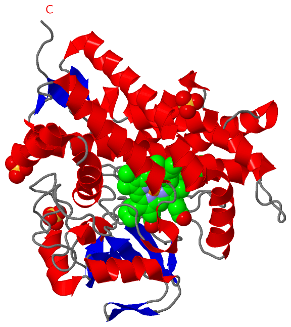
Ligands, Modified Residues, Ions (3, 5)| Asymmetric/Biological Unit (3, 5) |
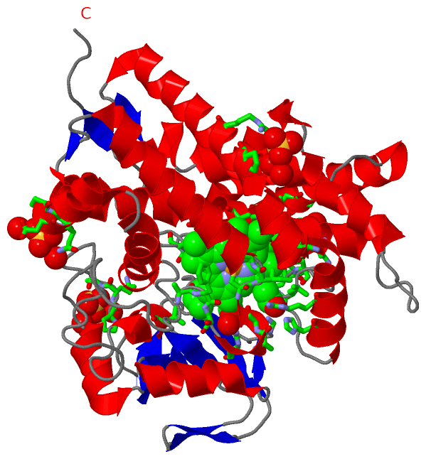
Sites (5, 5)
Asymmetric Unit (5, 5)
|
SS Bonds (0, 0)| (no "SS Bond" information available for 3ABA) |
Cis Peptide Bonds (2, 2)
Asymmetric/Biological Unit
|
||||||||||||
SAPs(SNPs)/Variants (0, 0)| (no "SAP(SNP)/Variant" information available for 3ABA) |
PROSITE Motifs (0, 0)| (no "PROSITE Motif" information available for 3ABA) |
Exons (0, 0)| (no "Exon" information available for 3ABA) |
Sequences/Alignments
Asymmetric/Biological UnitChain A from PDB Type:PROTEIN Length:397 aligned with Q93H81_STRAX | Q93H81 from UniProtKB/TrEMBL Length:399 Alignment length:397 399 16 26 36 46 56 66 76 86 96 106 116 126 136 146 156 166 176 186 196 206 216 226 236 246 256 266 276 286 296 306 316 326 336 346 356 366 376 386 396 | Q93H81_STRAX 7 DAPTVPKARSCPFLPPDGIADIRAAAPVTRATFTSGHEAWLVTGYEEVRALLRDSSFSVQVPHALHTQDGVVTQKPGRGSLLWQDEPEHTSDRKLLAKEFTVRRMQALRPNIQRIVDEHLDAIEARGGPVDLVKTFANAVPSMVISDLFGVPVERRAEFQDIAEAMMRVDQDAAATEAAGMRLGGLLYQLVQERRANPGDDLISALITTEDPDGVVDDMFLMNAAGTLLIAAHDTTACMIGLGTALLLDSPDQLALLREDPSLVGNAVEELLRYLTIGQFGGERVATRDVELGGVRIAKGEQVVAHVLAADFDPAFVEEPERFDITRRPAPHLAFGFGAHQCIGQQLARIELQIVFETLFRRLPGLRLAKPVEELRFRHDMVFYGVHELPVTW---- - SCOP domains d3abaa_ A: automated matches SCOP domains CATH domains 3abaA00 A:7-403 Cytochrome p450 CATH domains Pfam domains ------------------------------------------------------------------------------------------------------------------------------------------------------------------------------------------------------------------------------------------------------------------------------------------------------------------------------------------------------------------------------------------------------------- Pfam domains SAPs(SNPs) ------------------------------------------------------------------------------------------------------------------------------------------------------------------------------------------------------------------------------------------------------------------------------------------------------------------------------------------------------------------------------------------------------------- SAPs(SNPs) PROSITE ------------------------------------------------------------------------------------------------------------------------------------------------------------------------------------------------------------------------------------------------------------------------------------------------------------------------------------------------------------------------------------------------------------- PROSITE Transcript ------------------------------------------------------------------------------------------------------------------------------------------------------------------------------------------------------------------------------------------------------------------------------------------------------------------------------------------------------------------------------------------------------------- Transcript 3aba A 7 DAPTVPKARSCPFLPPDGIADIRAAAPVTRATFTSGHEAWLVTGYEEVRALLRDSSFSVQVPHALHTQDGVVTQKPGRGSLLWQDEPEHTSDRKLLAKEFTVRRMQALRPNIQRIVDEHLDAIEARGGPVDLVKTFANAVPSMVISDLFGVPVERRAEFQDIAEAMMRVDQDAAATEAAGMRLGGLLYQLVQERRANPGDDLISALITTEDPDGVVDDMFLMNAAGTLLIAAHDTTACMIGLGTALLLDSPDQLALLREDPSLVGNAVEELLRYLTIGQFGGERVATRDVELGGVRIAKGEQVVAHVLAADFDPAFVEEPERFDITRRPAPHLAFGFGAHQCIGQQLARIELQIVFETLFRRLPGLRLAKPVEELRFRHDMVFYGVHELPVTWHHHH 403 16 26 36 46 56 66 76 86 96 106 116 126 136 146 156 166 176 186 196 206 216 226 236 246 256 266 276 286 296 306 316 326 336 346 356 366 376 386 396
|
||||||||||||||||||||
SCOP Domains (1, 1)
Asymmetric/Biological Unit
 |
CATH Domains (1, 1)
Asymmetric/Biological Unit
|
Pfam Domains (0, 0)| (no "Pfam Domain" information available for 3ABA) |
Gene Ontology (7, 7)|
Asymmetric/Biological Unit(hide GO term definitions) Chain A (Q93H81_STRAX | Q93H81)
|
||||||||||||||||||||||||||||||||||||||||||||||||||||||
Interactive Views
|
||||||||||||||||||||||||||||||||||||||||||||||||||||||||||||||||||||||||||||||||||||||||||||||||||||||||||||||||||||||||||||||||||||||||||||||||||||||||||||||||||||||||
Still Images
|
||||||||||||||||
Databases
|
||||||||||||||||||||||||||||||||||||||||||||||||||||||||||||||||||||||||||||||||||||||||||||||||||||||||||||||||||||||||||||||||||||||||||||||||||||||||||||||||
Analysis Tools
|
|||||||||||||||||||||||||||||||||||||||||||||||||||||||||||||
Entries Sharing at Least One Protein Chain (UniProt ID)
Related Entries Specified in the PDB File
|
|