| molecular function |
|---|
| | GO:0003677 | | DNA binding | | Any molecular function by which a gene product interacts selectively and non-covalently with DNA (deoxyribonucleic acid). |
| | GO:0003682 | | chromatin binding | | Interacting selectively and non-covalently with chromatin, the network of fibers of DNA, protein, and sometimes RNA, that make up the chromosomes of the eukaryotic nucleus during interphase. |
| | GO:0005515 | | protein binding | | Interacting selectively and non-covalently with any protein or protein complex (a complex of two or more proteins that may include other nonprotein molecules). |
| | GO:0003697 | | single-stranded DNA binding | | Interacting selectively and non-covalently with single-stranded DNA. |
| biological process |
|---|
| | GO:0006260 | | DNA replication | | The cellular metabolic process in which a cell duplicates one or more molecules of DNA. DNA replication begins when specific sequences, known as origins of replication, are recognized and bound by initiation proteins, and ends when the original DNA molecule has been completely duplicated and the copies topologically separated. The unit of replication usually corresponds to the genome of the cell, an organelle, or a virus. The template for replication can either be an existing DNA molecule or RNA. |
| | GO:0070584 | | mitochondrion morphogenesis | | The process in which the anatomical structures of a mitochondrion are generated and organized. |
| | GO:0007005 | | mitochondrion organization | | A process that is carried out at the cellular level which results in the assembly, arrangement of constituent parts, or disassembly of a mitochondrion; includes mitochondrial morphogenesis and distribution, and replication of the mitochondrial genome as well as synthesis of new mitochondrial components. |
| | GO:0051096 | | positive regulation of helicase activity | | Any process that activates or increases the activity of a helicase. |
| cellular component |
|---|
| | GO:0070062 | | extracellular exosome | | A vesicle that is released into the extracellular region by fusion of the limiting endosomal membrane of a multivesicular body with the plasma membrane. Extracellular exosomes, also simply called exosomes, have a diameter of about 40-100 nm. |
| | GO:0005759 | | mitochondrial matrix | | The gel-like material, with considerable fine structure, that lies in the matrix space, or lumen, of a mitochondrion. It contains the enzymes of the tricarboxylic acid cycle and, in some organisms, the enzymes concerned with fatty acid oxidation. |
| | GO:0042645 | | mitochondrial nucleoid | | The region of a mitochondrion to which the DNA is confined. |
| | GO:0005739 | | mitochondrion | | A semiautonomous, self replicating organelle that occurs in varying numbers, shapes, and sizes in the cytoplasm of virtually all eukaryotic cells. It is notably the site of tissue respiration. |
| | GO:0005634 | | nucleus | | A membrane-bounded organelle of eukaryotic cells in which chromosomes are housed and replicated. In most cells, the nucleus contains all of the cell's chromosomes except the organellar chromosomes, and is the site of RNA synthesis and processing. In some species, or in specialized cell types, RNA metabolism or DNA replication may be absent. |
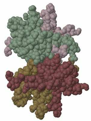
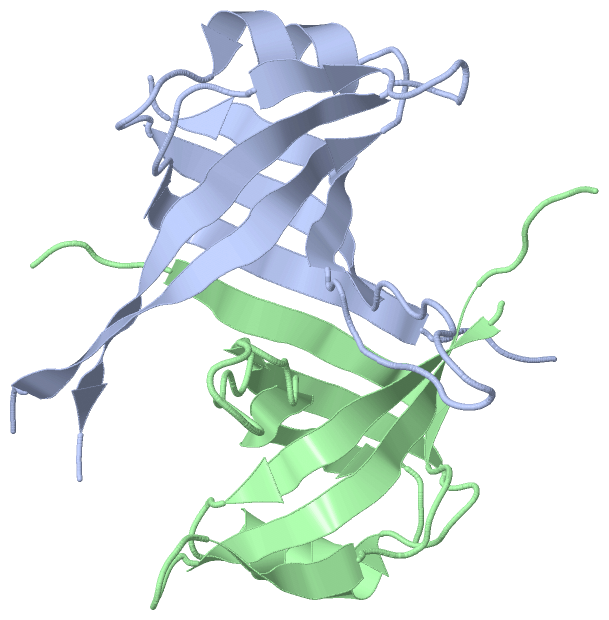
 Description
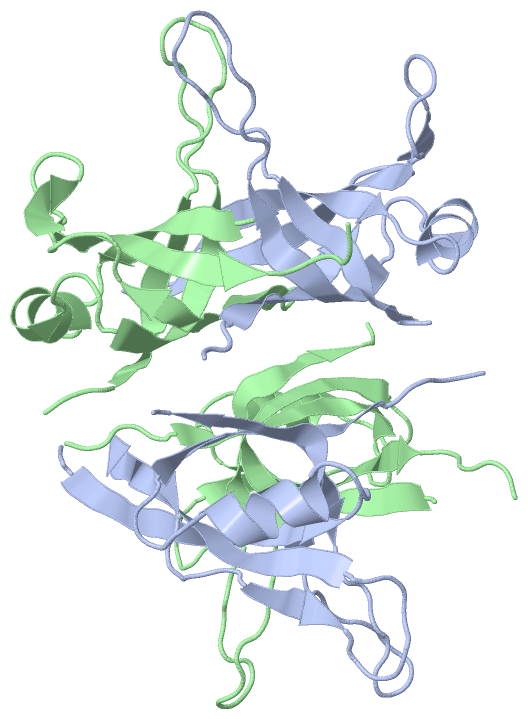