|
|
|
|
 Description
|
|
Compounds
|
||||||||||||||||||||||||||||||||||||||||
Chains, Units
Summary Information (see also Sequences/Alignments below) |
Ligands, Modified Residues, Ions (4, 11)| Asymmetric/Biological Unit (4, 11) |
Sites (9, 9)
Asymmetric Unit (9, 9)
|
SS Bonds (2, 2)
Asymmetric/Biological Unit
|
||||||||||||
Cis Peptide Bonds (0, 0)| (no "Cis Peptide Bond" information available for 3T0X) |
SAPs(SNPs)/Variants (0, 0)| (no "SAP(SNP)/Variant" information available for 3T0X) |
PROSITE Motifs (0, 0)| (no "PROSITE Motif" information available for 3T0X) |
Exons (0, 0)| (no "Exon" information available for 3T0X) |
Sequences/Alignments
Asymmetric/Biological Unit
Chain A from PDB Type:PROTEIN Length:111
SCOP domains d3t0xa_ A: automated matches SCOP domains
CATH domains --------------------------------------------------------------------------------------------------------------- CATH domains
Pfam domains --------------------------------------------------------------------------------------------------------------- Pfam domains
SAPs(SNPs) --------------------------------------------------------------------------------------------------------------- SAPs(SNPs)
PROSITE --------------------------------------------------------------------------------------------------------------- PROSITE
Transcript --------------------------------------------------------------------------------------------------------------- Transcript
3t0x A 1 xPVLTQSPSVSGTPGQKVTIFCSGSSSNVEDNSVYWYQQFPGTTPKVLIYNDDRRPSGVPDRFSGSKSGTSASLAISGLRSEDEADYYCLSWDDSLNGWVFGGGTKVTVLD 107
| 11 21 ||29 39 49 59 69 79 89 ||97 106A
| 9| 27A| 95A| 106A
1-PCA 11 27B 95B
Chain B from PDB Type:PROTEIN Length:109
SCOP domains d3t0xb_ B: automated matches SCOP domains
CATH domains ------------------------------------------------------------------------------------------------------------- CATH domains
Pfam domains ------------------------------------------------------------------------------------------------------------- Pfam domains
SAPs(SNPs) ------------------------------------------------------------------------------------------------------------- SAPs(SNPs)
PROSITE ------------------------------------------------------------------------------------------------------------- PROSITE
Transcript ------------------------------------------------------------------------------------------------------------- Transcript
3t0x B 1 xPVLTQSPSVSGTPGQKVTIFCSGSSSNVEDNSVYWYQQFPGTTPKVLIYNDDRRPSGVPDRFSGSKSGTSASLAISGLRSEDEADYYCLSWDDSLNGWVFGGGTKVTV 106
| 11 21 ||29 39 49 59 69 79 89 ||97
| 9| 27A| 95A|
1-PCA 11 27B 95B
|
||||||||||||||||||||
SCOP Domains (1, 2)
Asymmetric/Biological Unit
 |
CATH Domains (0, 0)| (no "CATH Domain" information available for 3T0X) |
Pfam Domains (0, 0)| (no "Pfam Domain" information available for 3T0X) |
Gene Ontology (0, 0)|
Asymmetric/Biological Unit(hide GO term definitions)
(no "Gene Ontology" information available for 3T0X)
|
Interactive Views
|
|||||||||||||||||||||||||||||||||||||||||||||||||||||||||||||||||||||||||||||||||||||||||||||||||||||||||||||||||||||||||||||||||||||||||||||||||||||||||||||||||||||||||||||||||||||||||||||||||||
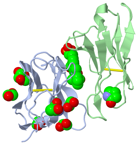
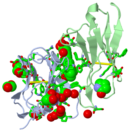
Still Images
|
||||||||||||||||
Databases
|
||||||||||||||||||||||||||||||||||||||||||||||||||||||||||||||||||||||||||||||||||||||||||||||||||||||||||||||||||||||||||||||||||||||||||||||||||||||||||||||||
Analysis Tools
|
|||||||||||||||||||||||||||||||||||||||||||||||||||||||||||||
Entries Sharing at Least One Protein Chain (UniProt ID)
Related Entries Specified in the PDB File
|
|