Show PDB file:   
         Plain Text   HTML   (compressed file size)
QuickSearch:   
by PDB,NDB,UniProt,PROSITE Code or Search Term(s)  
(-)Asymmetric Unit
(-)Asym. Unit - sites
(-)Biological Unit 1
(-)Biol. Unit 1 - sites
(-)Biological Unit 2
collapse expand < >
Image Asymmetric Unit
Asymmetric Unit  (Jmol Viewer)
Image Asym. Unit - sites
Asym. Unit - sites  (Jmol Viewer)
Image Biological Unit 1
Biological Unit 1  (Jmol Viewer)
Image Biol. Unit 1 - sites
Biol. Unit 1 - sites  (Jmol Viewer)
Image Biological Unit 2
Biological Unit 2  (Jmol Viewer)

(-) Description

Title :  CRYSTAL STRUCTURE OF THE CATALYTIC DOMAIN OF PLASMODIUM FALCIPARUM ARF GTPASE ACTIVATING PROTEIN
 
Authors :  W. J. Cook, D. Chattopadhyay
Date :  11 Jul 11  (Deposition) - 09 Nov 11  (Release) - 28 Mar 12  (Revision)
Method :  X-RAY DIFFRACTION
Resolution :  2.40
Chains :  Asym. Unit :  A,B
Biol. Unit 1:  A  (1x)
Biol. Unit 2:  B  (1x)
Keywords :  Gtpase-Activating Protein, Protein Trafficking, Hydrolase Activator (Keyword Search: [Gene Ontology, PubMed, Web (Google))
 
Reference :  W. J. Cook, O. Senkovich, D. Chattopadhyay
Structure Of The Catalytic Domain Of Plasmodium Falciparum Arf Gtpase-Activating Protein (Arfgap).
Acta Crystallogr. , Sect. F V. 67 1339 2011
PubMed-ID: 22102228  |  Reference-DOI: 10.1107/S1744309111032507

(-) Compounds

Molecule 1 - ADP-RIBOSYLATION FACTOR GTPASE-ACTIVATING PROTEIN
    ChainsA, B
    EngineeredYES
    Expression SystemESCHERICHIA COLI
    Expression System PlasmidPCR2.1
    Expression System Taxid562
    Expression System Vector TypePLASMID
    FragmentUNP RESIDUES 1-160
    GenePFL2140C
    Organism ScientificPLASMODIUM FALCIPARUM 3D7
    Organism Taxid36329

 Structural Features

(-) Chains, Units

  12
Asymmetric Unit AB
Biological Unit 1 (1x)A 
Biological Unit 2 (1x) B

Summary Information (see also Sequences/Alignments below)
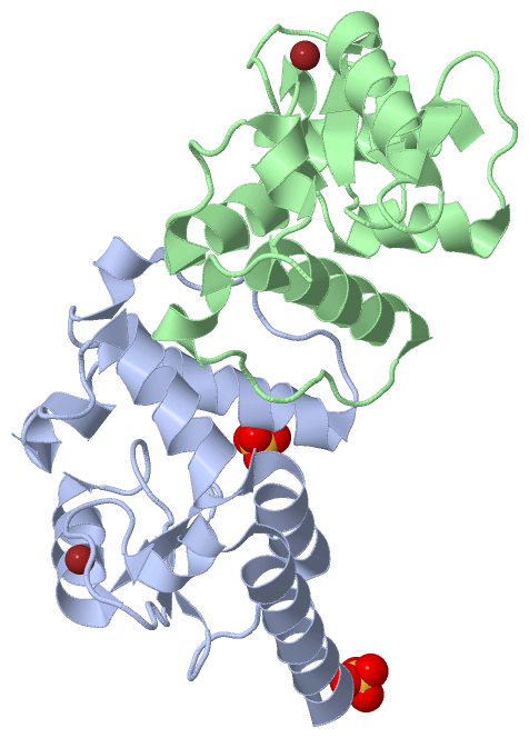
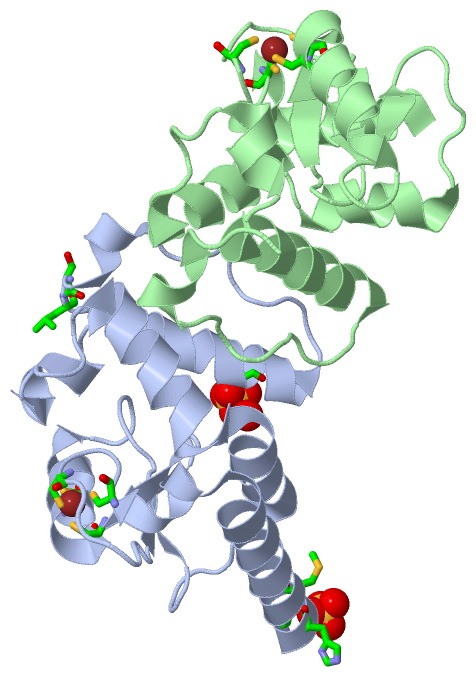
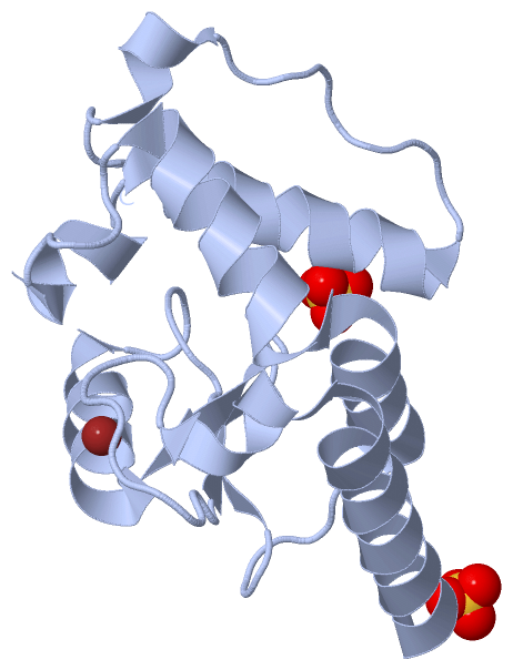
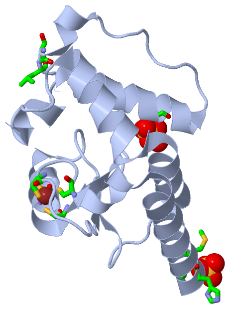
(-) Ligands, Modified Residues, Ions  (2, 4)

Asymmetric Unit (2, 4)
No.NameCountTypeFull Name
1SO42Ligand/IonSULFATE ION
2ZN2Ligand/IonZINC ION
Biological Unit 1 (1, 2)
No.NameCountTypeFull Name
1SO42Ligand/IonSULFATE ION
2ZN-1Ligand/IonZINC ION
Biological Unit 2 (0, 0)
No.NameCountTypeFull Name
1SO4-1Ligand/IonSULFATE ION
2ZN-1Ligand/IonZINC ION

(-) Sites  (4, 4)

Asymmetric Unit (4, 4)
No.NameEvidenceResiduesDescription
1AC1SOFTWARECYS A:22 , CYS A:25 , CYS A:42 , CYS A:45BINDING SITE FOR RESIDUE ZN A 161
2AC2SOFTWAREGLY A:-2 , HIS A:0 , MET A:1 , SER A:133 , LEU A:134BINDING SITE FOR RESIDUE SO4 A 162
3AC3SOFTWAREARG A:114 , HOH A:319BINDING SITE FOR RESIDUE SO4 A 163
4AC4SOFTWARECYS B:22 , CYS B:25 , CYS B:42 , CYS B:45BINDING SITE FOR RESIDUE ZN B 161

(-) SS Bonds  (0, 0)

(no "SS Bond" information available for 3SUB)

(-) Cis Peptide Bonds  (0, 0)

(no "Cis Peptide Bond" information available for 3SUB)

 Sequence-Structure Mapping

(-) SAPs(SNPs)/Variants  (0, 0)

(no "SAP(SNP)/Variant" information available for 3SUB)

(-) PROSITE Motifs  (0, 0)

(no "PROSITE Motif" information available for 3SUB)

(-) Exons   (0, 0)

(no "Exon" information available for 3SUB)

(-) Sequences/Alignments

Asymmetric Unit
   Reformat: Number of residues per line =  ('0' or empty: single-line sequence representation)
  Number of residues per labelling interval =   
  UniProt sequence: complete  aligned part    
   Show mapping: SCOP domains CATH domains Pfam domains Secondary structure (by author)
SAPs(SNPs) PROSITE motifs Exons
(details for a mapped element are shown in a popup box when the mouse pointer rests over it)
Chain A from PDB  Type:PROTEIN  Length:139
 aligned with Q8I4Y5_PLAF7 | Q8I4Y5 from UniProtKB/TrEMBL  Length:332

    Alignment length:139
                               1                                                                                                                                       
                               |     7        17        27        37        47        57        67        77        87        97       107       117       127         
         Q8I4Y5_PLAF7     - ---MNAAAVELINRLKKEDESNNKCFDCGISNPDWVSVNHGIFLCINCSGVHRSLGVHISIVRSIKMDIFTDEQLKYIDKGGNKKCQTYLENYGISDFIPERKYRTKAADHYRKILRSIVHNVDPPAPLPLDEGKSLIN 136
               SCOP domains ------------------------------------------------------------------------------------------------------------------------------------------- SCOP domains
               CATH domains ------------------------------------------------------------------------------------------------------------------------------------------- CATH domains
               Pfam domains ------------------------------------------------------------------------------------------------------------------------------------------- Pfam domains
         Sec.struct. author hhhhhhhhhhhhhhhhhhhhhhhh..........eee....eeehhhhhhhhhhh......eee......hhhhhhhhhhhhhhhhhhhhhhh.....hhhhhh.hhhhhhhhhhhhhhhh........hhhhh..... Sec.struct. author
                 SAPs(SNPs) ------------------------------------------------------------------------------------------------------------------------------------------- SAPs(SNPs)
                    PROSITE ------------------------------------------------------------------------------------------------------------------------------------------- PROSITE
                 Transcript ------------------------------------------------------------------------------------------------------------------------------------------- Transcript
                 3sub A  -2 GSHMNAAAVEFINRLKKEDESNNKCFDCGISNPDWVSVNHGIFLCINCSGVHRSLGVHISIVRSIKMDIFTDEQLKYIDKGGNKKCQTYLENYGISDFIPERKYRTKAADHYRKILRSIVHNVDPPAPLPLDEGKSLIN 136
                                     7        17        27        37        47        57        67        77        87        97       107       117       127         

Chain B from PDB  Type:PROTEIN  Length:131
 aligned with Q8I4Y5_PLAF7 | Q8I4Y5 from UniProtKB/TrEMBL  Length:332

    Alignment length:131
                                    14        24        34        44        54        64        74        84        94       104       114       124       134 
         Q8I4Y5_PLAF7     5 AVELINRLKKEDESNNKCFDCGISNPDWVSVNHGIFLCINCSGVHRSLGVHISIVRSIKMDIFTDEQLKYIDKGGNKKCQTYLENYGISDFIPERKYRTKAADHYRKILRSIVHNVDPPAPLPLDEGKSLI 135
               SCOP domains ----------------------------------------------------------------------------------------------------------------------------------- SCOP domains
               CATH domains ----------------------------------------------------------------------------------------------------------------------------------- CATH domains
               Pfam domains ----------------------------------------------------------------------------------------------------------------------------------- Pfam domains
         Sec.struct. author hhhhhhhhhhhhhhhhh..........eee....eeehhhhhhhhhhhh.....eee......hhhhhhhhhh.hhhhhhhhhhhh.....hhhhhh.hhhhhhhhhhhhhhhh........hhhhh.... Sec.struct. author
                 SAPs(SNPs) ----------------------------------------------------------------------------------------------------------------------------------- SAPs(SNPs)
                    PROSITE ----------------------------------------------------------------------------------------------------------------------------------- PROSITE
                 Transcript ----------------------------------------------------------------------------------------------------------------------------------- Transcript
                 3sub B   5 AVEFINRLKKEDESNNKCFDCGISNPDWVSVNHGIFLCINCSGVHRSLGVHISIVRSIKMDIFTDEQLKYIDKGGNKKCQTYLENYGISDFIPERKYRTKAADHYRKILRSIVHNVDPPAPLPLDEGKSLI 135
                                    14        24        34        44        54        64        74        84        94       104       114       124       134 

   Legend:   → Mismatch (orange background)
  - → Gap (green background, '-', border residues have a numbering label)
    → Modified Residue (blue background, lower-case, 'x' indicates undefined single-letter code, labelled with number + name)
  x → Chemical Group (purple background, 'x', labelled with number + name, e.g. ACE or NH2)
  extra numbering lines below/above indicate numbering irregularities and modified residue names etc., number ends below/above '|'

 Classification and Annotation

(-) SCOP Domains  (0, 0)

(no "SCOP Domain" information available for 3SUB)

(-) CATH Domains  (0, 0)

(no "CATH Domain" information available for 3SUB)

(-) Pfam Domains  (0, 0)

(no "Pfam Domain" information available for 3SUB)

(-) Gene Ontology  (4, 4)

Asymmetric Unit(hide GO term definitions)
Chain A,B   (Q8I4Y5_PLAF7 | Q8I4Y5)
molecular function
    GO:0003677    DNA binding    Any molecular function by which a gene product interacts selectively and non-covalently with DNA (deoxyribonucleic acid).
    GO:0005096    GTPase activator activity    Binds to and increases the activity of a GTPase, an enzyme that catalyzes the hydrolysis of GTP.
    GO:0046872    metal ion binding    Interacting selectively and non-covalently with any metal ion.
biological process
    GO:0043547    positive regulation of GTPase activity    Any process that activates or increases the activity of a GTPase.

 Visualization

(-) Interactive Views

Asymmetric Unit
  Complete Structure
    Jena3D(integrated viewing of ligand, site, SAP, PROSITE, SCOP information)
    WebMol | AstexViewer[tm]@PDBe
(Java Applets, require no local installation except for Java; loading may be slow)
    STRAP
(Java WebStart application, automatic local installation, requires Java; full application with system access!)
    RasMol
(require local installation)
    Molscript (VRML)
(requires installation of a VRML viewer; select preferred view via VRML and generate a mono or stereo PDF format file)
 
  Ligands, Modified Residues, Ions
    SO4  [ RasMol | Jena3D ]  +environment [ RasMol | Jena3D ]
    ZN  [ RasMol | Jena3D ]  +environment [ RasMol | Jena3D ]
 
  Sites
    AC1  [ RasMol ]  +environment [ RasMol ]
    AC2  [ RasMol ]  +environment [ RasMol ]
    AC3  [ RasMol ]  +environment [ RasMol ]
    AC4  [ RasMol ]  +environment [ RasMol ]
 
  Cis Peptide Bonds
(no "Cis Peptide Bonds" information available for 3sub)
 
Biological Units
  Complete Structure
    Biological Unit 1  [ Jena3D ]
    Biological Unit 2  [ Jena3D ]

(-) Still Images

Jmol
  protein: cartoon or spacefill or dots and stick; nucleic acid: cartoon and stick; ligands: spacefill; active site: stick
Molscript
  protein, nucleic acid: cartoon; ligands: spacefill; active site: ball and stick

 Databases and Analysis Tools

(-) Databases

Access by PDB/NDB ID
  3sub
    Family and Domain InformationProDom | SYSTERS
    General Structural InformationGlycoscienceDB | MMDB | NDB | OCA | PDB | PDBe | PDBj | PDBsum | PDBWiki | PQS | PROTEOPEDIA
    Orientation in MembranesOPM
    Protein SurfaceSURFACE
    Secondary StructureDSSP (structure derived) | HSSP (homology derived)
    Structural GenomicsGeneCensus
    Structural NeighboursCE | VAST
    Structure ClassificationCATH | Dali | SCOP
    Validation and Original DataBMRB Data View | BMRB Restraints Grid | EDS | PROCHECK | RECOORD | WHAT_CHECK
 
Access by UniProt ID/Accession number
  Q8I4Y5_PLAF7 | Q8I4Y5
    Comparative Protein Structure ModelsModBase
    Genomic InformationEnsembl
    Protein-protein InteractionDIP
    Sequence, Family and Domain InformationInterPro | Pfam | SMART | UniProtKB/TrEMBL
 
Access by Enzyme Classificator   (EC Number)
  (no 'Enzyme Classificator' available)
    General Enzyme InformationBRENDA | EC-PDB | Enzyme | IntEnz
    PathwayKEGG | MetaCyc
 
Access by Disease Identifier   (MIM ID)
  (no 'MIM ID' available)
    Disease InformationOMIM
 
Access by GenAge ID
  (no 'GenAge ID' available)
    Age Related InformationGenAge

(-) Analysis Tools

Access by PDB/NDB ID
    Domain InformationXDom
    Interatomic Contacts of Structural UnitsCSU
    Ligand-protein ContactsLPC
    Protein CavitiescastP
    Sequence and Secondary StructurePDBCartoon
    Structure AlignmentSTRAP(Java WebStart application, automatic local installation, requires Java; full application with system access!)
    Structure and Sequence BrowserSTING
 
Access by UniProt ID/Accession number
  Q8I4Y5_PLAF7 | Q8I4Y5
    Protein Disorder PredictionDisEMBL | FoldIndex | GLOBPLOT (for more information see DisProt)

 Related Entries

(-) Entries Sharing at Least One Protein Chain (UniProt ID)

(no "Entries Sharing at Least One Protein Chain" available for 3SUB)

(-) Related Entries Specified in the PDB File

(no "Related Entries Specified in the PDB File" available for 3SUB)