|
|
|
|
 Description
|
|
Compounds
|
||||||||||||||||||||||||||||||||||||||||||||||||||||||||||||||||||||||||||||||
Chains, Units
Summary Information (see also Sequences/Alignments below) |
Ligands, Modified Residues, Ions (0, 0)| (no "Ligand,Modified Residues,Ions" information available for 3SJ9) |
Sites (0, 0)| (no "Site" information available for 3SJ9) |
SS Bonds (0, 0)| (no "SS Bond" information available for 3SJ9) |
Cis Peptide Bonds (0, 0)| (no "Cis Peptide Bond" information available for 3SJ9) |
SAPs(SNPs)/Variants (0, 0)| (no "SAP(SNP)/Variant" information available for 3SJ9) |
PROSITE Motifs (0, 0)| (no "PROSITE Motif" information available for 3SJ9) |
Exons (0, 0)| (no "Exon" information available for 3SJ9) |
Sequences/Alignments
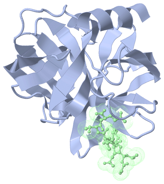
Asymmetric/Biological UnitChain A from PDB Type:PROTEIN Length:179 aligned with C8CIL7_9ENTO | C8CIL7 from UniProtKB/TrEMBL Length:645 Alignment length:179 13 23 33 43 53 63 73 83 93 103 113 123 133 143 153 163 173 C8CIL7_9ENTO 4 LDFALSLLRRNIRQVQTDQGHFTMLGVRDRLAILPRHSQPGKTIWVEHKLINVLDAVELVDEQGVNLELTLVTLDTNEKFRDVTKFIPETITGASDATLIINTEHMPSMFVPVGDVVQYGFLNLSGKPTHRTMMYNFPTKAGQCGGVVTSVGKIIGIHIGGNGRQGFCAGLKRGYFASE 182 SCOP domains d3sj9a_ A: 3C cysteine protease (picornain 3C) SCOP domains CATH domains ----------------------------------------------------------------------------------------------------------------------------------------------------------------------------------- CATH domains Pfam domains ----------------------------------------------------------------------------------------------------------------------------------------------------------------------------------- Pfam domains SAPs(SNPs) ----------------------------------------------------------------------------------------------------------------------------------------------------------------------------------- SAPs(SNPs) PROSITE ----------------------------------------------------------------------------------------------------------------------------------------------------------------------------------- PROSITE Transcript ----------------------------------------------------------------------------------------------------------------------------------------------------------------------------------- Transcript 3sj9 A 4 LDFALSLLRRNIRQVQTDQGHFTMLGVRDRLAILPRHSQPGKTIWVEHKLINVLDAVELVDEQGVNLELTLVTLDTNEKFRDVTKFIPETITGASDATLIINTEHMPSMFVPVGDVVQYGFLNLSGKPTHRTMMYNFPTKAGQAGGVVTSVGKIIGIHIGGNGRQGFCAGLKRGYFASE 182 13 23 33 43 53 63 73 83 93 103 113 123 133 143 153 163 173
Chain B from PDB Type:PROTEIN Length:8
SCOP domains -------- SCOP domains
CATH domains -------- CATH domains
Pfam domains -------- Pfam domains
SAPs(SNPs) -------- SAPs(SNPs)
PROSITE -------- PROSITE
Transcript -------- Transcript
3sj9 B 1 GLRQAVTQ 8
|
||||||||||||||||||||
SCOP Domains (1, 1)
Asymmetric/Biological Unit
 |
CATH Domains (0, 0)| (no "CATH Domain" information available for 3SJ9) |
Pfam Domains (0, 0)| (no "Pfam Domain" information available for 3SJ9) |
Gene Ontology (12, 12)|
Asymmetric/Biological Unit(hide GO term definitions) Chain A (C8CIL7_9ENTO | C8CIL7)
|
||||||||||||||||||||||||||||||||||||||||||||||||||||||||||||||||||||||||||||||||||||
Interactive Views
|
||||||||||||||||||||||||||||||||||||||||||||||||||||||||||||||||||||||||||||||||||||||||||||||||||||||||||||||||||||
Still Images
|
||||||||||||||||
Databases
|
||||||||||||||||||||||||||||||||||||||||||||||||||||||||||||||||||||||||||||||||||||||||||||||||||||||||||||||||||||||||||||||||||||||||||||||||||||||||||||||||
Analysis Tools
|
|||||||||||||||||||||||||||||||||||||||||||||||||||||||||||||
Entries Sharing at Least One Protein Chain (UniProt ID)
Related Entries Specified in the PDB File
|
|