Show PDB file:   
         Plain Text   HTML   (compressed file size)
QuickSearch:   
by PDB,NDB,UniProt,PROSITE Code or Search Term(s)  
(-)Biol.Unit 1 - manually
(-)Asymmetric Unit
(-)Asym. Unit - sites
(-)Biological Unit 1
(-)Biol. Unit 1 - sites
collapse expand < >
Image Biol.Unit 1 - manually
Biol.Unit 1 - manually  (Jmol Viewer)
Image Asymmetric Unit
Asymmetric Unit  (Jmol Viewer)
Image Asym. Unit - sites
Asym. Unit - sites  (Jmol Viewer)
Image Biological Unit 1
Biological Unit 1  (Jmol Viewer)
Image Biol. Unit 1 - sites
Biol. Unit 1 - sites  (Jmol Viewer)

(-) Description

Title :  CRYSTAL STRUCTURE OF URE3-BINDING PROTEIN, (D127A,N129A) MUTANT, IODIDE PHASED
 
Authors :  Seattle Structural Genomics Center For Infectious Disease (S P. Skubak
Date :  17 Jun 11  (Deposition) - 29 Jun 11  (Release) - 29 Jun 11  (Revision)
Method :  X-RAY DIFFRACTION
Resolution :  2.05
Chains :  Asym. Unit :  A
Biol. Unit 1:  A  (2x)
Keywords :  Dna Binding Protein, Ef-Hand, Structural Genomics, Seattle Structural Genomics Center For Infectious Disease, Ssgcid (Keyword Search: [Gene Ontology, PubMed, Web (Google))
 
Reference :  Seattle Structural Genomics Center For Infectious Disease (Ssgcid), A. Gardberg, T. Edwards, B. Staker, P. Skubak, L. Stewart, C. Gilchrist
Crystal Structure Of Ure3-Binding Protein
To Be Published
PubMed: search

(-) Compounds

Molecule 1 - URE3-BP SEQUENCE SPECIFIC DNA BINDING PROTEIN
    ChainsA
    EngineeredYES
    Expression SystemESCHERICHIA COLI
    Expression System PlasmidPET28A
    Expression System StrainBL21(DE3)
    Expression System Taxid562
    Expression System Vector TypePLASMID
    GeneURE3-BP
    MutationYES
    Organism ScientificENTAMOEBA HISTOLYTICA
    Organism Taxid5759

 Structural Features

(-) Chains, Units

  1
Asymmetric Unit A
Biological Unit 1 (2x)A

Summary Information (see also Sequences/Alignments below)

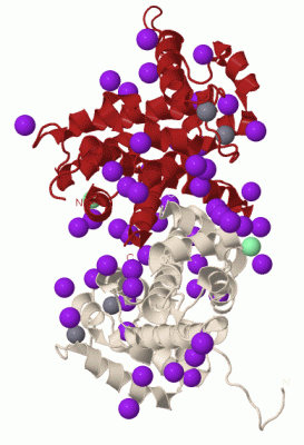
(-) Ligands, Modified Residues, Ions  (3, 29)

Asymmetric Unit (3, 29)
No.NameCountTypeFull Name
1CA2Ligand/IonCALCIUM ION
2CL1Ligand/IonCHLORIDE ION
3IOD26Ligand/IonIODIDE ION
Biological Unit 1 (1, 52)
No.NameCountTypeFull Name
1CA-1Ligand/IonCALCIUM ION
2CL-1Ligand/IonCHLORIDE ION
3IOD52Ligand/IonIODIDE ION

(-) Sites  (20, 20)

Asymmetric Unit (20, 20)
No.NameEvidenceResiduesDescription
01AC1SOFTWAREASP A:61 , ASP A:63 , SER A:65 , THR A:67 , GLU A:69 , GLU A:72BINDING SITE FOR RESIDUE CA A 221
02AC2SOFTWAREASP A:97 , ASP A:99 , ASN A:101 , HIS A:103 , GLU A:108 , HOH A:260BINDING SITE FOR RESIDUE CA A 222
03AC3SOFTWAREPRO A:30 , SER A:105 , HOH A:259BINDING SITE FOR RESIDUE IOD A 223
04AC4SOFTWAREARG A:185BINDING SITE FOR RESIDUE IOD A 225
05AC5SOFTWAREGLN A:17 , ASN A:43 , IOD A:236BINDING SITE FOR RESIDUE IOD A 226
06AC6SOFTWARELYS A:114BINDING SITE FOR RESIDUE IOD A 227
07AC7SOFTWARELEU A:118 , ASN A:121BINDING SITE FOR RESIDUE IOD A 228
08AC8SOFTWAREMET A:206 , IOD A:245BINDING SITE FOR RESIDUE IOD A 231
09AC9SOFTWAREGLY A:80BINDING SITE FOR RESIDUE IOD A 232
10BC1SOFTWARESER A:85 , HOH A:292BINDING SITE FOR RESIDUE IOD A 233
11BC2SOFTWAREPRO A:86BINDING SITE FOR RESIDUE IOD A 235
12BC3SOFTWAREGLY A:18 , SER A:131 , IOD A:226 , HOH A:279BINDING SITE FOR RESIDUE IOD A 236
13BC4SOFTWAREARG A:153BINDING SITE FOR RESIDUE IOD A 237
14BC5SOFTWAREMET A:190BINDING SITE FOR RESIDUE IOD A 240
15BC6SOFTWAREHOH A:289 , HOH A:308BINDING SITE FOR RESIDUE IOD A 244
16BC7SOFTWAREMET A:74 , ASN A:204 , MET A:206 , IOD A:231BINDING SITE FOR RESIDUE IOD A 245
17BC8SOFTWARESER A:32BINDING SITE FOR RESIDUE IOD A 246
18BC9SOFTWAREGLY A:22BINDING SITE FOR RESIDUE IOD A 247
19CC1SOFTWARETYR A:196 , HOH A:309 , HOH A:310BINDING SITE FOR RESIDUE IOD A 248
20CC2SOFTWAREGLN A:152BINDING SITE FOR RESIDUE CL A 249

(-) SS Bonds  (0, 0)

(no "SS Bond" information available for 3SIA)

(-) Cis Peptide Bonds  (0, 0)

(no "Cis Peptide Bond" information available for 3SIA)

 Sequence-Structure Mapping

(-) SAPs(SNPs)/Variants  (0, 0)

(no "SAP(SNP)/Variant" information available for 3SIA)

(-) PROSITE Motifs  (0, 0)

(no "PROSITE Motif" information available for 3SIA)

(-) Exons   (0, 0)

(no "Exon" information available for 3SIA)

(-) Sequences/Alignments

Asymmetric Unit
   Reformat: Number of residues per line =  ('0' or empty: single-line sequence representation)
  Number of residues per labelling interval =   
  UniProt sequence: complete  aligned part    
   Show mapping: SCOP domains CATH domains Pfam domains Secondary structure (by author)
SAPs(SNPs) PROSITE motifs Exons
(details for a mapped element are shown in a popup box when the mouse pointer rests over it)
Chain A from PDB  Type:PROTEIN  Length:212
 aligned with Q9GSV7_ENTHI | Q9GSV7 from UniProtKB/TrEMBL  Length:220

    Alignment length:214
                                    16        26        36        46        56        66        76        86        96       106       116       126       136       146       156       166       176       186       196       206       216    
         Q9GSV7_ENTHI     7 NFCLWNLQPIQGSWMGAACIYQMPPSVRNTWWFPLLNTIPLDQYTRIYQWFMGVDRDRSGTLEINELMMGQFPGGIRLSPQTALRMMRIFDTDFNGHISFYEFMAMYKFMELAYNLFVMNDRNRSGTLEPHEILPALQQLGFYINQRTSLLLHRLFARGMAFCDLNCWIAICAFAAQTRSAYQMIFMNPYYGPMKPFNPMEFGKFLDVVTSLLE 220
               SCOP domains ---------------------------------------------------------------------------------------------------------------------------------------------------------------------------------------------------------------------- SCOP domains
               CATH domains ---------------------------------------------------------------------------------------------------------------------------------------------------------------------------------------------------------------------- CATH domains
               Pfam domains ---------------------------------------------------------------------------------efhand-3siaA01 A:88-116      -------------------------------------------------------------------------------------------------------- Pfam domains
         Sec.struct. author ............hhhhhhhhhhhhhhhhh..hhhhhhhhhhhhhhhhhhhhhhhh.......hhhhhhhh.hhhhh..hhhhhhhhhhhhh.......hhhhhhhhhhhhhhhhhhhhhhh.......hhhhhhhhhhhhh...hhhhhhhhhhhhh--....hhhhhhhhhhhhhhhhhhhhhhhhhhhhh.....hhhhhhhhhhhhhhh.. Sec.struct. author
                 SAPs(SNPs) ---------------------------------------------------------------------------------------------------------------------------------------------------------------------------------------------------------------------- SAPs(SNPs)
                    PROSITE ---------------------------------------------------------------------------------------------------------------------------------------------------------------------------------------------------------------------- PROSITE
                 Transcript ---------------------------------------------------------------------------------------------------------------------------------------------------------------------------------------------------------------------- Transcript
                 3sia A   7 NFCLWNLQPIQGSWMGAACIYQMPPSVRNTWWFPLLNTIPLDQYTRIYQWFMGVDRDRSGTLEINELMMGQFPGGIRLSPQTALRMMRIFDTDFNGHISFYEFMAMYKFMELAYNLFVMNARARSGTLEPHEILPALQQLGFYINQRTSLLLHRLFA--MAFCDLNCWIAICAFAAQTRSAYQMIFMNPYYGPMKPFNPMEFGKFLDVVTSLLE 220
                                    16        26        36        46        56        66        76        86        96       106       116       126       136       146       156      |166       176       186       196       206       216    
                                                                                                                                                                                      163  |                                                      
                                                                                                                                                                                         166                                                      

   Legend:   → Mismatch (orange background)
  - → Gap (green background, '-', border residues have a numbering label)
    → Modified Residue (blue background, lower-case, 'x' indicates undefined single-letter code, labelled with number + name)
  x → Chemical Group (purple background, 'x', labelled with number + name, e.g. ACE or NH2)
  extra numbering lines below/above indicate numbering irregularities and modified residue names etc., number ends below/above '|'

 Classification and Annotation

(-) SCOP Domains  (0, 0)

(no "SCOP Domain" information available for 3SIA)

(-) CATH Domains  (0, 0)

(no "CATH Domain" information available for 3SIA)

(-) Pfam Domains  (1, 1)

Asymmetric Unit
(-)
Clan: EF_hand (270)

(-) Gene Ontology  (2, 2)

Asymmetric Unit(hide GO term definitions)
Chain A   (Q9GSV7_ENTHI | Q9GSV7)
molecular function
    GO:0005509    calcium ion binding    Interacting selectively and non-covalently with calcium ions (Ca2+).
    GO:0046872    metal ion binding    Interacting selectively and non-covalently with any metal ion.

 Visualization

(-) Interactive Views

Asymmetric Unit
  Complete Structure
    Jena3D(integrated viewing of ligand, site, SAP, PROSITE, SCOP information)
    WebMol | AstexViewer[tm]@PDBe
(Java Applets, require no local installation except for Java; loading may be slow)
    STRAP
(Java WebStart application, automatic local installation, requires Java; full application with system access!)
    RasMol
(require local installation)
    Molscript (VRML)
(requires installation of a VRML viewer; select preferred view via VRML and generate a mono or stereo PDF format file)
 
  Ligands, Modified Residues, Ions
    CA  [ RasMol | Jena3D ]  +environment [ RasMol | Jena3D ]
    CL  [ RasMol | Jena3D ]  +environment [ RasMol | Jena3D ]
    IOD  [ RasMol | Jena3D ]  +environment [ RasMol | Jena3D ]
 
  Sites
    AC1  [ RasMol ]  +environment [ RasMol ]
    AC2  [ RasMol ]  +environment [ RasMol ]
    AC3  [ RasMol ]  +environment [ RasMol ]
    AC4  [ RasMol ]  +environment [ RasMol ]
    AC5  [ RasMol ]  +environment [ RasMol ]
    AC6  [ RasMol ]  +environment [ RasMol ]
    AC7  [ RasMol ]  +environment [ RasMol ]
    AC8  [ RasMol ]  +environment [ RasMol ]
    AC9  [ RasMol ]  +environment [ RasMol ]
    BC1  [ RasMol ]  +environment [ RasMol ]
    BC2  [ RasMol ]  +environment [ RasMol ]
    BC3  [ RasMol ]  +environment [ RasMol ]
    BC4  [ RasMol ]  +environment [ RasMol ]
    BC5  [ RasMol ]  +environment [ RasMol ]
    BC6  [ RasMol ]  +environment [ RasMol ]
    BC7  [ RasMol ]  +environment [ RasMol ]
    BC8  [ RasMol ]  +environment [ RasMol ]
    BC9  [ RasMol ]  +environment [ RasMol ]
    CC1  [ RasMol ]  +environment [ RasMol ]
    CC2  [ RasMol ]  +environment [ RasMol ]
 
  Cis Peptide Bonds
(no "Cis Peptide Bonds" information available for 3sia)
 
Biological Unit
  Complete Structure
    Biological Unit 1  [ Jena3D ]

(-) Still Images

Jmol
  protein: cartoon or spacefill or dots and stick; nucleic acid: cartoon and stick; ligands: spacefill; active site: stick
Molscript
  protein, nucleic acid: cartoon; ligands: spacefill; active site: ball and stick

 Databases and Analysis Tools

(-) Databases

Access by PDB/NDB ID
  3sia
    Family and Domain InformationProDom | SYSTERS
    General Structural InformationGlycoscienceDB | MMDB | NDB | OCA | PDB | PDBe | PDBj | PDBsum | PDBWiki | PQS | PROTEOPEDIA
    Orientation in MembranesOPM
    Protein SurfaceSURFACE
    Secondary StructureDSSP (structure derived) | HSSP (homology derived)
    Structural GenomicsGeneCensus
    Structural NeighboursCE | VAST
    Structure ClassificationCATH | Dali | SCOP
    Validation and Original DataBMRB Data View | BMRB Restraints Grid | EDS | PROCHECK | RECOORD | WHAT_CHECK
 
Access by UniProt ID/Accession number
  Q9GSV7_ENTHI | Q9GSV7
    Comparative Protein Structure ModelsModBase
    Genomic InformationEnsembl
    Protein-protein InteractionDIP
    Sequence, Family and Domain InformationInterPro | Pfam | SMART | UniProtKB/TrEMBL
 
Access by Enzyme Classificator   (EC Number)
  (no 'Enzyme Classificator' available)
    General Enzyme InformationBRENDA | EC-PDB | Enzyme | IntEnz
    PathwayKEGG | MetaCyc
 
Access by Disease Identifier   (MIM ID)
  (no 'MIM ID' available)
    Disease InformationOMIM
 
Access by GenAge ID
  (no 'GenAge ID' available)
    Age Related InformationGenAge

(-) Analysis Tools

Access by PDB/NDB ID
    Domain InformationXDom
    Interatomic Contacts of Structural UnitsCSU
    Ligand-protein ContactsLPC
    Protein CavitiescastP
    Sequence and Secondary StructurePDBCartoon
    Structure AlignmentSTRAP(Java WebStart application, automatic local installation, requires Java; full application with system access!)
    Structure and Sequence BrowserSTING
 
Access by UniProt ID/Accession number
  Q9GSV7_ENTHI | Q9GSV7
    Protein Disorder PredictionDisEMBL | FoldIndex | GLOBPLOT (for more information see DisProt)

 Related Entries

(-) Entries Sharing at Least One Protein Chain (UniProt ID)

UniProtKB/Swiss-Prot
        Q9GSV7_ENTHI | Q9GSV73sjs
UniProtKB/TrEMBL
        Q9GSV7_ENTHI | Q9GSV73sib 4q04

(-) Related Entries Specified in the PDB File

3sib WILD-TYPE PROTEIN RELATED ID: ENHIA.01648.A RELATED DB: TARGETDB