Show PDB file:   
         Plain Text   HTML   (compressed file size)
QuickSearch:   
by PDB,NDB,UniProt,PROSITE Code or Search Term(s)  
(-)Biol.Unit 1 - manually
(-)Asymmetric Unit
(-)Asym. Unit - sites
(-)Biological Unit 1
(-)Biol. Unit 1 - sites
(-)Biological Unit 2
(-)Biological Unit 3
collapse expand < >
Image Biol.Unit 1 - manually
Biol.Unit 1 - manually  (Jmol Viewer)
Image Asymmetric Unit
Asymmetric Unit  (Jmol Viewer)
Image Asym. Unit - sites
Asym. Unit - sites  (Jmol Viewer)
Image Biological Unit 1
Biological Unit 1  (Jmol Viewer)
Image Biol. Unit 1 - sites
Biol. Unit 1 - sites  (Jmol Viewer)
Image Biological Unit 2
Biological Unit 2  (Jmol Viewer)
Image Biological Unit 3
Biological Unit 3  (Jmol Viewer)

(-) Description

Title :  CRYSTAL STRUCTURE OF A RIBOSE-5-PHOSPHATE ISOMERASE B RPIB FROM COCCIDIOIDES IMMITIS BOUND TO PHOSPHATE
 
Authors :  Seattle Structural Genomics Center For Infectious Disease (S
Date :  09 Jun 11  (Deposition) - 22 Jun 11  (Release) - 26 Oct 11  (Revision)
Method :  X-RAY DIFFRACTION
Resolution :  1.80
Chains :  Asym. Unit :  A
Biol. Unit 1:  A  (2x)
Biol. Unit 2:  A  (2x)
Biol. Unit 3:  A  (4x)
Keywords :  Structural Genomics, Seattle Structural Genomics Center For Infectious Disease, Ssgcid, Ribose-5-Phosphate, Rpib, Valley Fever, Coccidioidomycosis, Pathogenic Fungus, Dust-Borne Pathogen, Isomerase, Nucleotide And Co-Factor Biogenesis, Pentose Phosphate Pathway (Keyword Search: [Gene Ontology, PubMed, Web (Google))
 
Reference :  T. E. Edwards, A. B. Abramov, E. R. Smith, R. O. Baydo, J. T. Leonard, D. J. Leibly, K. B. Thompkins, M. C. Clifton, A. S. Gardberg, B. L. Staker, W. C. Van Voorhis, P. J. Myler, L. J. Stewart
Structural Characterization Of A Ribose-5-Phosphate Isomerase B From The Pathogenic Fungus Coccidioides Immitis
Bmc Struct. Biol. V. 11 39 2011
PubMed-ID: 21995815  |  Reference-DOI: 10.1186/1472-6807-11-39

(-) Compounds

Molecule 1 - RIBOSE 5-PHOSPHATE ISOMERASE
    ChainsA
    EC Number5.3.1.-
    EngineeredYES
    Expression SystemESCHERICHIA COLI
    Expression System Taxid562
    Expression System Vector TypePAVA0421
    GeneCIMG_07932
    Organism CommonVALLEY FEVER FUNGUS
    Organism ScientificCOCCIDIOIDES IMMITIS
    Organism Taxid246410
    StrainRS

 Structural Features

(-) Chains, Units

  1
Asymmetric Unit A
Biological Unit 1 (2x)A
Biological Unit 2 (2x)A
Biological Unit 3 (4x)A

Summary Information (see also Sequences/Alignments below)

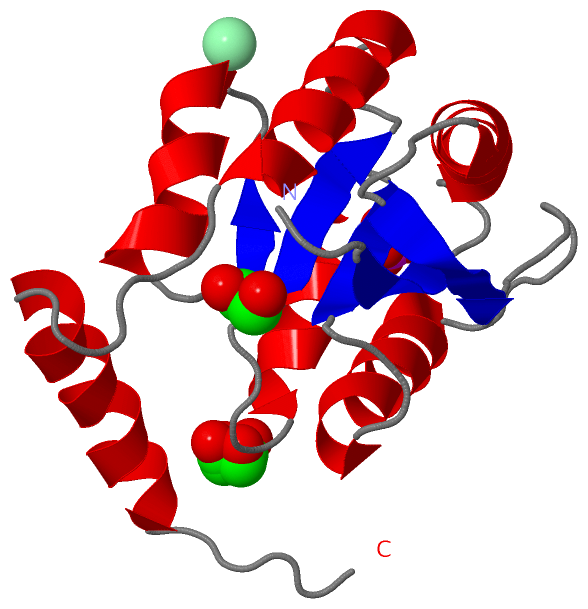
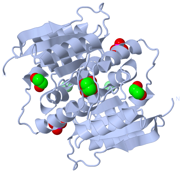
(-) Ligands, Modified Residues, Ions  (3, 4)

Asymmetric Unit (3, 4)
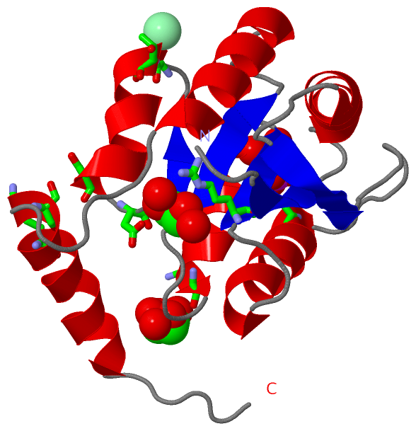
No.NameCountTypeFull Name
1CL1Ligand/IonCHLORIDE ION
2EDO2Ligand/Ion1,2-ETHANEDIOL
3PO41Ligand/IonPHOSPHATE ION
Biological Unit 1 (2, 6)
No.NameCountTypeFull Name
1CL-1Ligand/IonCHLORIDE ION
2EDO4Ligand/Ion1,2-ETHANEDIOL
3PO42Ligand/IonPHOSPHATE ION
Biological Unit 2 (2, 6)
No.NameCountTypeFull Name
1CL-1Ligand/IonCHLORIDE ION
2EDO4Ligand/Ion1,2-ETHANEDIOL
3PO42Ligand/IonPHOSPHATE ION
Biological Unit 3 (2, 12)
No.NameCountTypeFull Name
1CL-1Ligand/IonCHLORIDE ION
2EDO8Ligand/Ion1,2-ETHANEDIOL
3PO44Ligand/IonPHOSPHATE ION

(-) Sites  (4, 4)

Asymmetric Unit (4, 4)
No.NameEvidenceResiduesDescription
1AC1SOFTWARETYR A:53 , SER A:109 , SER A:145 , LYS A:148 , HOH A:171 , HOH A:315BINDING SITE FOR RESIDUE PO4 A 164
2AC2SOFTWAREARG A:71 , ASP A:111 , ALA A:112 , HOH A:191 , HOH A:251BINDING SITE FOR RESIDUE EDO A 165
3AC3SOFTWARELYS A:88 , HOH A:209BINDING SITE FOR RESIDUE EDO A 166
4AC4SOFTWAREASP A:99 , THR A:100BINDING SITE FOR RESIDUE CL A 167

(-) SS Bonds  (0, 0)

(no "SS Bond" information available for 3SDW)

(-) Cis Peptide Bonds  (1, 1)

Asymmetric Unit
No.Residues
1Gly A:43 -Val A:44

 Sequence-Structure Mapping

(-) SAPs(SNPs)/Variants  (0, 0)

(no "SAP(SNP)/Variant" information available for 3SDW)

(-) PROSITE Motifs  (0, 0)

(no "PROSITE Motif" information available for 3SDW)

(-) Exons   (0, 0)

(no "Exon" information available for 3SDW)

(-) Sequences/Alignments

Asymmetric Unit
   Reformat: Number of residues per line =  ('0' or empty: single-line sequence representation)
  Number of residues per labelling interval =   
  UniProt sequence: complete  aligned part    
   Show mapping: SCOP domains CATH domains Pfam domains Secondary structure (by author)
SAPs(SNPs) PROSITE motifs Exons
(details for a mapped element are shown in a popup box when the mouse pointer rests over it)
Chain A from PDB  Type:PROTEIN  Length:158
 aligned with RPIB_COCIM | P0CL19 from UniProtKB/Swiss-Prot  Length:163

    Alignment length:158
                                    15        25        35        45        55        65        75        85        95       105       115       125       135       145       155        
           RPIB_COCIM     6 LPPLRLAIACDDAGVSYKEALKAHLSDNPLVSSITDVGVTSTTDKTAYPHVAIQAAQLIKDGKVDRALMICGTGLGVAISANKVPGIRAVTAHDTFSVERAILSNDAQVLCFGQRVIGIELAKRLAGEWLTYRFDQKSASAQKVQAISDYEKKFVEVN 163
               SCOP domains d3sdwa_ A: automated matches                                                                                                                                   SCOP domains
               CATH domains -------------------------------------------------------------------------------------------------------------------------------------------------------------- CATH domains
               Pfam domains ----LacAB_rpiB-3sdwA01 A:10-153                                                                                                                     ---------- Pfam domains
         Sec.struct. author ...eeeeeee...hhhhhhhhhhhhh....eeeeee..........hhhhhhhhhhhhhhh....eeeeee..hhhhhhhhhh.....eee..hhhhhhhhhhh....eeeee....hhhhhhhhhhhhhh.......hhhhhhhhhhhhhh...... Sec.struct. author
                 SAPs(SNPs) -------------------------------------------------------------------------------------------------------------------------------------------------------------- SAPs(SNPs)
                    PROSITE -------------------------------------------------------------------------------------------------------------------------------------------------------------- PROSITE
                 Transcript -------------------------------------------------------------------------------------------------------------------------------------------------------------- Transcript
                 3sdw A   6 LPPLRLAIACDDAGVSYKEALKAHLSDNPLVSSITDVGVTSTTDKTAYPHVAIQAAQLIKDGKVDRALMICGTGLGVAISANKVPGIRAVTAHDTFSVERAILSNDAQVLCFGQRVIGIELAKRLAGEWLTYRFDQKSASAQKVQAISDYEKKFVEVN 163
                                    15        25        35        45        55        65        75        85        95       105       115       125       135       145       155        

   Legend:   → Mismatch (orange background)
  - → Gap (green background, '-', border residues have a numbering label)
    → Modified Residue (blue background, lower-case, 'x' indicates undefined single-letter code, labelled with number + name)
  x → Chemical Group (purple background, 'x', labelled with number + name, e.g. ACE or NH2)
  extra numbering lines below/above indicate numbering irregularities and modified residue names etc., number ends below/above '|'

 Classification and Annotation

(-) SCOP Domains  (1, 1)

Asymmetric Unit

(-) CATH Domains  (0, 0)

(no "CATH Domain" information available for 3SDW)

(-) Pfam Domains  (1, 1)

Asymmetric Unit

(-) Gene Ontology  (3, 3)

Asymmetric Unit(hide GO term definitions)
Chain A   (RPIB_COCIM | P0CL19)
molecular function
    GO:0016853    isomerase activity    Catalysis of the geometric or structural changes within one molecule. Isomerase is the systematic name for any enzyme of EC class 5.
    GO:0004751    ribose-5-phosphate isomerase activity    Catalysis of the reaction: D-ribose 5-phosphate = D-ribulose 5-phosphate.
biological process
    GO:0005975    carbohydrate metabolic process    The chemical reactions and pathways involving carbohydrates, any of a group of organic compounds based of the general formula Cx(H2O)y. Includes the formation of carbohydrate derivatives by the addition of a carbohydrate residue to another molecule.

 Visualization

(-) Interactive Views

Asymmetric Unit
  Complete Structure
    Jena3D(integrated viewing of ligand, site, SAP, PROSITE, SCOP information)
    WebMol | AstexViewer[tm]@PDBe
(Java Applets, require no local installation except for Java; loading may be slow)
    STRAP
(Java WebStart application, automatic local installation, requires Java; full application with system access!)
    RasMol
(require local installation)
    Molscript (VRML)
(requires installation of a VRML viewer; select preferred view via VRML and generate a mono or stereo PDF format file)
 
  Ligands, Modified Residues, Ions
    CL  [ RasMol | Jena3D ]  +environment [ RasMol | Jena3D ]
    EDO  [ RasMol | Jena3D ]  +environment [ RasMol | Jena3D ]
    PO4  [ RasMol | Jena3D ]  +environment [ RasMol | Jena3D ]
 
  Sites
    AC1  [ RasMol ]  +environment [ RasMol ]
    AC2  [ RasMol ]  +environment [ RasMol ]
    AC3  [ RasMol ]  +environment [ RasMol ]
    AC4  [ RasMol ]  +environment [ RasMol ]
 
  Cis Peptide Bonds
    Gly A:43 - Val A:44   [ RasMol ]  
 
Biological Units
  Complete Structure
    Biological Unit 1  [ Jena3D ]
    Biological Unit 2  [ Jena3D ]
    Biological Unit 3  [ Jena3D ]

(-) Still Images

Jmol
  protein: cartoon or spacefill or dots and stick; nucleic acid: cartoon and stick; ligands: spacefill; active site: stick
Molscript
  protein, nucleic acid: cartoon; ligands: spacefill; active site: ball and stick

 Databases and Analysis Tools

(-) Databases

Access by PDB/NDB ID
  3sdw
    Family and Domain InformationProDom | SYSTERS
    General Structural InformationGlycoscienceDB | MMDB | NDB | OCA | PDB | PDBe | PDBj | PDBsum | PDBWiki | PQS | PROTEOPEDIA
    Orientation in MembranesOPM
    Protein SurfaceSURFACE
    Secondary StructureDSSP (structure derived) | HSSP (homology derived)
    Structural GenomicsGeneCensus
    Structural NeighboursCE | VAST
    Structure ClassificationCATH | Dali | SCOP
    Validation and Original DataBMRB Data View | BMRB Restraints Grid | EDS | PROCHECK | RECOORD | WHAT_CHECK
 
Access by UniProt ID/Accession number
  RPIB_COCIM | P0CL19
    Comparative Protein Structure ModelsModBase
    Genomic InformationEnsembl
    Protein-protein InteractionDIP
    Sequence, Family and Domain InformationInterPro | Pfam | SMART | UniProtKB/SwissProt
 
Access by Enzyme Classificator   (EC Number)
  5.3.1.-
    General Enzyme InformationBRENDA | EC-PDB | Enzyme | IntEnz
    PathwayKEGG | MetaCyc
 
Access by Disease Identifier   (MIM ID)
  (no 'MIM ID' available)
    Disease InformationOMIM
 
Access by GenAge ID
  (no 'GenAge ID' available)
    Age Related InformationGenAge

(-) Analysis Tools

Access by PDB/NDB ID
    Domain InformationXDom
    Interatomic Contacts of Structural UnitsCSU
    Ligand-protein ContactsLPC
    Protein CavitiescastP
    Sequence and Secondary StructurePDBCartoon
    Structure AlignmentSTRAP(Java WebStart application, automatic local installation, requires Java; full application with system access!)
    Structure and Sequence BrowserSTING
 
Access by UniProt ID/Accession number
  RPIB_COCIM | P0CL19
    Protein Disorder PredictionDisEMBL | FoldIndex | GLOBPLOT (for more information see DisProt)

 Related Entries

(-) Entries Sharing at Least One Protein Chain (UniProt ID)

UniProtKB/Swiss-Prot
        RPIB_COCIM | P0CL193qd5 3sgw

(-) Related Entries Specified in the PDB File

3qd5 SAME PROTEIN SOLVED BY IODIDE ION SAD PHASING WITH OXIDIZED CYSTEINE RESIDUE IN ACTIVE SITE
3s5p RPIB FROM GIARDIA LAMBLIA RELATED ID: COIMA.00584.A RELATED DB: TARGETDB