Show PDB file:   
         Plain Text   HTML   (compressed file size)
QuickSearch:   
by PDB,NDB,UniProt,PROSITE Code or Search Term(s)  
(-)Asymmetric Unit
(-)Asym. Unit - sites
(-)Biological Unit 1
(-)Biol. Unit 1 - sites
(-)Biological Unit 2
(-)Biological Unit 3
collapse expand < >
Image Asymmetric Unit
Asymmetric Unit  (Jmol Viewer)
Image Asym. Unit - sites
Asym. Unit - sites  (Jmol Viewer)
Image Biological Unit 1
Biological Unit 1  (Jmol Viewer)
Image Biol. Unit 1 - sites
Biol. Unit 1 - sites  (Jmol Viewer)
Image Biological Unit 2
Biological Unit 2  (Jmol Viewer)
Image Biological Unit 3
Biological Unit 3  (Jmol Viewer)

(-) Description

Title :  CRYSTAL STRUCTURE OF CYTIDYLATE KINASE FROM MYCOBACTERIUM SMEGMATIS
 
Authors :  Seattle Structural Genomics Center For Infectious Disease (S
Date :  11 Mar 11  (Deposition) - 13 Apr 11  (Release) - 12 Oct 16  (Revision)
Method :  X-RAY DIFFRACTION
Resolution :  2.00
Chains :  Asym. Unit :  A
Biol. Unit 1:  A  (1x)
Biol. Unit 2:  A  (3x)
Biol. Unit 3:  A  (2x)
Keywords :  Structural Genomics, Seattle Structural Genomics Center For Infectious Disease, Ssgcid, Cytidylate Kinase, Adp, Dcmp, Dcdp, Transferase (Keyword Search: [Gene Ontology, PubMed, Web (Google))
 
Reference :  L. Baugh, I. Phan, D. W. Begley, M. C. Clifton, B. Armour, D. M. Dranow, B. M. Taylor, M. M. Muruthi, J. Abendroth, J. W. Fairman, D. Fox, S. H. Dieterich, B. L. Staker, A. S. Gardberg, R. Choi, S. N. Hewitt, A. J. Napuli, J. Myers, L. K. Barrett, Y. Zhang, M. Ferrell, E. Mundt, K. Thompkins, N. Tran, S. Lyons-Abbott, A. Abramov, A. Sekar, D. Serbzhinskiy, D. Lorimer, G. W. Buchko, R. Stacy, L. J. Stewart, T. E. Edwards, W. C. Van Voorhis, P. J. Myler
Increasing The Structural Coverage Of Tuberculosis Drug Targets.
Tuberculosis (Edinb) V. 95 142 2015
PubMed-ID: 25613812  |  Reference-DOI: 10.1016/J.TUBE.2014.12.003
(for further references see the PDB file header)

(-) Compounds

Molecule 1 - CYTIDYLATE KINASE
    ChainsA
    EC Number2.7.4.14
    EngineeredYES
    Expression SystemESCHERICHIA COLI
    Expression System Taxid562
    Expression System Vector TypeAVA0421
    GeneCMK, MSMEG_3739
    Organism ScientificMYCOBACTERIUM SMEGMATIS
    Organism Taxid246196
    StrainATCC 700084 / MC(2)155
    SynonymCK, CYTIDINE MONOPHOSPHATE KINASE

 Structural Features

(-) Chains, Units

  1
Asymmetric Unit A
Biological Unit 1 (1x)A
Biological Unit 2 (3x)A
Biological Unit 3 (2x)A

Summary Information (see also Sequences/Alignments below)

(-) Ligands, Modified Residues, Ions  (4, 4)

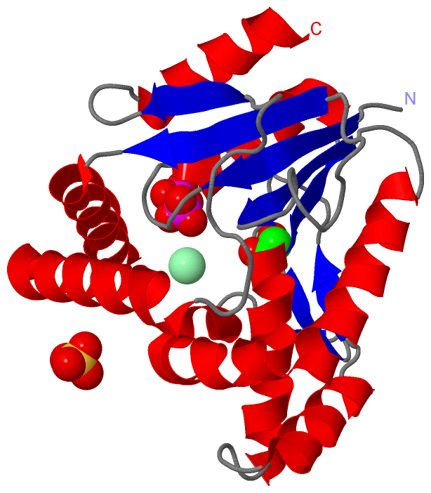
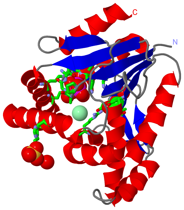
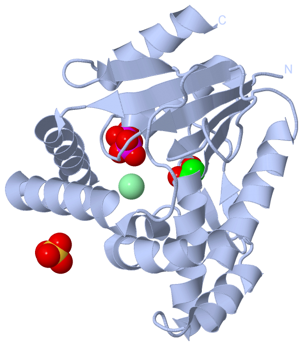
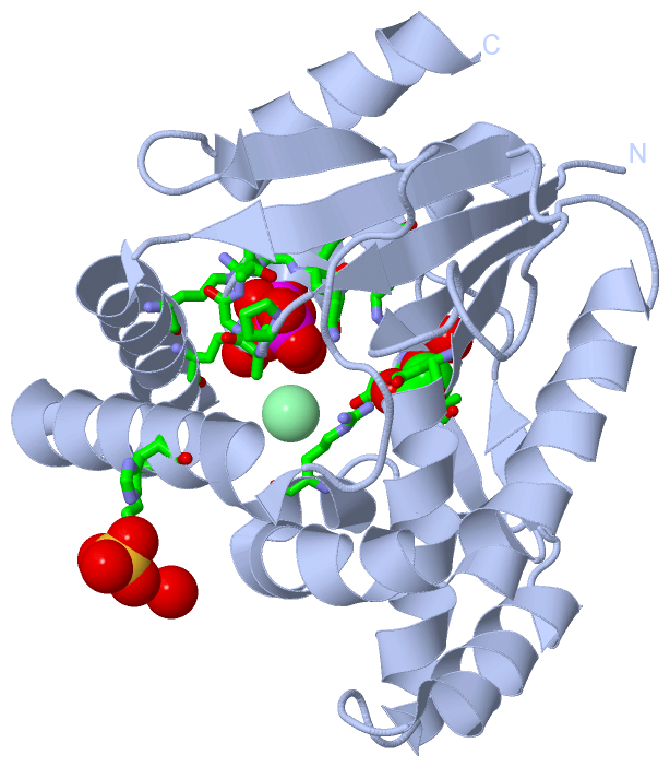
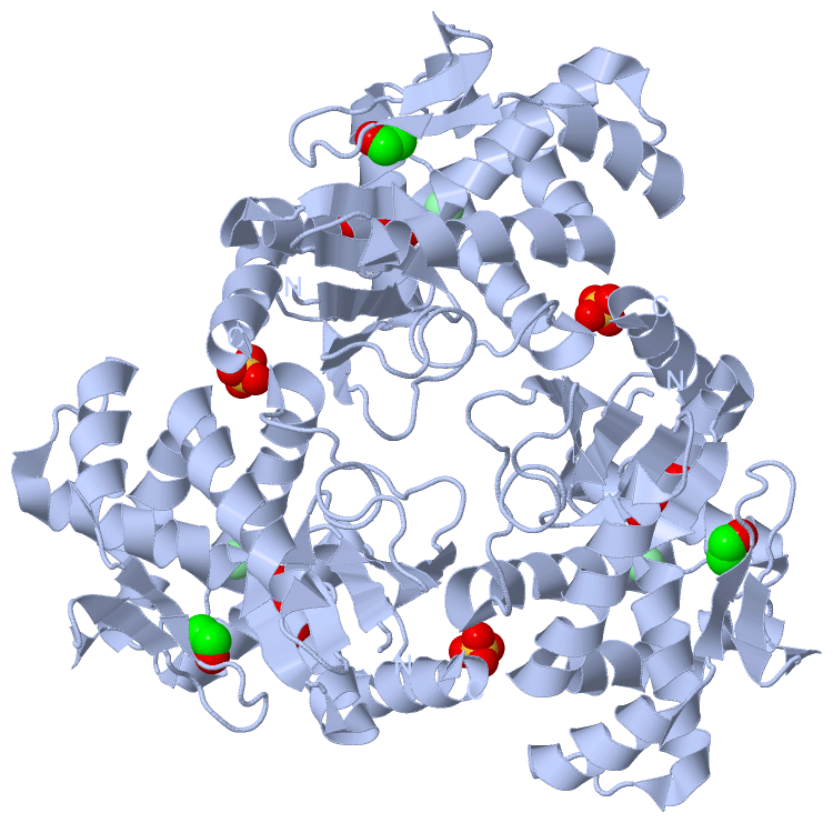
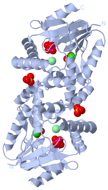
Asymmetric Unit (4, 4)
No.NameCountTypeFull Name
1CL1Ligand/IonCHLORIDE ION
2DPO1Ligand/IonDIPHOSPHATE
3EDO1Ligand/Ion1,2-ETHANEDIOL
4SO41Ligand/IonSULFATE ION
Biological Unit 1 (3, 3)
No.NameCountTypeFull Name
1CL-1Ligand/IonCHLORIDE ION
2DPO1Ligand/IonDIPHOSPHATE
3EDO1Ligand/Ion1,2-ETHANEDIOL
4SO41Ligand/IonSULFATE ION
Biological Unit 2 (3, 9)
No.NameCountTypeFull Name
1CL-1Ligand/IonCHLORIDE ION
2DPO3Ligand/IonDIPHOSPHATE
3EDO3Ligand/Ion1,2-ETHANEDIOL
4SO43Ligand/IonSULFATE ION
Biological Unit 3 (3, 6)
No.NameCountTypeFull Name
1CL-1Ligand/IonCHLORIDE ION
2DPO2Ligand/IonDIPHOSPHATE
3EDO2Ligand/Ion1,2-ETHANEDIOL
4SO42Ligand/IonSULFATE ION

(-) Sites  (3, 3)

Asymmetric Unit (3, 3)
No.NameEvidenceResiduesDescription
1AC1SOFTWAREPRO A:13 , ALA A:14 , GLY A:15 , THR A:16 , GLY A:17 , LYS A:18 , SER A:19 , SER A:20 , ARG A:158 , GLN A:162 , HOH A:296 , HOH A:410 , HOH A:411BINDING SITE FOR RESIDUE DPO A 230
2AC2SOFTWAREARG A:23 , ASP A:35 , VAL A:77 , ASP A:78 , ALA A:79 , ARG A:92 , HOH A:266BINDING SITE FOR RESIDUE EDO A 231
3AC3SOFTWAREARG A:181 , HIS A:184 , HOH A:426BINDING SITE FOR RESIDUE SO4 A 233

(-) SS Bonds  (0, 0)

(no "SS Bond" information available for 3R20)

(-) Cis Peptide Bonds  (0, 0)

(no "Cis Peptide Bond" information available for 3R20)

 Sequence-Structure Mapping

(-) SAPs(SNPs)/Variants  (0, 0)

(no "SAP(SNP)/Variant" information available for 3R20)

(-) PROSITE Motifs  (0, 0)

(no "PROSITE Motif" information available for 3R20)

(-) Exons   (0, 0)

(no "Exon" information available for 3R20)

(-) Sequences/Alignments

Asymmetric Unit
   Reformat: Number of residues per line =  ('0' or empty: single-line sequence representation)
  Number of residues per labelling interval =   
  UniProt sequence: complete  aligned part    
   Show mapping: SCOP domains CATH domains Pfam domains Secondary structure (by author)
SAPs(SNPs) PROSITE motifs Exons
(details for a mapped element are shown in a popup box when the mouse pointer rests over it)
Chain A from PDB  Type:PROTEIN  Length:217
 aligned with A0QYQ0_MYCS2 | A0QYQ0 from UniProtKB/TrEMBL  Length:228

    Alignment length:217
                                    13        23        33        43        53        63        73        83        93       103       113       123       133       143       153       163       173       183       193       203       213       
         A0QYQ0_MYCS2     4 SLVVAVDGPAGTGKSSVSRGLARALGARYLDTGAMYRIATLAVLRAGADLTDPAAIEKAAADAEIGVGSDPDVDAAFLAGEDVSSEIRGDAVTGAVSAVSAVPAVRTRLVDIQRKLATEGGRVVVEGRDIGTVVLPDADVKIFLTASAEERARRRNAQNVANGLPDDYATVLADVQRRDHLDSTRPVSPLRAADDALVVDTSDMDQAQVIAHLLDLV 220
               SCOP domains d3r20a_ A: automated matches                                                                                                                                                                                              SCOP domains
               CATH domains ------------------------------------------------------------------------------------------------------------------------------------------------------------------------------------------------------------------------- CATH domains
               Pfam domains --------------------------------------------------------Cytidylate_kin-3r20A01 A:61-219                                                                                                                                -- Pfam domains
         Sec.struct. author ..eeeee.....hhhhhhhhhhhhhh.eeeehhhhhhhhhhhhhhhh....hhhhhhhhhhh..eee........eee..ee..hhhhhhhhhhhhhhhhhhhhhhhhhhhhhhhhhh....eeeee............eeeeee.hhhhhhhhhhhhhhhh....hhhhhhhhhhhhhhhhhhh...........eeee....hhhhhhhhhhhhh Sec.struct. author
                 SAPs(SNPs) ------------------------------------------------------------------------------------------------------------------------------------------------------------------------------------------------------------------------- SAPs(SNPs)
                    PROSITE ------------------------------------------------------------------------------------------------------------------------------------------------------------------------------------------------------------------------- PROSITE
                 Transcript ------------------------------------------------------------------------------------------------------------------------------------------------------------------------------------------------------------------------- Transcript
                 3r20 A   5 SLVVAVDGPAGTGKSSVSRGLARALGARYLDTGAMYRIATLAVLRAGADLTDPAAIEKAAADAEIGVGSDPDVDAAFLAGEDVSSEIRGDAVTGAVSAVSAVPAVRTRLVDIQRKLATEGGRVVVEGRDIGTVVLPDADVKIFLTASAEERARRRNAQNVANGLPDDYATVLADVQRRDHLDSTRPVSPLRAADDALVVDTSDMDQAQVIAHLLDLV 221
                                    14        24        34        44        54        64        74        84        94       104       114       124       134       144       154       164       174       184       194       204       214       

   Legend:   → Mismatch (orange background)
  - → Gap (green background, '-', border residues have a numbering label)
    → Modified Residue (blue background, lower-case, 'x' indicates undefined single-letter code, labelled with number + name)
  x → Chemical Group (purple background, 'x', labelled with number + name, e.g. ACE or NH2)
  extra numbering lines below/above indicate numbering irregularities and modified residue names etc., number ends below/above '|'

 Classification and Annotation

(-) SCOP Domains  (1, 1)

Asymmetric Unit

(-) CATH Domains  (0, 0)

(no "CATH Domain" information available for 3R20)

(-) Pfam Domains  (1, 1)

Asymmetric Unit

(-) Gene Ontology  (10, 10)

Asymmetric Unit(hide GO term definitions)
Chain A   (A0QYQ0_MYCS2 | A0QYQ0)
molecular function
    GO:0005524    ATP binding    Interacting selectively and non-covalently with ATP, adenosine 5'-triphosphate, a universally important coenzyme and enzyme regulator.
    GO:0004127    cytidylate kinase activity    Catalysis of the reaction: ATP + (d)CMP = ADP + (d)CDP.
    GO:0016301    kinase activity    Catalysis of the transfer of a phosphate group, usually from ATP, to a substrate molecule.
    GO:0000166    nucleotide binding    Interacting selectively and non-covalently with a nucleotide, any compound consisting of a nucleoside that is esterified with (ortho)phosphate or an oligophosphate at any hydroxyl group on the ribose or deoxyribose.
    GO:0016740    transferase activity    Catalysis of the transfer of a group, e.g. a methyl group, glycosyl group, acyl group, phosphorus-containing, or other groups, from one compound (generally regarded as the donor) to another compound (generally regarded as the acceptor). Transferase is the systematic name for any enzyme of EC class 2.
biological process
    GO:0006139    nucleobase-containing compound metabolic process    Any cellular metabolic process involving nucleobases, nucleosides, nucleotides and nucleic acids.
    GO:0046939    nucleotide phosphorylation    The process of introducing one or more phosphate groups into a nucleotide to produce a phosphorylated nucleoside.
    GO:0016310    phosphorylation    The process of introducing a phosphate group into a molecule, usually with the formation of a phosphoric ester, a phosphoric anhydride or a phosphoric amide.
    GO:0006220    pyrimidine nucleotide metabolic process    The chemical reactions and pathways involving a pyrimidine nucleotide, a compound consisting of nucleoside (a pyrimidine base linked to a deoxyribose or ribose sugar) esterified with a phosphate group at either the 3' or 5'-hydroxyl group of the sugar.
cellular component
    GO:0005737    cytoplasm    All of the contents of a cell excluding the plasma membrane and nucleus, but including other subcellular structures.

 Visualization

(-) Interactive Views

Asymmetric Unit
  Complete Structure
    Jena3D(integrated viewing of ligand, site, SAP, PROSITE, SCOP information)
    WebMol | AstexViewer[tm]@PDBe
(Java Applets, require no local installation except for Java; loading may be slow)
    STRAP
(Java WebStart application, automatic local installation, requires Java; full application with system access!)
    RasMol
(require local installation)
    Molscript (VRML)
(requires installation of a VRML viewer; select preferred view via VRML and generate a mono or stereo PDF format file)
 
  Ligands, Modified Residues, Ions
    CL  [ RasMol | Jena3D ]  +environment [ RasMol | Jena3D ]
    DPO  [ RasMol | Jena3D ]  +environment [ RasMol | Jena3D ]
    EDO  [ RasMol | Jena3D ]  +environment [ RasMol | Jena3D ]
    SO4  [ RasMol | Jena3D ]  +environment [ RasMol | Jena3D ]
 
  Sites
    AC1  [ RasMol ]  +environment [ RasMol ]
    AC2  [ RasMol ]  +environment [ RasMol ]
    AC3  [ RasMol ]  +environment [ RasMol ]
 
  Cis Peptide Bonds
(no "Cis Peptide Bonds" information available for 3r20)
 
Biological Units
  Complete Structure
    Biological Unit 1  [ Jena3D ]
    Biological Unit 2  [ Jena3D ]
    Biological Unit 3  [ Jena3D ]

(-) Still Images

Jmol
  protein: cartoon or spacefill or dots and stick; nucleic acid: cartoon and stick; ligands: spacefill; active site: stick
Molscript
  protein, nucleic acid: cartoon; ligands: spacefill; active site: ball and stick

 Databases and Analysis Tools

(-) Databases

Access by PDB/NDB ID
  3r20
    Family and Domain InformationProDom | SYSTERS
    General Structural InformationGlycoscienceDB | MMDB | NDB | OCA | PDB | PDBe | PDBj | PDBsum | PDBWiki | PQS | PROTEOPEDIA
    Orientation in MembranesOPM
    Protein SurfaceSURFACE
    Secondary StructureDSSP (structure derived) | HSSP (homology derived)
    Structural GenomicsGeneCensus
    Structural NeighboursCE | VAST
    Structure ClassificationCATH | Dali | SCOP
    Validation and Original DataBMRB Data View | BMRB Restraints Grid | EDS | PROCHECK | RECOORD | WHAT_CHECK
 
Access by UniProt ID/Accession number
  A0QYQ0_MYCS2 | A0QYQ0
    Comparative Protein Structure ModelsModBase
    Genomic InformationEnsembl
    Protein-protein InteractionDIP
    Sequence, Family and Domain InformationInterPro | Pfam | SMART | UniProtKB/TrEMBL
 
Access by Enzyme Classificator   (EC Number)
  2.7.4.14
    General Enzyme InformationBRENDA | EC-PDB | Enzyme | IntEnz
    PathwayKEGG | MetaCyc
 
Access by Disease Identifier   (MIM ID)
  (no 'MIM ID' available)
    Disease InformationOMIM
 
Access by GenAge ID
  (no 'GenAge ID' available)
    Age Related InformationGenAge

(-) Analysis Tools

Access by PDB/NDB ID
    Domain InformationXDom
    Interatomic Contacts of Structural UnitsCSU
    Ligand-protein ContactsLPC
    Protein CavitiescastP
    Sequence and Secondary StructurePDBCartoon
    Structure AlignmentSTRAP(Java WebStart application, automatic local installation, requires Java; full application with system access!)
    Structure and Sequence BrowserSTING
 
Access by UniProt ID/Accession number
  A0QYQ0_MYCS2 | A0QYQ0
    Protein Disorder PredictionDisEMBL | FoldIndex | GLOBPLOT (for more information see DisProt)

 Related Entries

(-) Entries Sharing at Least One Protein Chain (UniProt ID)

(no "Entries Sharing at Least One Protein Chain" available for 3R20)

(-) Related Entries Specified in the PDB File

(no "Related Entries Specified in the PDB File" available for 3R20)