|
|
|
|
 Description
|
|
Compounds
|
||||||||||||||||||||||||||||||||||||||||||||||||||||
Chains, Units
Summary Information (see also Sequences/Alignments below) |
Ligands, Modified Residues, Ions (0, 0)| (no "Ligand,Modified Residues,Ions" information available for 3PHS) |
Sites (0, 0)| (no "Site" information available for 3PHS) |
SS Bonds (0, 0)| (no "SS Bond" information available for 3PHS) |
Cis Peptide Bonds (0, 0)| (no "Cis Peptide Bond" information available for 3PHS) |
SAPs(SNPs)/Variants (0, 0)| (no "SAP(SNP)/Variant" information available for 3PHS) |
PROSITE Motifs (0, 0)| (no "PROSITE Motif" information available for 3PHS) |
Exons (0, 0)| (no "Exon" information available for 3PHS) |
Sequences/Alignments
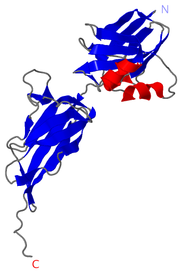
Asymmetric/Biological UnitChain A from PDB Type:PROTEIN Length:238 aligned with Q8E0S8_STRA5 | Q8E0S8 from UniProtKB/TrEMBL Length:307 Alignment length:238 39 49 59 69 79 89 99 109 119 129 139 149 159 169 179 189 199 209 219 229 239 249 259 Q8E0S8_STRA5 30 THQLTIVHLEARDIDRPNPQLEIAPKEGTPIEGVLYQLYQLKSTEDGDLLAHWNSLTITELKKQAQQVFEATTNQQGKATFNQLPDGIYYGLAVKAGEKNRNVSAFLVDLSEDKVIYPKIIWSTGELDLLKVGVDGDTKKPLAGVVFELYEKNGRTPIRVKNGVHSQDIDAAKHLETDSSGHIRISGLIHGDYVLKEIETQSGYQIGQAETAVTIEKSKTVTVTIENKKVPTPKVPSR 267 SCOP domains ---------------------------------------------------------------------------------------------------------------------------------------------------------------------------------------------------------------------------------------------- SCOP domains CATH domains ---------------------------------------------------------------------------------------------------------------------------------------------------------------------------------------------------------------------------------------------- CATH domains Pfam domains ---------------------------------------------------------------------------------------------------------------------------------------------Cna_B-3phsA01 A:171-253 -------------- Pfam domains SAPs(SNPs) ---------------------------------------------------------------------------------------------------------------------------------------------------------------------------------------------------------------------------------------------- SAPs(SNPs) PROSITE ---------------------------------------------------------------------------------------------------------------------------------------------------------------------------------------------------------------------------------------------- PROSITE Transcript ---------------------------------------------------------------------------------------------------------------------------------------------------------------------------------------------------------------------------------------------- Transcript 3phs A 30 SHQLTIVHLEARDIDRPNPQLEIAPKEGTPIEGVLYQLYQLKSTEDGDLLAHWNSLTITELKKQAQQVFEATTNQQGKATFNQLPDGIYYGLAVKAGEKNRNVSAFLVDLSEDKVIYPKIIWSTGELDLLKVGVDGDTKKPLAGVVFELYEKNGRTPIRVKNGVHSQDIDAAKHLETDSSGHIRISGLIHGDYVLKEIETQSGYQIGQAETAVTIEKSKTVTVTIENKKVPTPKVPSR 267 39 49 59 69 79 89 99 109 119 129 139 149 159 169 179 189 199 209 219 229 239 249 259
|
||||||||||||||||||||
SCOP Domains (0, 0)| (no "SCOP Domain" information available for 3PHS) |
CATH Domains (0, 0)| (no "CATH Domain" information available for 3PHS) |
Pfam Domains (1, 1)| Asymmetric/Biological Unit |
Gene Ontology (2, 2)|
Asymmetric/Biological Unit(hide GO term definitions) Chain A (Q8E0S8_STRA5 | Q8E0S8)
|
||||||||||||||||||
Interactive Views
|
||||||||||||||||||||||||||||||||||||||||||||||||||||||||||||||||||||||||||||||||||||||||||||||||||||||||||||||||||||
Still Images
|
||||||||||||||||
Databases
|
||||||||||||||||||||||||||||||||||||||||||||||||||||||||||||||||||||||||||||||||||||||||||||||||||||||||||||||||||||||||||||||||||||||||||||||||||||||||||||||||
Analysis Tools
|
|||||||||||||||||||||||||||||||||||||||||||||||||||||||||||||
Entries Sharing at Least One Protein Chain (UniProt ID)
Related Entries Specified in the PDB File
|
|