|
|
|
|
 Description
|
|
Compounds
|
||||||||||||||||||||||||
Chains, Units
Summary Information (see also Sequences/Alignments below) |
Ligands, Modified Residues, Ions (2, 3)| NMR Structure (2, 3) |
Sites (4, 4)
NMR Structure (4, 4)
|
SS Bonds (0, 0)| (no "SS Bond" information available for 3PAT) |
Cis Peptide Bonds (0, 0)| (no "Cis Peptide Bond" information available for 3PAT) |
SAPs(SNPs)/Variants (2, 2)
NMR Structure (2, 2)
|
|||||||||||||||||||||||||||||||||||||||||||||||||||||||||||||||||||||||
PROSITE Motifs (2, 4)
NMR Structure (2, 4)
|
||||||||||||||||||||||||||||||||
Exons (0, 0)| (no "Exon" information available for 3PAT) |
Sequences/Alignments
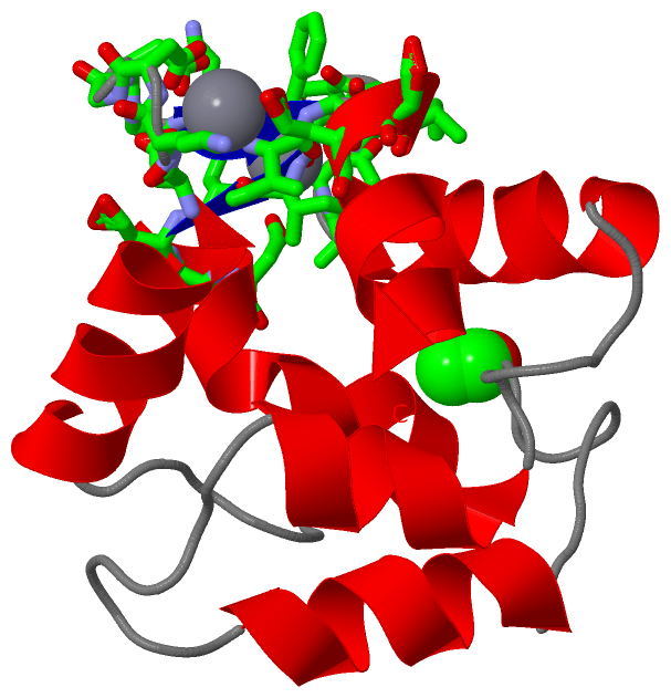
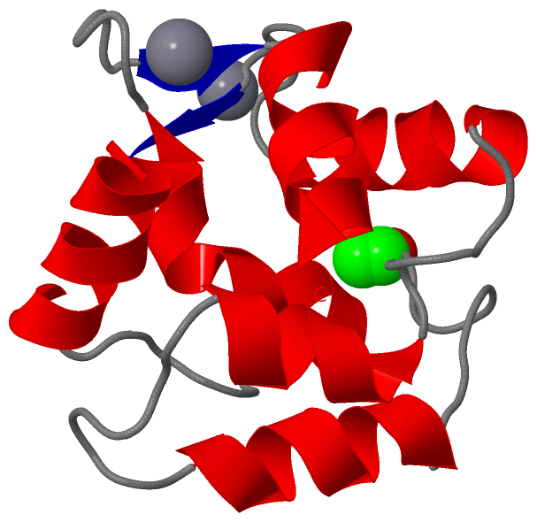
NMR StructureChain A from PDB Type:PROTEIN Length:110 aligned with PRVA_ESOLU | P02628 from UniProtKB/Swiss-Prot Length:108 Alignment length:110 1 | 8 18 28 38 48 58 68 78 88 98 108 PRVA_ESOLU - --AKDLLKADDIKKALDAVKAEGSFNHKKFFALVGLKAMSANDVKKVFKAIDADASGFIEEEELKFVLKSFAADGRDLTDAETKAFLKAADKDGDGKIGIDEFETLVHEA 108 SCOP domains d3pata_ A: Parvalbumin SCOP domains CATH domains -3patA00 A:1-109 EF-hand CATH domains Pfam domains ------------------------------------------efhand-3patA01 A:42-70 --------------------------------------- Pfam domains SAPs(SNPs) ----------------------------A---K----------------------------------------------------------------------------- SAPs(SNPs) PROSITE (1) --------------------------------------EF_HAND_2 PDB: A:38-73 ---EF_HAND_2 PDB: A:77-109 PROSITE (1) PROSITE (2) ---------------------------------------------------EF_HAND_1 --------------------------EF_HAND_1 ------- PROSITE (2) Transcript -------------------------------------------------------------------------------------------------------------- Transcript 3pat A 0 xAAKDLLKADDIKKALDAVKAEGSFNHKKFFALVGLKAMSANDVKKVFKAIDADASGFIEEEELKFVLKSFAADGRDLTDAETKAFLKAADKDGDGKIGIDEFETLVHEA 109 | 9 19 29 39 49 59 69 79 89 99 109 | 0-ACE
|
||||||||||||||||||||
SCOP Domains (1, 1)
NMR Structure
 |
CATH Domains (1, 1)
NMR Structure
|
Pfam Domains (1, 1)
NMR Structure
|
Gene Ontology (2, 2)|
NMR Structure(hide GO term definitions) Chain A (PRVA_ESOLU | P02628)
|
||||||||||||||||||
Interactive Views
|
||||||||||||||||||||||||||||||||||||||||||||||||||||||||||||||||||||||||||||||||||||||||||||||||||||||||||||||||||||||||||||||||||||||||||||||||||
Still Images
|
||||||||||||||||
Databases
|
||||||||||||||||||||||||||||||||||||||||||||||||||||||||||||||||||||||||||||||||||||||||||||||||||||||||||||||||||||||||||||||||||||||||||||||||||||||||||||||||
Analysis Tools
|
|||||||||||||||||||||||||||||||||||||||||||||||||||||||||||||
Entries Sharing at Least One Protein Chain (UniProt ID)
Related Entries Specified in the PDB File
|
|