|
|
|
|
 Description
|
|
Compounds
|
||||||||||||||||||||||||||||||||||||||||||||||||
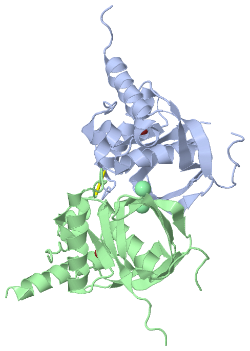
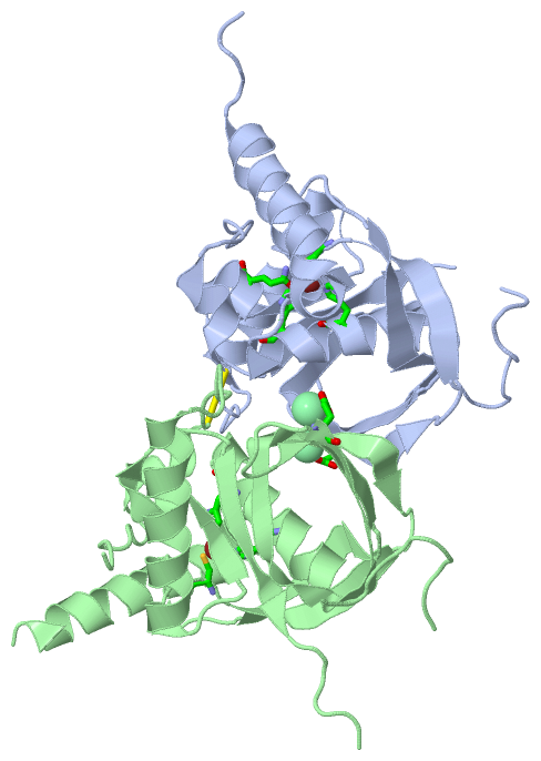
Chains, Units
Summary Information (see also Sequences/Alignments below) |
Ligands, Modified Residues, Ions (2, 4)| Asymmetric/Biological Unit (2, 4) |
Sites (4, 4)
Asymmetric Unit (4, 4)
|
SS Bonds (1, 1)
Asymmetric/Biological Unit
|
||||||||
Cis Peptide Bonds (4, 4)
Asymmetric/Biological Unit
|
||||||||||||||||||||
SAPs(SNPs)/Variants (0, 0)| (no "SAP(SNP)/Variant" information available for 3OCA) |
PROSITE Motifs (0, 0)| (no "PROSITE Motif" information available for 3OCA) |
Exons (0, 0)| (no "Exon" information available for 3OCA) |
Sequences/Alignments
Asymmetric/Biological UnitChain A from PDB Type:PROTEIN Length:179 aligned with Q2GI30_EHRCR | Q2GI30 from UniProtKB/TrEMBL Length:188 Alignment length:187 11 21 31 41 51 61 71 81 91 101 111 121 131 141 151 161 171 181 Q2GI30_EHRCR 2 SVLSIVTVPDKRLSLCSEEVEKVDQSIRKLVDDMFETMHANQGLGLAAVQVGVHKRILVMNVPEEFEDSEDIENVEDKIEGYELYGGPYCIINPKIVDISQEKVKLKEGCLSVPGYFDYIVRPQRIAVQYLDYNGNECIIKAQGWLARCLQHEIDHLNGTVFLKYLSKFKRDFAIEKVKKKERTDLI 188 SCOP domains d3ocaa_ A: automated matches SCOP domains CATH domains ------------------------------------------------------------------------------------------------------------------------------------------------------------------------------------------- CATH domains Pfam domains ------------------------------------------------------------------------------------------------------------------------------------------------------------------------------------------- Pfam domains SAPs(SNPs) ------------------------------------------------------------------------------------------------------------------------------------------------------------------------------------------- SAPs(SNPs) PROSITE ------------------------------------------------------------------------------------------------------------------------------------------------------------------------------------------- PROSITE Transcript ------------------------------------------------------------------------------------------------------------------------------------------------------------------------------------------- Transcript 3oca A 2 SVLSIVTVPDKRLSLCSEEVEKVDQSIRKLVDDMFETMHANQGLGLAAVQVGVHKRILVMNVPE--------ENVEDKIEGYELYGGPYCIINPKIVDISQEKVKLKEGCLSVPGYFDYIVRPQRIAVQYLDYNGNECIIKAQGWLARCLQHEIDHLNGTVFLKYLSKFKRDFAIEKVKKKERTDLI 188 11 21 31 41 51 61 | - | 81 91 101 111 121 131 141 151 161 171 181 65 74 Chain B from PDB Type:PROTEIN Length:179 aligned with Q2GI30_EHRCR | Q2GI30 from UniProtKB/TrEMBL Length:188 Alignment length:187 11 21 31 41 51 61 71 81 91 101 111 121 131 141 151 161 171 181 Q2GI30_EHRCR 2 SVLSIVTVPDKRLSLCSEEVEKVDQSIRKLVDDMFETMHANQGLGLAAVQVGVHKRILVMNVPEEFEDSEDIENVEDKIEGYELYGGPYCIINPKIVDISQEKVKLKEGCLSVPGYFDYIVRPQRIAVQYLDYNGNECIIKAQGWLARCLQHEIDHLNGTVFLKYLSKFKRDFAIEKVKKKERTDLI 188 SCOP domains d3ocab_ B: automated matches SCOP domains CATH domains ------------------------------------------------------------------------------------------------------------------------------------------------------------------------------------------- CATH domains Pfam domains (1) -Pep_deformylase-3ocaB01 B:3-173 --------------- Pfam domains (1) Pfam domains (2) -Pep_deformylase-3ocaB02 B:3-173 --------------- Pfam domains (2) SAPs(SNPs) ------------------------------------------------------------------------------------------------------------------------------------------------------------------------------------------- SAPs(SNPs) PROSITE ------------------------------------------------------------------------------------------------------------------------------------------------------------------------------------------- PROSITE Transcript ------------------------------------------------------------------------------------------------------------------------------------------------------------------------------------------- Transcript 3oca B 2 SVLSIVTVPDKRLSLCSEEVEKVDQSIRKLVDDMFETMHANQGLGLAAVQVGVHKRILVMNVPEEFED--------DKIEGYELYGGPYCIINPKIVDISQEKVKLKEGCLSVPGYFDYIVRPQRIAVQYLDYNGNECIIKAQGWLARCLQHEIDHLNGTVFLKYLSKFKRDFAIEKVKKKERTDLI 188 11 21 31 41 51 61 | - | 81 91 101 111 121 131 141 151 161 171 181 69 78
|
||||||||||||||||||||
SCOP Domains (1, 2)
Asymmetric/Biological Unit
 |
CATH Domains (0, 0)| (no "CATH Domain" information available for 3OCA) |
Pfam Domains (1, 2)
Asymmetric/Biological Unit
|
Gene Ontology (5, 5)|
Asymmetric/Biological Unit(hide GO term definitions) Chain A,B (Q2GI30_EHRCR | Q2GI30)
|
||||||||||||||||||||||||||||||||||||||||||
Interactive Views
|
||||||||||||||||||||||||||||||||||||||||||||||||||||||||||||||||||||||||||||||||||||||||||||||||||||||||||||||||||||||||||||||||||||||||||||||||||||||||||||||||||||||||
Still Images
|
||||||||||||||||
Databases
|
||||||||||||||||||||||||||||||||||||||||||||||||||||||||||||||||||||||||||||||||||||||||||||||||||||||||||||||||||||||||||||||||||||||||||||||||||||||||||||||||
Analysis Tools
|
|||||||||||||||||||||||||||||||||||||||||||||||||||||||||||||
Entries Sharing at Least One Protein Chain (UniProt ID)
Related Entries Specified in the PDB File
|
|