|
|
|
|
 Description
|
|
Compounds
|
||||||||||||||||||||||||||||||||||||||||||||||||
Chains, Units
Summary Information (see also Sequences/Alignments below) |
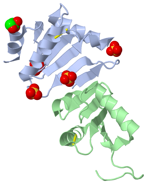
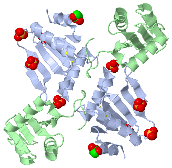
Ligands, Modified Residues, Ions (2, 5)| Asymmetric Unit (2, 5) Biological Unit 1 (2, 10) Biological Unit 2 (2, 10) |
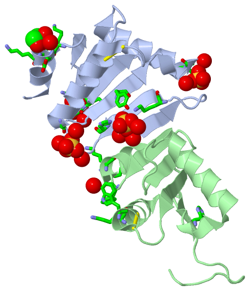
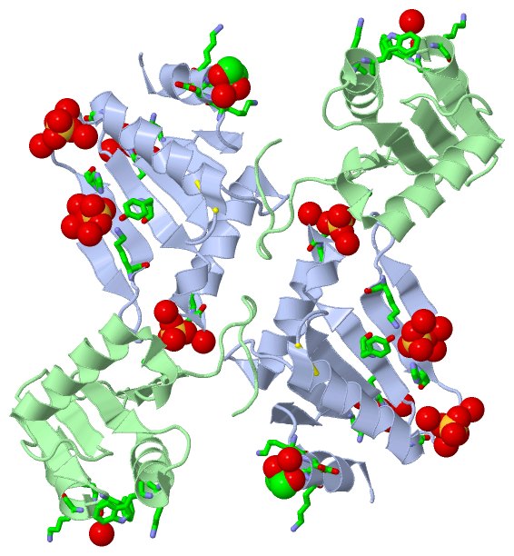
Sites (5, 5)
Asymmetric Unit (5, 5)
|
SS Bonds (2, 2)
Asymmetric Unit
|
||||||||||||
Cis Peptide Bonds (2, 2)
Asymmetric Unit
|
||||||||||||
SAPs(SNPs)/Variants (0, 0)| (no "SAP(SNP)/Variant" information available for 3NZN) |
PROSITE Motifs (0, 0)| (no "PROSITE Motif" information available for 3NZN) |
Exons (0, 0)| (no "Exon" information available for 3NZN) |
Sequences/Alignments
Asymmetric UnitChain A from PDB Type:PROTEIN Length:103 aligned with Q8PS17_METMA | Q8PS17 from UniProtKB/TrEMBL Length:100 Alignment length:103 1 | 7 17 27 37 47 57 67 77 87 97 Q8PS17_METMA - ---MNLFGQKDRGNHVSGVDRGKVIMYGLSTCVWCKKTKKLLTDLGVDFDYVYVDRLEGKEEEEAVEEVRRFNPSVSFPTTIINDEKAIVGFKEKEIRESLGF 100 SCOP domains ------------------------------------------------------------------------------------------------------- SCOP domains CATH domains ------------------------------------------------------------------------------------------------------- CATH domains Pfam domains ------------------------------------------------------------------------------------------------------- Pfam domains SAPs(SNPs) ------------------------------------------------------------------------------------------------------- SAPs(SNPs) PROSITE ------------------------------------------------------------------------------------------------------- PROSITE Transcript ------------------------------------------------------------------------------------------------------- Transcript 3nzn A 1 SNAVNLFGQKDRGNHVSGVDRGKVIMYGLSTCVWCKKTKKLLTDLGVDFDYVYVDRLEGKEEEEAVEEVRRFNPSVSFPTTIINDEKAIVGFKEKEIRESLGF 103 10 20 30 40 50 60 70 80 90 100 Chain B from PDB Type:PROTEIN Length:103 aligned with Q8PS17_METMA | Q8PS17 from UniProtKB/TrEMBL Length:100 Alignment length:103 1 | 7 17 27 37 47 57 67 77 87 97 Q8PS17_METMA - ---MNLFGQKDRGNHVSGVDRGKVIMYGLSTCVWCKKTKKLLTDLGVDFDYVYVDRLEGKEEEEAVEEVRRFNPSVSFPTTIINDEKAIVGFKEKEIRESLGF 100 SCOP domains ------------------------------------------------------------------------------------------------------- SCOP domains CATH domains ------------------------------------------------------------------------------------------------------- CATH domains Pfam domains (1) -----------------------Glutaredoxin-3nznB01 B:24-88 --------------- Pfam domains (1) Pfam domains (2) -----------------------Glutaredoxin-3nznB02 B:24-88 --------------- Pfam domains (2) SAPs(SNPs) ------------------------------------------------------------------------------------------------------- SAPs(SNPs) PROSITE ------------------------------------------------------------------------------------------------------- PROSITE Transcript ------------------------------------------------------------------------------------------------------- Transcript 3nzn B 1 SNAVNLFGQKDRGNHVSGVDRGKVIMYGLSTCVWCKKTKKLLTDLGVDFDYVYVDRLEGKEEEEAVEEVRRFNPSVSFPTTIINDEKAIVGFKEKEIRESLGF 103 10 20 30 40 50 60 70 80 90 100
|
||||||||||||||||||||
SCOP Domains (0, 0)| (no "SCOP Domain" information available for 3NZN) |
CATH Domains (0, 0)| (no "CATH Domain" information available for 3NZN) |
Pfam Domains (1, 2)
Asymmetric Unit
 |
Gene Ontology (5, 5)|
Asymmetric Unit(hide GO term definitions) Chain A,B (Q8PS17_METMA | Q8PS17)
|
||||||||||||||||||||||||||||||||||||||||||||||||
Interactive Views
|
||||||||||||||||||||||||||||||||||||||||||||||||||||||||||||||||||||||||||||||||||||||||||||||||||||||||||||||||||||||||||||||||||||||||||||||||||||||||||||||||||||||||||||||||||||||||
Still Images
|
||||||||||||||||
Databases
|
||||||||||||||||||||||||||||||||||||||||||||||||||||||||||||||||||||||||||||||||||||||||||||||||||||||||||||||||||||||||||||||||||||||||||||||||||||||||||||||||
Analysis Tools
|
|||||||||||||||||||||||||||||||||||||||||||||||||||||||||||||
Entries Sharing at Least One Protein Chain (UniProt ID)
Related Entries Specified in the PDB File
|
|