Show PDB file:   
         Plain Text   HTML   (compressed file size)
QuickSearch:   
by PDB,NDB,UniProt,PROSITE Code or Search Term(s)  
(-)Asym./Biol. Unit
(-)Asym./Biol. Unit - sites
collapse expand < >
Image Asym./Biol. Unit
Asym./Biol. Unit  (Jmol Viewer)
Image Asym./Biol. Unit - sites
Asym./Biol. Unit - sites  (Jmol Viewer)

(-) Description

Title :  CRYSTAL STRUCTURE OF SM1, AN ELICITOR OF PLANT DEFENCE RESPONSES FROM TRICHODERMA VIRENS.
 
Authors :  I. V. Krieger, W. A. Vargas, C. M. Kenerley, J. C. Sacchettini
Date :  09 Mar 10  (Deposition) - 09 Feb 11  (Release) - 09 Feb 11  (Revision)
Method :  X-RAY DIFFRACTION
Resolution :  1.39
Chains :  Asym./Biol. Unit :  A
Keywords :  Fungal, Plant Defense, Fungus, Polysaccharide-Binding Protein (Keyword Search: [Gene Ontology, PubMed, Web (Google))
 
Reference :  I. V. Krieger, W. A. Vargas, C. M. Kenerley, J. C. Sacchettini
Crystal Structure Of Sm1, An Elicitor Of Plant Defence Responses From Trichoderma Virens.
To Be Published
PubMed: search

(-) Compounds

Molecule 1 - EPL1 PROTEIN
    ChainsA
    Organism CommonTRICHODERMA VIRENS
    Organism ScientificHYPOCREA VIRENS
    Organism Taxid413071
    Other DetailsSECRETED
    StrainGV29-8
    SynonymEXTRACELLULAR SMALL PROTEIN 1

 Structural Features

(-) Chains, Units

  1
Asymmetric/Biological Unit A

Summary Information (see also Sequences/Alignments below)

(-) Ligands, Modified Residues, Ions  (3, 16)

Asymmetric/Biological Unit (3, 16)
No.NameCountTypeFull Name
1BR2Ligand/IonBROMIDE ION
2EDO13Ligand/Ion1,2-ETHANEDIOL
3ZN1Ligand/IonZINC ION

(-) Sites  (16, 16)

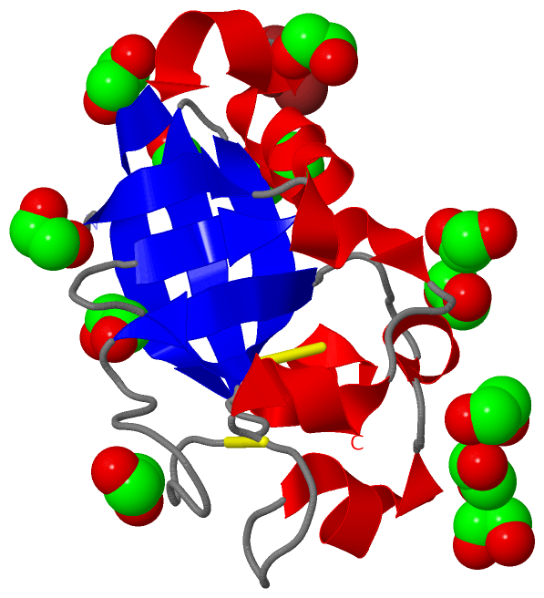
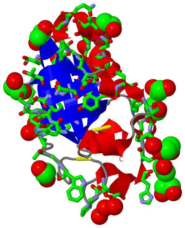
Asymmetric Unit (16, 16)
No.NameEvidenceResiduesDescription
01AC1SOFTWAREHIS A:89 , HIS A:98 , ZN A:139BINDING SITE FOR RESIDUE BR A 1
02AC2SOFTWAREEDO A:6 , HIS A:89 , ZN A:139BINDING SITE FOR RESIDUE BR A 2
03AC3SOFTWAREBR A:1 , BR A:2 , HIS A:89 , HIS A:98BINDING SITE FOR RESIDUE ZN A 139
04AC4SOFTWAREEDO A:8 , ASP A:19 , THR A:20 , GLY A:102BINDING SITE FOR RESIDUE EDO A 140
05AC5SOFTWAREEDO A:3 , EDO A:4 , SER A:31 , HIS A:89 , THR A:90 , ILE A:91 , ALA A:112BINDING SITE FOR RESIDUE EDO A 141
06AC6SOFTWAREEDO A:4 , ASP A:28 , GLY A:30 , SER A:101 , EDO A:141 , HOH A:178BINDING SITE FOR RESIDUE EDO A 3
07AC7SOFTWAREEDO A:3 , GLY A:88 , HIS A:89 , THR A:90 , ALA A:99 , ALA A:100 , SER A:101 , ASN A:104 , EDO A:141BINDING SITE FOR RESIDUE EDO A 4
08AC8SOFTWAREASN A:111 , GLY A:116 , ALA A:118 , VAL A:119 , GLN A:120BINDING SITE FOR RESIDUE EDO A 5
09AC9SOFTWAREBR A:2 , EDO A:11 , PRO A:43 , SER A:53 , THR A:54 , GLN A:57 , ALA A:71 , HOH A:175 , HOH A:196BINDING SITE FOR RESIDUE EDO A 6
10BC1SOFTWAREASP A:24 , TYR A:27 , TYR A:86 , SER A:87 , GLY A:88 , ASN A:104BINDING SITE FOR RESIDUE EDO A 7
11BC2SOFTWAREGLY A:56 , PRO A:59 , PHE A:61 , ALA A:68 , SER A:101 , GLY A:102 , EDO A:140BINDING SITE FOR RESIDUE EDO A 8
12BC3SOFTWAREASP A:41 , THR A:48 , ALA A:71 , GLY A:72 , TRP A:73 , HOH A:187 , HOH A:189 , HOH A:196BINDING SITE FOR RESIDUE EDO A 9
13BC4SOFTWAREEDO A:11 , ASP A:41 , HIS A:51BINDING SITE FOR RESIDUE EDO A 10
14BC5SOFTWAREEDO A:6 , EDO A:10 , ASP A:41 , GLY A:42 , HIS A:51 , SER A:53 , GLN A:57 , ALA A:71 , HOH A:187 , HOH A:196BINDING SITE FOR RESIDUE EDO A 11
15BC6SOFTWARETYR A:86 , LEU A:121 , SER A:125BINDING SITE FOR RESIDUE EDO A 12
16BC7SOFTWAREASP A:19 , ALA A:68 , ALA A:69 , TRP A:82 , PHE A:103 , HOH A:193BINDING SITE FOR RESIDUE EDO A 13

(-) SS Bonds  (2, 2)

Asymmetric/Biological Unit
No.Residues
1A:39 -A:78
2A:81 -A:136

(-) Cis Peptide Bonds  (1, 1)

Asymmetric/Biological Unit
No.Residues
1Phe A:61 -Pro A:62

 Sequence-Structure Mapping

(-) SAPs(SNPs)/Variants  (0, 0)

(no "SAP(SNP)/Variant" information available for 3M3G)

(-) PROSITE Motifs  (0, 0)

(no "PROSITE Motif" information available for 3M3G)

(-) Exons   (0, 0)

(no "Exon" information available for 3M3G)

(-) Sequences/Alignments

Asymmetric/Biological Unit
   Reformat: Number of residues per line =  ('0' or empty: single-line sequence representation)
  Number of residues per labelling interval =   
  UniProt sequence: complete  aligned part    
   Show mapping: SCOP domains CATH domains Pfam domains Secondary structure (by author)
SAPs(SNPs) PROSITE motifs Exons
(details for a mapped element are shown in a popup box when the mouse pointer rests over it)
Chain A from PDB  Type:PROTEIN  Length:120
 aligned with Q0R411_HYPVI | Q0R411 from UniProtKB/TrEMBL  Length:138

    Alignment length:120
                                    28        38        48        58        68        78        88        98       108       118       128       138
         Q0R411_HYPVI    19 DTVSYDTGYDNGSRSLNDVSCSDGPNGLETRYHWSTQGQIPRFPYIGGAAAVAGWNSASCGTCWKLQYSGHTIYVLAVDHAASGFNIALDAMNALTGGQAVQLGRVSATATQVPVKNCGL 138
               SCOP domains ------------------------------------------------------------------------------------------------------------------------ SCOP domains
               CATH domains ------------------------------------------------------------------------------------------------------------------------ CATH domains
               Pfam domains Cerato-platanin-3m3gA01 A:19-138                                                                                         Pfam domains
         Sec.struct. author .eeee.hhhhh...hhhhh.......hhhhhhh..hhhhh.....eeee.............eeeeee..eeeeeeeeee....eeehhhhhhhhhh.hhhhhheee.eeeeehhhhhh. Sec.struct. author
                 SAPs(SNPs) ------------------------------------------------------------------------------------------------------------------------ SAPs(SNPs)
                    PROSITE ------------------------------------------------------------------------------------------------------------------------ PROSITE
                 Transcript ------------------------------------------------------------------------------------------------------------------------ Transcript
                 3m3g A  19 DTVSYDTGYDNGSRSLNDVSCSDGPNGLETRYHWSTQGQIPRFPYIGGAAAVAGWNSASCGTCWKLQYSGHTIYVLAVDHAASGFNIALDAMNALTGGQAVQLGRVSATATQVPVKNCGL 138
                                    28        38        48        58        68        78        88        98       108       118       128       138

   Legend:   → Mismatch (orange background)
  - → Gap (green background, '-', border residues have a numbering label)
    → Modified Residue (blue background, lower-case, 'x' indicates undefined single-letter code, labelled with number + name)
  x → Chemical Group (purple background, 'x', labelled with number + name, e.g. ACE or NH2)
  extra numbering lines below/above indicate numbering irregularities and modified residue names etc., number ends below/above '|'

 Classification and Annotation

(-) SCOP Domains  (0, 0)

(no "SCOP Domain" information available for 3M3G)

(-) CATH Domains  (0, 0)

(no "CATH Domain" information available for 3M3G)

(-) Pfam Domains  (1, 1)

Asymmetric/Biological Unit
(-)
Clan: DPBB (9)

(-) Gene Ontology  (0, 0)

Asymmetric/Biological Unit(hide GO term definitions)
    (no "Gene Ontology" information available for 3M3G)

 Visualization

(-) Interactive Views

Asymmetric/Biological Unit
  Complete Structure
    Jena3D(integrated viewing of ligand, site, SAP, PROSITE, SCOP information)
    WebMol | AstexViewer[tm]@PDBe
(Java Applets, require no local installation except for Java; loading may be slow)
    STRAP
(Java WebStart application, automatic local installation, requires Java; full application with system access!)
    RasMol
(require local installation)
    Molscript (VRML)
(requires installation of a VRML viewer; select preferred view via VRML and generate a mono or stereo PDF format file)
 
  Ligands, Modified Residues, Ions
    BR  [ RasMol | Jena3D ]  +environment [ RasMol | Jena3D ]
    EDO  [ RasMol | Jena3D ]  +environment [ RasMol | Jena3D ]
    ZN  [ RasMol | Jena3D ]  +environment [ RasMol | Jena3D ]
 
  Sites
    AC1  [ RasMol ]  +environment [ RasMol ]
    AC2  [ RasMol ]  +environment [ RasMol ]
    AC3  [ RasMol ]  +environment [ RasMol ]
    AC4  [ RasMol ]  +environment [ RasMol ]
    AC5  [ RasMol ]  +environment [ RasMol ]
    AC6  [ RasMol ]  +environment [ RasMol ]
    AC7  [ RasMol ]  +environment [ RasMol ]
    AC8  [ RasMol ]  +environment [ RasMol ]
    AC9  [ RasMol ]  +environment [ RasMol ]
    BC1  [ RasMol ]  +environment [ RasMol ]
    BC2  [ RasMol ]  +environment [ RasMol ]
    BC3  [ RasMol ]  +environment [ RasMol ]
    BC4  [ RasMol ]  +environment [ RasMol ]
    BC5  [ RasMol ]  +environment [ RasMol ]
    BC6  [ RasMol ]  +environment [ RasMol ]
    BC7  [ RasMol ]  +environment [ RasMol ]
 
  Cis Peptide Bonds
    Phe A:61 - Pro A:62   [ RasMol ]  
 

(-) Still Images

Jmol
  protein: cartoon or spacefill or dots and stick; nucleic acid: cartoon and stick; ligands: spacefill; active site: stick
Molscript
  protein, nucleic acid: cartoon; ligands: spacefill; active site: ball and stick

 Databases and Analysis Tools

(-) Databases

Access by PDB/NDB ID
  3m3g
    Family and Domain InformationProDom | SYSTERS
    General Structural InformationGlycoscienceDB | MMDB | NDB | OCA | PDB | PDBe | PDBj | PDBsum | PDBWiki | PQS | PROTEOPEDIA
    Orientation in MembranesOPM
    Protein SurfaceSURFACE
    Secondary StructureDSSP (structure derived) | HSSP (homology derived)
    Structural GenomicsGeneCensus
    Structural NeighboursCE | VAST
    Structure ClassificationCATH | Dali | SCOP
    Validation and Original DataBMRB Data View | BMRB Restraints Grid | EDS | PROCHECK | RECOORD | WHAT_CHECK
 
Access by UniProt ID/Accession number
  Q0R411_HYPVI | Q0R411
    Comparative Protein Structure ModelsModBase
    Genomic InformationEnsembl
    Protein-protein InteractionDIP
    Sequence, Family and Domain InformationInterPro | Pfam | SMART | UniProtKB/TrEMBL
 
Access by Enzyme Classificator   (EC Number)
  (no 'Enzyme Classificator' available)
    General Enzyme InformationBRENDA | EC-PDB | Enzyme | IntEnz
    PathwayKEGG | MetaCyc
 
Access by Disease Identifier   (MIM ID)
  (no 'MIM ID' available)
    Disease InformationOMIM
 
Access by GenAge ID
  (no 'GenAge ID' available)
    Age Related InformationGenAge

(-) Analysis Tools

Access by PDB/NDB ID
    Domain InformationXDom
    Interatomic Contacts of Structural UnitsCSU
    Ligand-protein ContactsLPC
    Protein CavitiescastP
    Sequence and Secondary StructurePDBCartoon
    Structure AlignmentSTRAP(Java WebStart application, automatic local installation, requires Java; full application with system access!)
    Structure and Sequence BrowserSTING
 
Access by UniProt ID/Accession number
  Q0R411_HYPVI | Q0R411
    Protein Disorder PredictionDisEMBL | FoldIndex | GLOBPLOT (for more information see DisProt)

 Related Entries

(-) Entries Sharing at Least One Protein Chain (UniProt ID)

(no "Entries Sharing at Least One Protein Chain" available for 3M3G)

(-) Related Entries Specified in the PDB File

(no "Related Entries Specified in the PDB File" available for 3M3G)