|
|
|
|
 Description
|
|
Compounds
|
||||||||||||||||||||||||||||||||||||||||||||||||||||
Chains, Units
Summary Information (see also Sequences/Alignments below) |
Ligands, Modified Residues, Ions (2, 6)| Asymmetric/Biological Unit (2, 6) |
Sites (3, 3)
Asymmetric Unit (3, 3)
|
SS Bonds (0, 0)| (no "SS Bond" information available for 3LWX) |
Cis Peptide Bonds (0, 0)| (no "Cis Peptide Bond" information available for 3LWX) |
SAPs(SNPs)/Variants (0, 0)| (no "SAP(SNP)/Variant" information available for 3LWX) |
PROSITE Motifs (0, 0)| (no "PROSITE Motif" information available for 3LWX) |
Exons (0, 0)| (no "Exon" information available for 3LWX) |
Sequences/Alignments
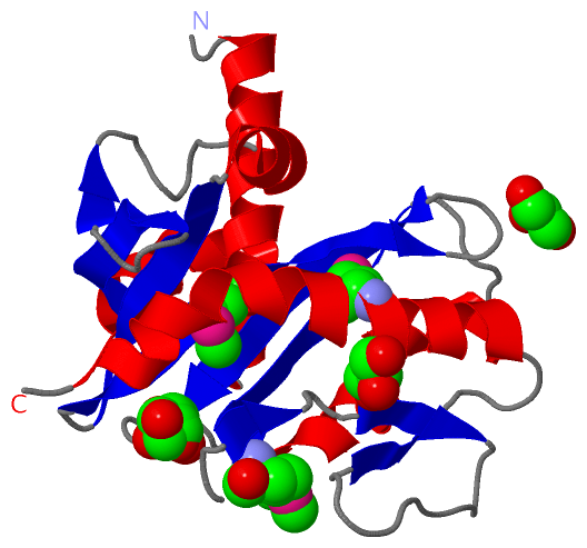
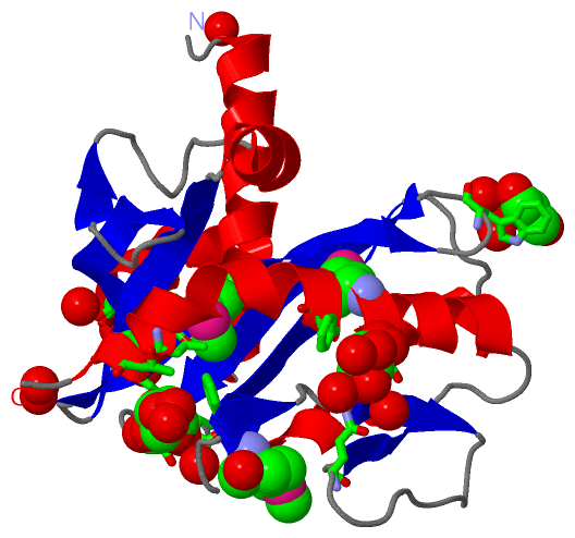
Asymmetric/Biological UnitChain A from PDB Type:PROTEIN Length:199 aligned with A6LB22_PARD8 | A6LB22 from UniProtKB/TrEMBL Length:270 Alignment length:199 81 91 101 111 121 131 141 151 161 171 181 191 201 211 221 231 241 251 261 A6LB22_PARD8 72 SQSLRSFQKQNEDNDKRQQILRSINVNVSSSEAETKYNELIKEAFLVNENGEKVEGDAFATDVVKAATEHQYPVFVANVDGQPKYIMALHGAGLWGPLWGYISVDSDKNTIYGADFSHQGETPGLGAEISKPVFSNEFKGKKIFMSGEFKSVAVVKPGKSVAGQDYVDGISGGTITSKGVDEMLFNSLSGYVKFLTSQN 270 SCOP domains ------------------------------------------------------------------------------------------------------------------------------------------------------------------------------------------------------- SCOP domains CATH domains ------------------------------------------------------------------------------------------------------------------------------------------------------------------------------------------------------- CATH domains Pfam domains --------------------------------------------------------------------------------------------FMN_bind-3lwxA01 A:164-262 -------- Pfam domains SAPs(SNPs) ------------------------------------------------------------------------------------------------------------------------------------------------------------------------------------------------------- SAPs(SNPs) PROSITE ------------------------------------------------------------------------------------------------------------------------------------------------------------------------------------------------------- PROSITE Transcript ------------------------------------------------------------------------------------------------------------------------------------------------------------------------------------------------------- Transcript 3lwx A 0 GQSLRSFQKQNEDNDKRQQILRSINVNVSSSEAETKYNELIKEAFLVNENGEKVEGDAFATDVVKAATEHQYPVFVANVDGQPKYImALHGAGLWGPLWGYISVDSDKNTIYGADFSHQGETPGLGAEISKPVFSNEFKGKKIFmSGEFKSVAVVKPGKSVAGQDYVDGISGGTITSKGVDEmLFNSLSGYVKFLTSQN 270 || 81 91 101 111 121 131 141 151 |161 171 181 191 201 211 | 221 231 241 251 | 261 || 158-MSE 216-MSE 254-MSE 0| 73
|
||||||||||||||||||||
SCOP Domains (0, 0)| (no "SCOP Domain" information available for 3LWX) |
CATH Domains (0, 0)| (no "CATH Domain" information available for 3LWX) |
Pfam Domains (1, 1)
Asymmetric/Biological Unit
|
Gene Ontology (12, 12)|
Asymmetric/Biological Unit(hide GO term definitions) Chain A (A6LB22_PARD8 | A6LB22)
|
||||||||||||||||||||||||||||||||||||||||||||||||||||||||||||||||||||||||||||||||||||||||||
Interactive Views
|
|||||||||||||||||||||||||||||||||||||||||||||||||||||||||||||||||||||||||||||||||||||||||||||||||||||||||||||||||||||||||||||||||||||||||||
Still Images
|
||||||||||||||||
Databases
|
||||||||||||||||||||||||||||||||||||||||||||||||||||||||||||||||||||||||||||||||||||||||||||||||||||||||||||||||||||||||||||||||||||||||||||||||||||||||||||||||
Analysis Tools
|
|||||||||||||||||||||||||||||||||||||||||||||||||||||||||||||
Entries Sharing at Least One Protein Chain (UniProt ID)
Related Entries Specified in the PDB File
|
|